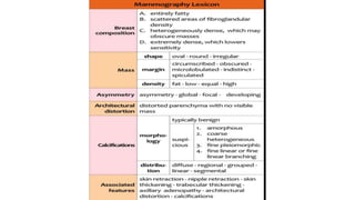
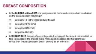
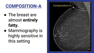
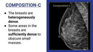
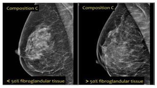
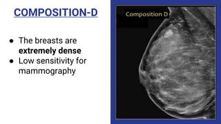
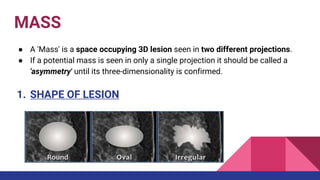
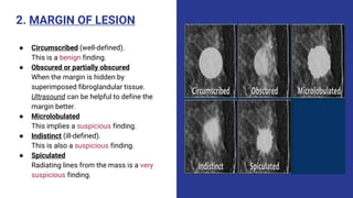
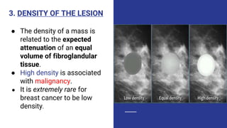
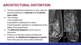
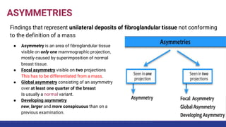
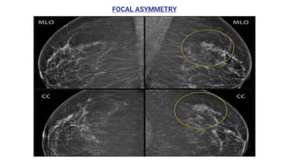
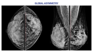
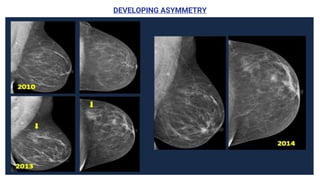
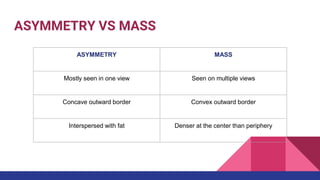
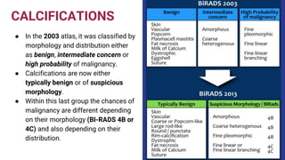
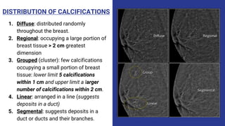
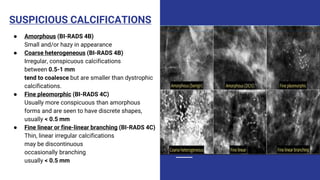
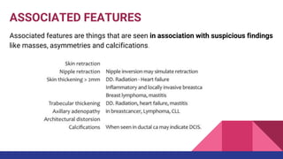
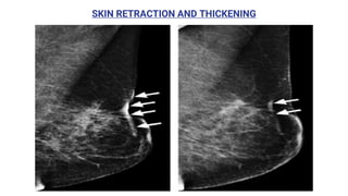
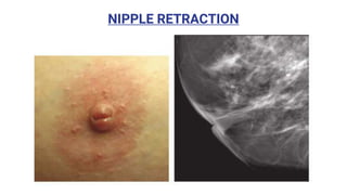
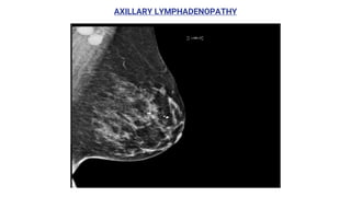
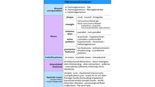
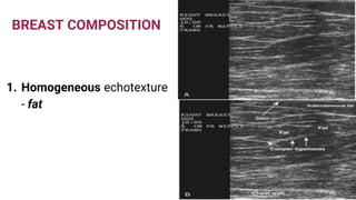
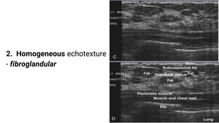
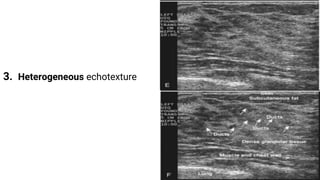
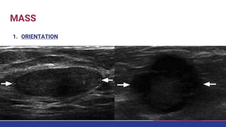
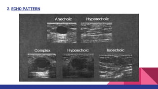
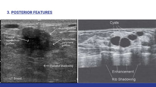
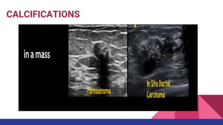
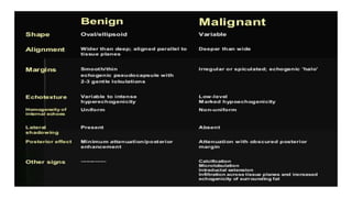
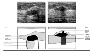
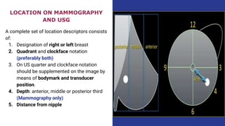
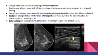
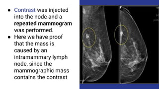
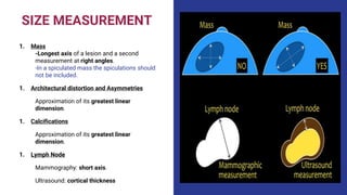
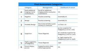
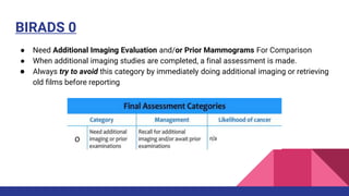
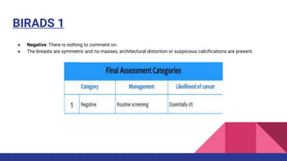
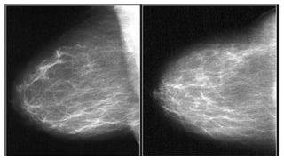
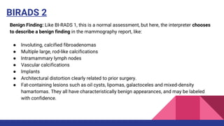
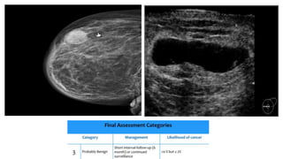
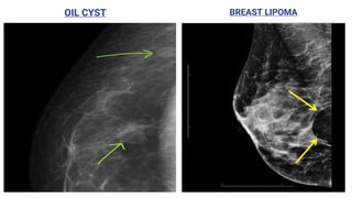
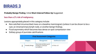
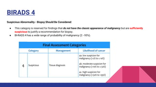
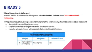
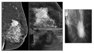
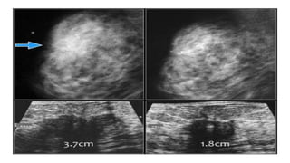
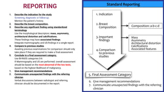
SlideShare a Scribd company logo
BIRADS FOR MAMMO AND
SONOMAMMOGRAM
PRESENTED BY: DR.GEORGINA GEORGE
MODERATOR: DR. MAHESH MIJAR
INTRODUCTION
● BI-RADS® is designed to standardize breast
imaging reporting and to reduce confusion
in breast imaging interpretations.
● It also facilitates outcome monitoring and
quality assessment.
● It contains a lexicon for standardized
terminology (descriptors) for
mammography, breast US and MRI, as well
as chapters on Report Organization and
Guidance Chapters for use in daily practice.
MAMMOGRAPHY LEXICON
BREAST COMPOSITION
● In the BI-RADS edition 2003 the assignment of the breast composition was based
on the overall density resulting in
❖ category 1 ( <25% fibroglandular tissue)
❖ category 2 ( 25-50%)
❖ category 3 (50-75%)
❖ category 4 (>75%).
● In BI-RADS 2013 the use of percentages is discouraged, because it is important to
take into account the chance that a mass can be obscured by fibroglandular
tissue than the percentage of breast density as an indicator.
COMPOSITION-A
● The breast are
almost entirely
fatty.
● Mammography is
highly sensitive in
this setting
COMPOSITION-B
● There are scattered
areas of fibroglandular
density.
● The term density
describes the degree of
x-ray attenuation of
breast tissue but not
discrete mammographic
findings.
COMPOSITION-C
● The breasts are
heterogeneously
dense.
● Some areas in the
breasts are
sufficiently dense to
obscure small
masses.
COMPOSITION-D
● The breasts are
extremely dense
● Low sensitivity for
mammography
MASS
● A 'Mass' is a space occupying 3D lesion seen in two different projections.
● If a potential mass is seen in only a single projection it should be called a
'asymmetry' until its three-dimensionality is confirmed.
1. SHAPE OF LESION
2. MARGIN OF LESION
● Circumscribed (well-defined).
This is a benign finding.
● Obscured or partially obscured
When the margin is hidden by
superimposed fibroglandular tissue.
Ultrasound can be helpful to define the
margin better.
● Microlobulated
This implies a suspicious finding.
● Indistinct (ill-defined).
This is also a suspicious finding.
● Spiculated
Radiating lines from the mass is a very
suspicious finding.
3. DENSITY OF THE LESION
● The density of a mass is
related to the expected
attenuation of an equal
volume of fibroglandular
tissue.
● High density is associated
with malignancy.
● It is extremely rare for
breast cancer to be low
density.
ARCHITECTURAL DISTORTION
● The term architectural distortion is used, when the
normal architecture is distorted with no definite
mass visible.
● This includes:
1.thin straight lines
2.spiculations radiating from a point
3.focal retraction, distortion or straightening at the
edges of the parenchyma.
● The differential diagnosis is scar tissue or
carcinoma.
● If there is a mass that causes architectural
distortion, the likelihood of malignancy is greater
than in the case of a mass without distortion.
ASYMMETRIES
Findings that represent unilateral deposits of fibroglandular tissue not conforming
to the definition of a mass
● Asymmetry is an area of fibroglandular tissue
visible on only one mammographic projection,
mostly caused by superimposition of normal
breast tissue.
● Focal asymmetry visible on two projections
This has to be differentiated from a mass.
● Global asymmetry consisting of an asymmetry
over at least one quarter of the breast
Is usually a normal variant.
● Developing asymmetry
new, larger and more conspicuous than on a
previous examination.
FOCAL ASYMMETRY
GLOBAL ASYMMETRY
DEVELOPING ASYMMETRY
ASYMMETRY VS MASS
ASYMMETRY MASS
Mostly seen in one view Seen on multiple views
Concave outward border Convex outward border
Interspersed with fat Denser at the center than periphery
CALCIFICATIONS
● In the 2003 atlas, it was classified by
morphology and distribution either
as benign, intermediate concern or
high probability of malignancy.
● Calcifications are now either
typically benign or of suspicious
morphology.
● Within this last group the chances of
malignancy are different depending
on their morphology (BI-RADS 4B or
4C) and also depending on their
distribution.
DISTRIBUTION OF CALCIFICATIONS
1. Diffuse: distributed randomly
throughout the breast.
2. Regional: occupying a large portion of
breast tissue > 2 cm greatest
dimension
3. Grouped (cluster): few calcifications
occupying a small portion of breast
tissue: lower limit 5 calcifications
within 1 cm and upper limit a larger
number of calcifications within 2 cm.
4. Linear: arranged in a line (suggests
deposits in a duct)
5. Segmental: suggests deposits in a
duct or ducts and their branches.
TYPICALLY BENIGN CALCIFICATIONS
There is one exception to
the rule:
An isolated group of
punctate calcifications that
is new, increasing, linear, or
segmental in distribution, or
adjacent to a known cancer
can be assigned as probably
benign or suspicious.
SUSPICIOUS CALCIFICATIONS
● Amorphous (BI-RADS 4B)
Small and/or hazy in appearance
● Coarse heterogeneous (BI-RADS 4B)
Irregular, conspicuous calcifications
between 0.5-1 mm
tend to coalesce but are smaller than dystrophic
calcifications.
● Fine pleomorphic (BI-RADS 4C)
Usually more conspicuous than amorphous
forms and are seen to have discrete shapes,
usually < 0.5 mm
● Fine linear or fine-linear branching (BI-RADS 4C)
Thin, linear irregular calcifications
may be discontinuous
occasionally branching
usually < 0.5 mm
ASSOCIATED FEATURES
Associated features are things that are seen in association with suspicious findings
like masses, asymmetries and calcifications.
SKIN RETRACTION AND THICKENING
NIPPLE RETRACTION
AXILLARY LYMPHADENOPATHY
ULTRASOUND LEXICON
BREAST COMPOSITION
1. Homogeneous echotexture
- fat
2. Homogeneous echotexture
- fibroglandular
3. Heterogeneous echotexture
MASS
1. ORIENTATION
2. ECHO PATTERN
3. POSTERIOR FEATURES
CALCIFICATIONS
LOCATION ON MAMMO AND
SONOMAMMOGRAPHY
LOCATION ON MAMMOGRAPHY
AND USG
A complete set of location descriptors consists
of:
1. Designation of right or left breast
2. Quadrant and clockface notation
(preferably both)
3. On US quarter and clockface notation
should be supplemented on the image by
means of bodymark and transducer
position.
4. Depth: anterior, middle or posterior third
(Mammography only)
5. Distance from nipple
● Always make sure, that you are dealing with the same lesion.
For instance a lesion found with US does not have to be the same as the mammographic or physical
finding.
● Sometimes repeated mammographic imaging with markers on the lesion found with US can be helpful.
● Cysts can be aspirated or filled with air after aspiration to make sure that the lesion found on the
mammogram is caused by a cyst.
● Solid lesions can be injected with contrast or a marker can be placed in difficult cases.
● Contrast was injected
into the node and a
repeated mammogram
was performed.
● Here we have proof
that the mass is
caused by an
intramammary lymph
node, since the
mammographic mass
contains the contrast
SIZE MEASUREMENT
1. Mass
-Longest axis of a lesion and a second
measurement at right angles.
-In a spiculated mass the spiculations should
not be included.
1. Architectural distortion and Asymmetries
Approximation of its greatest linear
dimension.
1. Calcifications
Approximation of its greatest linear
dimension.
1. Lymph Node
Mammography: short axis.
Ultrasound: cortical thickness
ANNUAL MAMMOGRAMS VS. ANNUAL ULTRASOUNDS
FINAL ASSESSMENT
CATEGORIES
BIRADS 0
● Need Additional Imaging Evaluation and/or Prior Mammograms For Comparison
● When additional imaging studies are completed, a final assessment is made.
● Always try to avoid this category by immediately doing additional imaging or retrieving
old films before reporting.
BIRADS 1
● Negative: There is nothing to comment on.
● The breasts are symmetric and no masses, architectural distortion or suspicious calcifications are present.
BIRADS 2
Benign Finding: Like BI-RADS 1, this is a normal assessment, but here, the interpreter chooses
to describe a benign finding in the mammography report, like:
● Involuting, calcified fibroadenomas
● Multiple large, rod-like calcifications
● Intramammary lymph nodes
● Vascular calcifications
● Implants
● Architectural distortion clearly related to prior surgery.
● Fat-containing lesions such as oil cysts, lipomas, galactoceles and mixed-density
hamartomas. They all have characteristically benign appearances, and may be labeled
with confidence.
OIL CYST BREAST LIPOMA
BIRADS 3
Probably Benign Finding- Initial Short-Interval Follow-Up Suggested:
less than a 2% risk of malignancy.
Lesions appropriately placed in this category include:
● Non-calcified circumscribed mass on a baseline mammogram (unless it can be shown to be a
cyst, an intramammary lymph node, or another benign finding),
● Focal asymmetry which becomes less dense on spot compression view
● Solitary group of punctate calcifications
BIRADS 4
Suspicious Abnormality - Biopsy Should Be Considered:
● This category is reserved for findings that do not have the classic appearance of malignancy but are sufficiently
suspicious to justify a recommendation for biopsy.
● BI-RADS 4 has a wide range of probability of malignancy (2 - 95%).
BIRADS 5
Highly Suggestive of Malignancy.
BI-RADS 5 must be reserved for findings that are classic breast cancers, with a >95% likelihood of
malignancy.
if the percutaneous tissue diagnosis is nonmalignant, this automatically should be considered as discordant.
● Spiculated, irregular high density mass.
● Segmental or linear arrangement of fine linear calcifications.
● Irregular spiculated mass with associated pleomorphic calcifications.
BIRADS 6
● Use after incomplete excision
● Use after monitoring response to neoadjuvant chemotherapy
REPORTING
1. Describe the indication for the study.
Screening, diagnostic or follow-up.
Mention the patient's history.
2. Describe the breast composition.
3. Describe any significant finding using standardized
terminology.
Use the morphological descriptors: mass, asymmetry,
architectural distortion and calcifications.
These findings may have associated findings.
Integrate mammography and US-findings in a single report.
4. Compare to previous studies.
Awaiting previous examinations for comparison should only
take place if they are required to make a final assessment
5. Conclude to a final assessment category.
Use BI-RADS categories 0-6
If Mammography and US are performed: overall assessment
should be based on the most abnormal of the two tests,
based on the highest likelihood of malignancy.
1. Give management recommendations.
2. Communicate unsuspected findings with the referring
clinician.
Verbal discussions between radiologist and referring
clinician should be documented in the report.
THANK YOU

More Related Content

What's hot

Mammography
MammographyMammography
Mammography
DORIKYADAV2
 
Sono Mammography part 1- Dr Chandni Wadhwani
Sono Mammography part 1- Dr Chandni WadhwaniSono Mammography part 1- Dr Chandni Wadhwani
Sono Mammography part 1- Dr Chandni Wadhwani
Chandni Wadhwani
 
Presentation1, radiological imaging of popliteal fossa masses.
Presentation1, radiological imaging of popliteal fossa masses.Presentation1, radiological imaging of popliteal fossa masses.
Presentation1, radiological imaging of popliteal fossa masses.
Abdellah Nazeer
 
Presentation1.pptx, radiological imaging of uterine lesions.
Presentation1.pptx, radiological imaging of uterine lesions.Presentation1.pptx, radiological imaging of uterine lesions.
Presentation1.pptx, radiological imaging of uterine lesions.
Abdellah Nazeer
 
PTBD (Percutaneus trans-hepatic biliary drainage) PPT,PDF PK
PTBD (Percutaneus trans-hepatic biliary drainage) PPT,PDF PKPTBD (Percutaneus trans-hepatic biliary drainage) PPT,PDF PK
PTBD (Percutaneus trans-hepatic biliary drainage) PPT,PDF PK
Dr pradeep Kumar
 
Git lymphomas
Git lymphomasGit lymphomas
Git lymphomas
airwave12
 
Presentation2.pptx, radiological imaging of the rectal diseases.
Presentation2.pptx, radiological imaging of the rectal diseases.Presentation2.pptx, radiological imaging of the rectal diseases.
Presentation2.pptx, radiological imaging of the rectal diseases.
Abdellah Nazeer
 
Radiology Spotters
Radiology Spotters Radiology Spotters
Radiology Spotters
Anish Choudhary
 
Doppler ultrasound of the portal system - Normal findings
Doppler ultrasound of the portal system - Normal findingsDoppler ultrasound of the portal system - Normal findings
Doppler ultrasound of the portal system - Normal findings
Samir Haffar
 
Imaging breast mammogram
Imaging breast mammogramImaging breast mammogram
Imaging breast mammogram
REKHAKHARE
 
Ultrasound of breast
Ultrasound of  breastUltrasound of  breast
Ultrasound of breast
LALIT KARKI
 
Imaging of benign focal hepatic lesions
Imaging of benign focal hepatic lesionsImaging of benign focal hepatic lesions
Imaging of benign focal hepatic lesions
Dev Lakhera
 
Doppler ultrasound of lower limb arteries
Doppler ultrasound of lower limb arteriesDoppler ultrasound of lower limb arteries
Doppler ultrasound of lower limb arteries
Samir Haffar
 
Presentation1.pptx, radiological imaging of malignant breast diseases.
Presentation1.pptx, radiological imaging of malignant breast diseases.Presentation1.pptx, radiological imaging of malignant breast diseases.
Presentation1.pptx, radiological imaging of malignant breast diseases.
Abdellah Nazeer
 
Trus biopsy prostate
Trus biopsy prostateTrus biopsy prostate
Trus biopsy prostate
Parth Nathwani
 
Ultrasonography of hepatic mass lesion
Ultrasonography of hepatic mass lesionUltrasonography of hepatic mass lesion
Ultrasonography of hepatic mass lesion
Abhilasha Singh
 
radiology Spotters mixed bag
radiology Spotters mixed bagradiology Spotters mixed bag
radiology Spotters mixed bag
Anish Choudhary
 
Sonographic evaluation of breast Dr. Muhammad Bin Zulfiqar
Sonographic evaluation of breast Dr. Muhammad Bin ZulfiqarSonographic evaluation of breast Dr. Muhammad Bin Zulfiqar
Sonographic evaluation of breast Dr. Muhammad Bin Zulfiqar
Dr. Muhammad Bin Zulfiqar
 
Coronary CT
Coronary CTCoronary CT
Coronary CT
Dr.Suhas Basavaiah
 
Jy seror breast cyst benign or malignant jfim hanoi 2015
Jy seror breast cyst benign or malignant jfim hanoi 2015Jy seror breast cyst benign or malignant jfim hanoi 2015
Jy seror breast cyst benign or malignant jfim hanoi 2015
JFIM - Journées Francophones d'Imagerie Médicale
 

What's hot (20)

Mammography
MammographyMammography
Mammography
 
Sono Mammography part 1- Dr Chandni Wadhwani
Sono Mammography part 1- Dr Chandni WadhwaniSono Mammography part 1- Dr Chandni Wadhwani
Sono Mammography part 1- Dr Chandni Wadhwani
 
Presentation1, radiological imaging of popliteal fossa masses.
Presentation1, radiological imaging of popliteal fossa masses.Presentation1, radiological imaging of popliteal fossa masses.
Presentation1, radiological imaging of popliteal fossa masses.
 
Presentation1.pptx, radiological imaging of uterine lesions.
Presentation1.pptx, radiological imaging of uterine lesions.Presentation1.pptx, radiological imaging of uterine lesions.
Presentation1.pptx, radiological imaging of uterine lesions.
 
PTBD (Percutaneus trans-hepatic biliary drainage) PPT,PDF PK
PTBD (Percutaneus trans-hepatic biliary drainage) PPT,PDF PKPTBD (Percutaneus trans-hepatic biliary drainage) PPT,PDF PK
PTBD (Percutaneus trans-hepatic biliary drainage) PPT,PDF PK
 
Git lymphomas
Git lymphomasGit lymphomas
Git lymphomas
 
Presentation2.pptx, radiological imaging of the rectal diseases.
Presentation2.pptx, radiological imaging of the rectal diseases.Presentation2.pptx, radiological imaging of the rectal diseases.
Presentation2.pptx, radiological imaging of the rectal diseases.
 
Radiology Spotters
Radiology Spotters Radiology Spotters
Radiology Spotters
 
Doppler ultrasound of the portal system - Normal findings
Doppler ultrasound of the portal system - Normal findingsDoppler ultrasound of the portal system - Normal findings
Doppler ultrasound of the portal system - Normal findings
 
Imaging breast mammogram
Imaging breast mammogramImaging breast mammogram
Imaging breast mammogram
 
Ultrasound of breast
Ultrasound of  breastUltrasound of  breast
Ultrasound of breast
 
Imaging of benign focal hepatic lesions
Imaging of benign focal hepatic lesionsImaging of benign focal hepatic lesions
Imaging of benign focal hepatic lesions
 
Doppler ultrasound of lower limb arteries
Doppler ultrasound of lower limb arteriesDoppler ultrasound of lower limb arteries
Doppler ultrasound of lower limb arteries
 
Presentation1.pptx, radiological imaging of malignant breast diseases.
Presentation1.pptx, radiological imaging of malignant breast diseases.Presentation1.pptx, radiological imaging of malignant breast diseases.
Presentation1.pptx, radiological imaging of malignant breast diseases.
 
Trus biopsy prostate
Trus biopsy prostateTrus biopsy prostate
Trus biopsy prostate
 
Ultrasonography of hepatic mass lesion
Ultrasonography of hepatic mass lesionUltrasonography of hepatic mass lesion
Ultrasonography of hepatic mass lesion
 
radiology Spotters mixed bag
radiology Spotters mixed bagradiology Spotters mixed bag
radiology Spotters mixed bag
 
Sonographic evaluation of breast Dr. Muhammad Bin Zulfiqar
Sonographic evaluation of breast Dr. Muhammad Bin ZulfiqarSonographic evaluation of breast Dr. Muhammad Bin Zulfiqar
Sonographic evaluation of breast Dr. Muhammad Bin Zulfiqar
 
Coronary CT
Coronary CTCoronary CT
Coronary CT
 
Jy seror breast cyst benign or malignant jfim hanoi 2015
Jy seror breast cyst benign or malignant jfim hanoi 2015Jy seror breast cyst benign or malignant jfim hanoi 2015
Jy seror breast cyst benign or malignant jfim hanoi 2015
 

Similar to Birads sono-mammography

Mammogram and BI-RADS classification .pptx
Mammogram and BI-RADS classification .pptxMammogram and BI-RADS classification .pptx
Mammogram and BI-RADS classification .pptx
Dr. LT
 
Imaging of breast pathologies
Imaging of breast pathologiesImaging of breast pathologies
Imaging of breast pathologies
Archana Koshy
 
Breast imaging power point
Breast imaging power pointBreast imaging power point
Breast imaging power point
karim sharifi
 
Breast prognostic factors,imaging,diagnosis,staging
Breast prognostic factors,imaging,diagnosis,stagingBreast prognostic factors,imaging,diagnosis,staging
Breast prognostic factors,imaging,diagnosis,staging
Nilesh Kucha
 
Imaging in breast cancer
Imaging in breast cancerImaging in breast cancer
Imaging in breast cancer
fahad shafi
 
Mammography
MammographyMammography
Mammography
Milan Silwal
 
approach to mammogram.ppt
approach to mammogram.pptapproach to mammogram.ppt
approach to mammogram.ppt
Bosan Khalid
 
BREAST IMAGING MRI ppt DR ABHIJIT R SINGH
BREAST IMAGING MRI ppt DR ABHIJIT R SINGHBREAST IMAGING MRI ppt DR ABHIJIT R SINGH
BREAST IMAGING MRI ppt DR ABHIJIT R SINGH
DrABHIJITRSINGH
 
Approach to mammogram
Approach to mammogramApproach to mammogram
Approach to mammogram
Arif S
 
Birads mamo n usg dr sumit
Birads mamo n usg dr sumitBirads mamo n usg dr sumit
Birads mamo n usg dr sumit
SUMIT KUMAR
 
Spiculate breast mass
Spiculate breast massSpiculate breast mass
Spiculate breast mass
airwave12
 
105723.ppt, mammography examinationradio
105723.ppt, mammography examinationradio105723.ppt, mammography examinationradio
105723.ppt, mammography examinationradio
Ritupanta1
 
Radiological approach for malignant breast lesions
Radiological approach for malignant breast lesionsRadiological approach for malignant breast lesions
Radiological approach for malignant breast lesions
Nazia Ashraf
 
Microcalcifications in Carcinoma Breast
Microcalcifications in Carcinoma BreastMicrocalcifications in Carcinoma Breast
Microcalcifications in Carcinoma Breast
Dr.Bhavin Vadodariya
 
BREAST IMAGING IN DIFFERENT CONDITIONS OF BREAST
BREAST IMAGING IN DIFFERENT CONDITIONS OF BREASTBREAST IMAGING IN DIFFERENT CONDITIONS OF BREAST
BREAST IMAGING IN DIFFERENT CONDITIONS OF BREAST
DharmendraThakur26
 
A Review of Segmentation of Mammographic Images Based on Breast Density
A Review of Segmentation of Mammographic Images Based on Breast DensityA Review of Segmentation of Mammographic Images Based on Breast Density
A Review of Segmentation of Mammographic Images Based on Breast Density
IJERA Editor
 
Breast cancer
Breast cancerBreast cancer
Breast cancer
kanikabhatia27
 
FINAL MAMMO.pptx
FINAL MAMMO.pptxFINAL MAMMO.pptx
FINAL MAMMO.pptx
JayaAditya6
 
Gross pathology of breast tumors (3)
Gross pathology of breast tumors (3)Gross pathology of breast tumors (3)
Gross pathology of breast tumors (3)
Neema Tiwari
 
Basic Principles of Oncoplastic breast surgery
Basic Principles of Oncoplastic breast surgeryBasic Principles of Oncoplastic breast surgery
Basic Principles of Oncoplastic breast surgery
Dr.Bhavin Vadodariya
 

Similar to Birads sono-mammography (20)

Mammogram and BI-RADS classification .pptx
Mammogram and BI-RADS classification .pptxMammogram and BI-RADS classification .pptx
Mammogram and BI-RADS classification .pptx
 
Imaging of breast pathologies
Imaging of breast pathologiesImaging of breast pathologies
Imaging of breast pathologies
 
Breast imaging power point
Breast imaging power pointBreast imaging power point
Breast imaging power point
 
Breast prognostic factors,imaging,diagnosis,staging
Breast prognostic factors,imaging,diagnosis,stagingBreast prognostic factors,imaging,diagnosis,staging
Breast prognostic factors,imaging,diagnosis,staging
 
Imaging in breast cancer
Imaging in breast cancerImaging in breast cancer
Imaging in breast cancer
 
Mammography
MammographyMammography
Mammography
 
approach to mammogram.ppt
approach to mammogram.pptapproach to mammogram.ppt
approach to mammogram.ppt
 
BREAST IMAGING MRI ppt DR ABHIJIT R SINGH
BREAST IMAGING MRI ppt DR ABHIJIT R SINGHBREAST IMAGING MRI ppt DR ABHIJIT R SINGH
BREAST IMAGING MRI ppt DR ABHIJIT R SINGH
 
Approach to mammogram
Approach to mammogramApproach to mammogram
Approach to mammogram
 
Birads mamo n usg dr sumit
Birads mamo n usg dr sumitBirads mamo n usg dr sumit
Birads mamo n usg dr sumit
 
Spiculate breast mass
Spiculate breast massSpiculate breast mass
Spiculate breast mass
 
105723.ppt, mammography examinationradio
105723.ppt, mammography examinationradio105723.ppt, mammography examinationradio
105723.ppt, mammography examinationradio
 
Radiological approach for malignant breast lesions
Radiological approach for malignant breast lesionsRadiological approach for malignant breast lesions
Radiological approach for malignant breast lesions
 
Microcalcifications in Carcinoma Breast
Microcalcifications in Carcinoma BreastMicrocalcifications in Carcinoma Breast
Microcalcifications in Carcinoma Breast
 
BREAST IMAGING IN DIFFERENT CONDITIONS OF BREAST
BREAST IMAGING IN DIFFERENT CONDITIONS OF BREASTBREAST IMAGING IN DIFFERENT CONDITIONS OF BREAST
BREAST IMAGING IN DIFFERENT CONDITIONS OF BREAST
 
A Review of Segmentation of Mammographic Images Based on Breast Density
A Review of Segmentation of Mammographic Images Based on Breast DensityA Review of Segmentation of Mammographic Images Based on Breast Density
A Review of Segmentation of Mammographic Images Based on Breast Density
 
Breast cancer
Breast cancerBreast cancer
Breast cancer
 
FINAL MAMMO.pptx
FINAL MAMMO.pptxFINAL MAMMO.pptx
FINAL MAMMO.pptx
 
Gross pathology of breast tumors (3)
Gross pathology of breast tumors (3)Gross pathology of breast tumors (3)
Gross pathology of breast tumors (3)
 
Basic Principles of Oncoplastic breast surgery
Basic Principles of Oncoplastic breast surgeryBasic Principles of Oncoplastic breast surgery
Basic Principles of Oncoplastic breast surgery
 

Recently uploaded

CHEMOTHERAPY_RDP_CHAPTER 6_Anti Malarial Drugs.pdf
CHEMOTHERAPY_RDP_CHAPTER 6_Anti Malarial Drugs.pdfCHEMOTHERAPY_RDP_CHAPTER 6_Anti Malarial Drugs.pdf
CHEMOTHERAPY_RDP_CHAPTER 6_Anti Malarial Drugs.pdf
rishi2789
 
Cardiac Assessment for B.sc Nursing Student.pdf
Cardiac Assessment for B.sc Nursing Student.pdfCardiac Assessment for B.sc Nursing Student.pdf
Cardiac Assessment for B.sc Nursing Student.pdf
shivalingatalekar1
 
Chapter 11 Nutrition and Chronic Diseases.pptx
Chapter 11 Nutrition and Chronic Diseases.pptxChapter 11 Nutrition and Chronic Diseases.pptx
Chapter 11 Nutrition and Chronic Diseases.pptx
Earlene McNair
 
Tests for analysis of different pharmaceutical.pptx
Tests for analysis of different pharmaceutical.pptxTests for analysis of different pharmaceutical.pptx
Tests for analysis of different pharmaceutical.pptx
taiba qazi
 
REGULATION FOR COMBINATION PRODUCTS AND MEDICAL DEVICES.pptx
REGULATION FOR COMBINATION PRODUCTS AND MEDICAL DEVICES.pptxREGULATION FOR COMBINATION PRODUCTS AND MEDICAL DEVICES.pptx
REGULATION FOR COMBINATION PRODUCTS AND MEDICAL DEVICES.pptx
LaniyaNasrink
 
Top 10 Best Ayurvedic Kidney Stone Syrups in India
Top 10 Best Ayurvedic Kidney Stone Syrups in IndiaTop 10 Best Ayurvedic Kidney Stone Syrups in India
Top 10 Best Ayurvedic Kidney Stone Syrups in India
Swastik Ayurveda
 
CHEMOTHERAPY_RDP_CHAPTER 4_ANTI VIRAL DRUGS.pdf
CHEMOTHERAPY_RDP_CHAPTER 4_ANTI VIRAL DRUGS.pdfCHEMOTHERAPY_RDP_CHAPTER 4_ANTI VIRAL DRUGS.pdf
CHEMOTHERAPY_RDP_CHAPTER 4_ANTI VIRAL DRUGS.pdf
rishi2789
 
Osteoporosis - Definition , Evaluation and Management .pdf
Osteoporosis - Definition , Evaluation and Management .pdfOsteoporosis - Definition , Evaluation and Management .pdf
Osteoporosis - Definition , Evaluation and Management .pdf
Jim Jacob Roy
 
Best Ayurvedic medicine for Gas and Indigestion
Best Ayurvedic medicine for Gas and IndigestionBest Ayurvedic medicine for Gas and Indigestion
Best Ayurvedic medicine for Gas and Indigestion
Swastik Ayurveda
 
Abortion PG Seminar Power point presentation
Abortion PG Seminar Power point presentationAbortion PG Seminar Power point presentation
Abortion PG Seminar Power point presentation
AksshayaRajanbabu
 
share - Lions, tigers, AI and health misinformation, oh my!.pptx
share - Lions, tigers, AI and health misinformation, oh my!.pptxshare - Lions, tigers, AI and health misinformation, oh my!.pptx
share - Lions, tigers, AI and health misinformation, oh my!.pptx
Tina Purnat
 
TEST BANK For Basic and Clinical Pharmacology, 14th Edition by Bertram G. Kat...
TEST BANK For Basic and Clinical Pharmacology, 14th Edition by Bertram G. Kat...TEST BANK For Basic and Clinical Pharmacology, 14th Edition by Bertram G. Kat...
TEST BANK For Basic and Clinical Pharmacology, 14th Edition by Bertram G. Kat...
rightmanforbloodline
 
Muscles of Mastication by Dr. Rabia Inam Gandapore.pptx
Muscles of Mastication by Dr. Rabia Inam Gandapore.pptxMuscles of Mastication by Dr. Rabia Inam Gandapore.pptx
Muscles of Mastication by Dr. Rabia Inam Gandapore.pptx
Dr. Rabia Inam Gandapore
 
CHEMOTHERAPY_RDP_CHAPTER 1_ANTI TB DRUGS.pdf
CHEMOTHERAPY_RDP_CHAPTER 1_ANTI TB DRUGS.pdfCHEMOTHERAPY_RDP_CHAPTER 1_ANTI TB DRUGS.pdf
CHEMOTHERAPY_RDP_CHAPTER 1_ANTI TB DRUGS.pdf
rishi2789
 
Journal Article Review on Rasamanikya
Journal Article Review on RasamanikyaJournal Article Review on Rasamanikya
Journal Article Review on Rasamanikya
Dr. Jyothirmai Paindla
 
Netter's Atlas of Human Anatomy 7.ed.pdf
Netter's Atlas of Human Anatomy 7.ed.pdfNetter's Atlas of Human Anatomy 7.ed.pdf
Netter's Atlas of Human Anatomy 7.ed.pdf
BrissaOrtiz3
 
CBL Seminar 2024_Preliminary Program.pdf
CBL Seminar 2024_Preliminary Program.pdfCBL Seminar 2024_Preliminary Program.pdf
CBL Seminar 2024_Preliminary Program.pdf
suvadeepdas911
 
Ear and its clinical correlations By Dr. Rabia Inam Gandapore.pptx
Ear and its clinical correlations By Dr. Rabia Inam Gandapore.pptxEar and its clinical correlations By Dr. Rabia Inam Gandapore.pptx
Ear and its clinical correlations By Dr. Rabia Inam Gandapore.pptx
Dr. Rabia Inam Gandapore
 
CHEMOTHERAPY_RDP_CHAPTER 3_ANTIFUNGAL AGENT.pdf
CHEMOTHERAPY_RDP_CHAPTER 3_ANTIFUNGAL AGENT.pdfCHEMOTHERAPY_RDP_CHAPTER 3_ANTIFUNGAL AGENT.pdf
CHEMOTHERAPY_RDP_CHAPTER 3_ANTIFUNGAL AGENT.pdf
rishi2789
 
Clinic ^%[+27633867063*Abortion Pills For Sale In Tembisa Central
Clinic ^%[+27633867063*Abortion Pills For Sale In Tembisa CentralClinic ^%[+27633867063*Abortion Pills For Sale In Tembisa Central
Clinic ^%[+27633867063*Abortion Pills For Sale In Tembisa Central
19various
 

Recently uploaded (20)

CHEMOTHERAPY_RDP_CHAPTER 6_Anti Malarial Drugs.pdf
CHEMOTHERAPY_RDP_CHAPTER 6_Anti Malarial Drugs.pdfCHEMOTHERAPY_RDP_CHAPTER 6_Anti Malarial Drugs.pdf
CHEMOTHERAPY_RDP_CHAPTER 6_Anti Malarial Drugs.pdf
 
Cardiac Assessment for B.sc Nursing Student.pdf
Cardiac Assessment for B.sc Nursing Student.pdfCardiac Assessment for B.sc Nursing Student.pdf
Cardiac Assessment for B.sc Nursing Student.pdf
 
Chapter 11 Nutrition and Chronic Diseases.pptx
Chapter 11 Nutrition and Chronic Diseases.pptxChapter 11 Nutrition and Chronic Diseases.pptx
Chapter 11 Nutrition and Chronic Diseases.pptx
 
Tests for analysis of different pharmaceutical.pptx
Tests for analysis of different pharmaceutical.pptxTests for analysis of different pharmaceutical.pptx
Tests for analysis of different pharmaceutical.pptx
 
REGULATION FOR COMBINATION PRODUCTS AND MEDICAL DEVICES.pptx
REGULATION FOR COMBINATION PRODUCTS AND MEDICAL DEVICES.pptxREGULATION FOR COMBINATION PRODUCTS AND MEDICAL DEVICES.pptx
REGULATION FOR COMBINATION PRODUCTS AND MEDICAL DEVICES.pptx
 
Top 10 Best Ayurvedic Kidney Stone Syrups in India
Top 10 Best Ayurvedic Kidney Stone Syrups in IndiaTop 10 Best Ayurvedic Kidney Stone Syrups in India
Top 10 Best Ayurvedic Kidney Stone Syrups in India
 
CHEMOTHERAPY_RDP_CHAPTER 4_ANTI VIRAL DRUGS.pdf
CHEMOTHERAPY_RDP_CHAPTER 4_ANTI VIRAL DRUGS.pdfCHEMOTHERAPY_RDP_CHAPTER 4_ANTI VIRAL DRUGS.pdf
CHEMOTHERAPY_RDP_CHAPTER 4_ANTI VIRAL DRUGS.pdf
 
Osteoporosis - Definition , Evaluation and Management .pdf
Osteoporosis - Definition , Evaluation and Management .pdfOsteoporosis - Definition , Evaluation and Management .pdf
Osteoporosis - Definition , Evaluation and Management .pdf
 
Best Ayurvedic medicine for Gas and Indigestion
Best Ayurvedic medicine for Gas and IndigestionBest Ayurvedic medicine for Gas and Indigestion
Best Ayurvedic medicine for Gas and Indigestion
 
Abortion PG Seminar Power point presentation
Abortion PG Seminar Power point presentationAbortion PG Seminar Power point presentation
Abortion PG Seminar Power point presentation
 
share - Lions, tigers, AI and health misinformation, oh my!.pptx
share - Lions, tigers, AI and health misinformation, oh my!.pptxshare - Lions, tigers, AI and health misinformation, oh my!.pptx
share - Lions, tigers, AI and health misinformation, oh my!.pptx
 
TEST BANK For Basic and Clinical Pharmacology, 14th Edition by Bertram G. Kat...
TEST BANK For Basic and Clinical Pharmacology, 14th Edition by Bertram G. Kat...TEST BANK For Basic and Clinical Pharmacology, 14th Edition by Bertram G. Kat...
TEST BANK For Basic and Clinical Pharmacology, 14th Edition by Bertram G. Kat...
 
Muscles of Mastication by Dr. Rabia Inam Gandapore.pptx
Muscles of Mastication by Dr. Rabia Inam Gandapore.pptxMuscles of Mastication by Dr. Rabia Inam Gandapore.pptx
Muscles of Mastication by Dr. Rabia Inam Gandapore.pptx
 
CHEMOTHERAPY_RDP_CHAPTER 1_ANTI TB DRUGS.pdf
CHEMOTHERAPY_RDP_CHAPTER 1_ANTI TB DRUGS.pdfCHEMOTHERAPY_RDP_CHAPTER 1_ANTI TB DRUGS.pdf
CHEMOTHERAPY_RDP_CHAPTER 1_ANTI TB DRUGS.pdf
 
Journal Article Review on Rasamanikya
Journal Article Review on RasamanikyaJournal Article Review on Rasamanikya
Journal Article Review on Rasamanikya
 
Netter's Atlas of Human Anatomy 7.ed.pdf
Netter's Atlas of Human Anatomy 7.ed.pdfNetter's Atlas of Human Anatomy 7.ed.pdf
Netter's Atlas of Human Anatomy 7.ed.pdf
 
CBL Seminar 2024_Preliminary Program.pdf
CBL Seminar 2024_Preliminary Program.pdfCBL Seminar 2024_Preliminary Program.pdf
CBL Seminar 2024_Preliminary Program.pdf
 
Ear and its clinical correlations By Dr. Rabia Inam Gandapore.pptx
Ear and its clinical correlations By Dr. Rabia Inam Gandapore.pptxEar and its clinical correlations By Dr. Rabia Inam Gandapore.pptx
Ear and its clinical correlations By Dr. Rabia Inam Gandapore.pptx
 
CHEMOTHERAPY_RDP_CHAPTER 3_ANTIFUNGAL AGENT.pdf
CHEMOTHERAPY_RDP_CHAPTER 3_ANTIFUNGAL AGENT.pdfCHEMOTHERAPY_RDP_CHAPTER 3_ANTIFUNGAL AGENT.pdf
CHEMOTHERAPY_RDP_CHAPTER 3_ANTIFUNGAL AGENT.pdf
 
Clinic ^%[+27633867063*Abortion Pills For Sale In Tembisa Central
Clinic ^%[+27633867063*Abortion Pills For Sale In Tembisa CentralClinic ^%[+27633867063*Abortion Pills For Sale In Tembisa Central
Clinic ^%[+27633867063*Abortion Pills For Sale In Tembisa Central
 

Birads sono-mammography

  • 1. BIRADS FOR MAMMO AND SONOMAMMOGRAM PRESENTED BY: DR.GEORGINA GEORGE MODERATOR: DR. MAHESH MIJAR
  • 2. INTRODUCTION ● BI-RADS® is designed to standardize breast imaging reporting and to reduce confusion in breast imaging interpretations. ● It also facilitates outcome monitoring and quality assessment. ● It contains a lexicon for standardized terminology (descriptors) for mammography, breast US and MRI, as well as chapters on Report Organization and Guidance Chapters for use in daily practice.
  • 4.
  • 5. BREAST COMPOSITION ● In the BI-RADS edition 2003 the assignment of the breast composition was based on the overall density resulting in ❖ category 1 ( <25% fibroglandular tissue) ❖ category 2 ( 25-50%) ❖ category 3 (50-75%) ❖ category 4 (>75%). ● In BI-RADS 2013 the use of percentages is discouraged, because it is important to take into account the chance that a mass can be obscured by fibroglandular tissue than the percentage of breast density as an indicator.
  • 6. COMPOSITION-A ● The breast are almost entirely fatty. ● Mammography is highly sensitive in this setting
  • 7. COMPOSITION-B ● There are scattered areas of fibroglandular density. ● The term density describes the degree of x-ray attenuation of breast tissue but not discrete mammographic findings.
  • 8. COMPOSITION-C ● The breasts are heterogeneously dense. ● Some areas in the breasts are sufficiently dense to obscure small masses.
  • 9.
  • 10. COMPOSITION-D ● The breasts are extremely dense ● Low sensitivity for mammography
  • 11. MASS ● A 'Mass' is a space occupying 3D lesion seen in two different projections. ● If a potential mass is seen in only a single projection it should be called a 'asymmetry' until its three-dimensionality is confirmed. 1. SHAPE OF LESION
  • 12. 2. MARGIN OF LESION ● Circumscribed (well-defined). This is a benign finding. ● Obscured or partially obscured When the margin is hidden by superimposed fibroglandular tissue. Ultrasound can be helpful to define the margin better. ● Microlobulated This implies a suspicious finding. ● Indistinct (ill-defined). This is also a suspicious finding. ● Spiculated Radiating lines from the mass is a very suspicious finding.
  • 13. 3. DENSITY OF THE LESION ● The density of a mass is related to the expected attenuation of an equal volume of fibroglandular tissue. ● High density is associated with malignancy. ● It is extremely rare for breast cancer to be low density.
  • 14. ARCHITECTURAL DISTORTION ● The term architectural distortion is used, when the normal architecture is distorted with no definite mass visible. ● This includes: 1.thin straight lines 2.spiculations radiating from a point 3.focal retraction, distortion or straightening at the edges of the parenchyma. ● The differential diagnosis is scar tissue or carcinoma. ● If there is a mass that causes architectural distortion, the likelihood of malignancy is greater than in the case of a mass without distortion.
  • 15. ASYMMETRIES Findings that represent unilateral deposits of fibroglandular tissue not conforming to the definition of a mass ● Asymmetry is an area of fibroglandular tissue visible on only one mammographic projection, mostly caused by superimposition of normal breast tissue. ● Focal asymmetry visible on two projections This has to be differentiated from a mass. ● Global asymmetry consisting of an asymmetry over at least one quarter of the breast Is usually a normal variant. ● Developing asymmetry new, larger and more conspicuous than on a previous examination.
  • 19. ASYMMETRY VS MASS ASYMMETRY MASS Mostly seen in one view Seen on multiple views Concave outward border Convex outward border Interspersed with fat Denser at the center than periphery
  • 20. CALCIFICATIONS ● In the 2003 atlas, it was classified by morphology and distribution either as benign, intermediate concern or high probability of malignancy. ● Calcifications are now either typically benign or of suspicious morphology. ● Within this last group the chances of malignancy are different depending on their morphology (BI-RADS 4B or 4C) and also depending on their distribution.
  • 21. DISTRIBUTION OF CALCIFICATIONS 1. Diffuse: distributed randomly throughout the breast. 2. Regional: occupying a large portion of breast tissue > 2 cm greatest dimension 3. Grouped (cluster): few calcifications occupying a small portion of breast tissue: lower limit 5 calcifications within 1 cm and upper limit a larger number of calcifications within 2 cm. 4. Linear: arranged in a line (suggests deposits in a duct) 5. Segmental: suggests deposits in a duct or ducts and their branches.
  • 23. There is one exception to the rule: An isolated group of punctate calcifications that is new, increasing, linear, or segmental in distribution, or adjacent to a known cancer can be assigned as probably benign or suspicious.
  • 24. SUSPICIOUS CALCIFICATIONS ● Amorphous (BI-RADS 4B) Small and/or hazy in appearance ● Coarse heterogeneous (BI-RADS 4B) Irregular, conspicuous calcifications between 0.5-1 mm tend to coalesce but are smaller than dystrophic calcifications. ● Fine pleomorphic (BI-RADS 4C) Usually more conspicuous than amorphous forms and are seen to have discrete shapes, usually < 0.5 mm ● Fine linear or fine-linear branching (BI-RADS 4C) Thin, linear irregular calcifications may be discontinuous occasionally branching usually < 0.5 mm
  • 25. ASSOCIATED FEATURES Associated features are things that are seen in association with suspicious findings like masses, asymmetries and calcifications.
  • 26. SKIN RETRACTION AND THICKENING
  • 30.
  • 38.
  • 39.
  • 40. LOCATION ON MAMMO AND SONOMAMMOGRAPHY
  • 41. LOCATION ON MAMMOGRAPHY AND USG A complete set of location descriptors consists of: 1. Designation of right or left breast 2. Quadrant and clockface notation (preferably both) 3. On US quarter and clockface notation should be supplemented on the image by means of bodymark and transducer position. 4. Depth: anterior, middle or posterior third (Mammography only) 5. Distance from nipple
  • 42. ● Always make sure, that you are dealing with the same lesion. For instance a lesion found with US does not have to be the same as the mammographic or physical finding. ● Sometimes repeated mammographic imaging with markers on the lesion found with US can be helpful. ● Cysts can be aspirated or filled with air after aspiration to make sure that the lesion found on the mammogram is caused by a cyst. ● Solid lesions can be injected with contrast or a marker can be placed in difficult cases.
  • 43. ● Contrast was injected into the node and a repeated mammogram was performed. ● Here we have proof that the mass is caused by an intramammary lymph node, since the mammographic mass contains the contrast
  • 44. SIZE MEASUREMENT 1. Mass -Longest axis of a lesion and a second measurement at right angles. -In a spiculated mass the spiculations should not be included. 1. Architectural distortion and Asymmetries Approximation of its greatest linear dimension. 1. Calcifications Approximation of its greatest linear dimension. 1. Lymph Node Mammography: short axis. Ultrasound: cortical thickness
  • 45. ANNUAL MAMMOGRAMS VS. ANNUAL ULTRASOUNDS
  • 47.
  • 48. BIRADS 0 ● Need Additional Imaging Evaluation and/or Prior Mammograms For Comparison ● When additional imaging studies are completed, a final assessment is made. ● Always try to avoid this category by immediately doing additional imaging or retrieving old films before reporting.
  • 49.
  • 50. BIRADS 1 ● Negative: There is nothing to comment on. ● The breasts are symmetric and no masses, architectural distortion or suspicious calcifications are present.
  • 51.
  • 52. BIRADS 2 Benign Finding: Like BI-RADS 1, this is a normal assessment, but here, the interpreter chooses to describe a benign finding in the mammography report, like: ● Involuting, calcified fibroadenomas ● Multiple large, rod-like calcifications ● Intramammary lymph nodes ● Vascular calcifications ● Implants ● Architectural distortion clearly related to prior surgery. ● Fat-containing lesions such as oil cysts, lipomas, galactoceles and mixed-density hamartomas. They all have characteristically benign appearances, and may be labeled with confidence.
  • 53.
  • 54. OIL CYST BREAST LIPOMA
  • 55. BIRADS 3 Probably Benign Finding- Initial Short-Interval Follow-Up Suggested: less than a 2% risk of malignancy. Lesions appropriately placed in this category include: ● Non-calcified circumscribed mass on a baseline mammogram (unless it can be shown to be a cyst, an intramammary lymph node, or another benign finding), ● Focal asymmetry which becomes less dense on spot compression view ● Solitary group of punctate calcifications
  • 56.
  • 57.
  • 58.
  • 59. BIRADS 4 Suspicious Abnormality - Biopsy Should Be Considered: ● This category is reserved for findings that do not have the classic appearance of malignancy but are sufficiently suspicious to justify a recommendation for biopsy. ● BI-RADS 4 has a wide range of probability of malignancy (2 - 95%).
  • 60.
  • 61.
  • 62. BIRADS 5 Highly Suggestive of Malignancy. BI-RADS 5 must be reserved for findings that are classic breast cancers, with a >95% likelihood of malignancy. if the percutaneous tissue diagnosis is nonmalignant, this automatically should be considered as discordant. ● Spiculated, irregular high density mass. ● Segmental or linear arrangement of fine linear calcifications. ● Irregular spiculated mass with associated pleomorphic calcifications.
  • 63.
  • 64. BIRADS 6 ● Use after incomplete excision ● Use after monitoring response to neoadjuvant chemotherapy
  • 65.
  • 66. REPORTING 1. Describe the indication for the study. Screening, diagnostic or follow-up. Mention the patient's history. 2. Describe the breast composition. 3. Describe any significant finding using standardized terminology. Use the morphological descriptors: mass, asymmetry, architectural distortion and calcifications. These findings may have associated findings. Integrate mammography and US-findings in a single report. 4. Compare to previous studies. Awaiting previous examinations for comparison should only take place if they are required to make a final assessment 5. Conclude to a final assessment category. Use BI-RADS categories 0-6 If Mammography and US are performed: overall assessment should be based on the most abnormal of the two tests, based on the highest likelihood of malignancy. 1. Give management recommendations. 2. Communicate unsuspected findings with the referring clinician. Verbal discussions between radiologist and referring clinician should be documented in the report.

Editor's Notes

  1. First describe the breast composition. When there is a significant finding use the descriptors in the table.
  2. left example the composition is c - heterogeneously dense, although the volume of fibroglandular tissue is less than 50%. The fibroglandular tissue in the upper part is sufficiently dense to obscure small masses. So it is called c, because small masses can be obscured. Historically this would have been called an ACR 2: 25-50% density. The example on the right has more than 50% glandular tissue and is also called composition c.
  3. The shape of a mass is either round, oval or irregular. Always make sure that a mass that is found on physical examination is the same as the mass that is found with mammography or ultrasound. oval (may include 2 or 3 lobulations), round or irregular
  4. Notice the distortion of the normal breast architecture on oblique view (yellow circle) and magnification view. A resection was performed and only scar tissue was found in the specimen.
  5. an example of a focal asymmetry seen on MLO and CC-view. Local compression views and ultrasound did not show any mass.
  6. This is an example of global asymmetry in the breast. The entire right breast is denser than the opposite breast. This finding has been present and stable for 4 years. No evidence of solid nodules in the ultrasound study (not shown), only simple mammary cysts were visualised bilaterally, compatible with BI-RADS II (benign findings).
  7. Developing Asymmetry. 40 yo woman. 2010 normal mammogram. Levonorgestrel- releasing intrauterine device hormonal treatment started in 2011. 2013: Developing asymmetry on mammogram. Normal US. Discontinue treatment, and follow-up. 2014. Focal asymmetry persists less dense Diagnosis. Normal breast parenchyma, hormonal response.
  8. The use of the term "density" is confusing, as the term "density" should only be used to describe the x-ray attenuation of a mass compared to an equal volume of fibroglandular tissue.
  9. Since calcifications of intermediate concern and of high probability of malignancy all are being treated the same way, which usually means biopsy, it is logic to group them together. Within this last group the chances of malignancy are different depending on their morphology (BI-RADS 4B or 4C) and also depending on their distribution.
  10. Skin, vascular, coarse, large rodlike, round or punctate (< 1mm), rim, dystrophic, milk of calcium and suture calcifications are typically benign.
  11. Moderate post-APBI focal skin thickening. (a) Mediolateral oblique mammogram of the left breast demonstrates moderate skin thickening (3-5 mm) (arrows), along with skin retraction. (b) Follow-up mediolateral oblique mammogram obtained 1 year later demonstrates regression of the skin thickening (arrows), which is now mild (<3 mm), as well as skin retraction. Note the maturation of the scar, with an interval decrease in regional density and contracture of the scar.
  12. Photograph of a woman's breast shows slitlike retraction of the nipple, a finding that is typically benign and often bilateral. Spot compression mammogram show slitlike retraction of the nipple in profile. No mass or other possible cause of retraction is visible. The clip in b is from a previous core-needle biopsy.
  13. The ultrasound lexicon has many similarities to the mammography lexicon, but there are some descriptors that are specific for ultrasound.
  14. Many descriptors for ultrasound are the same as for mammography, like when we describe the shape or margin of a mass ultrasounds of a normal fatty breast show the thin, white, superficial skin line (arrow); dark subcutaneous fat; Note that the fat is uniformly gray throughout the image, and multiple fatty lobules are interspersed between the thin, linear Cooper ligaments. the muscle and chest wall at the posterior aspect of the image. the white glandular tissue is thicker than the Cooper ligaments seen in A and how the fatty islands might be mistaken for breast masses.
  15. normal breast ultrasounds of fibroglandular and fatty breast tissue show the white glandular tissue interspersed by dark hypoechoic fatty lobules. Note how the white glandular tissue is thicker than the Cooper ligaments seen in A and how the fatty islands might be mistaken for breast masses.
  16. ultrasounds of a normal dense breast show mostly white glandular tissue with scant amounts of fat and hypoechoic ducts over an area of thickening in a young patient.
  17. unique to US-imaging, and defined as parallel (benign) or not parallel (suspicious finding) to the skin.
  18. anechoic, hypoechoic, complex cystic and solid, isoechoic, hyperechoic Echogenicity can contribute to the assessment of a lesion, together with other feature categories. Alone it has little specificity.
  19. enhancement, shadowing. Posterior features represent the attenuation characteristics of a mass with respect to its acoustic transmission, also of additional value. Alone it has little specificity.
  20. On US poorly characterized compared with mammography, but can be recognized as echogenic foci, particularly when in a mass
  21. There may be variability within breast imaging practices, members of a group practice should agree upon a consistent policy for documenting. A mass is seen in the outer lower quadrant of the left breast at 4 o' clock in the posterior portion of the breast at 4cm distance from the nipple.
  22. They are of a patient with a new lesion found at screening. With ultrasound an intramammary lymph node was found, but we weren't sure whether this was the same as the mass on the mammogram.
  23. This patient presented with a mass on the mammogram at screening, which was assigned as BI-RADS 0 (needs additional imaging evaluation). Additional ultrasound demonstrated that the mass was caused by an intramammary lymph node. The final assessment is BI-RADS 2 (benign finding).
  24. BI-RADS 1 (normal). There is nothing to comment on.
  25. Mass seen on mammogram proved to be a cyst
  26. Oil cysts in breast imaging refer to benign breast lesions where an area of focal fat necrosis becomes walled off by fibrous tissue
  27. It is not expected to change over the follow-up interval, but the radiologist would prefer to establish its stability.
  28. Here a non-palpable sharply defined mass with a group of punctate calcifications. The mass was categorized as BI-RADS 3. Continue with follow up images. The initial short-term follow-up of a BI-RADS 3 lesion is a unilateral mammogram at 6 months, then a bilateral follow-up examination at 12 months. Assuming stability perform a follow-up after one year and optionally after another year. Follow-up at 6, 12 and 24 months showed no change and the final assessment was changed into a Category 2. PA: benign vascular malformation.
  29. If a BI-RADS 3 lesion shows any change during follow up, it will change into a BI-RADS 4 or 5 and biopsy should be performed. The upper image shows a few amorphous calcifications initially classified as BI-RADS 3. At 12 month follow up more than five calcifications were noted in a group. The findings were now classified as BI-RADS 4. This proved to be DCIS with invasive carcinoma
  30. MAMMOGRAM SHOWED A 1.5CM MASS AND USG WAS ADVICED WHICH SHOWED A PARELLEL SOILD WELL CIRCUMSCRIBED MASS THAT IS PROBABLY BENIGN
  31. By subdividing Category 4 into 4A, 4B and 4C , it is encouraged that relevant probabilities for malignancy be indicated within this category so the patient and her physician can make an informed decision on the ultimate course of action
  32. The CC mammographic image shows a finding This finding is sufficiently suspicious to justify biopsy. A benign lesion, although unlikely, is a possibility. This could be for instance ectopic glandular tissue within a heterogeneously dense breast. This lesion is categorized as BI-RADS 4 Here another BI-RADS 4 abnormality. The pathologist could report to you that it is sclerosing adenosis or ductal carcinoma in situ. Both diagnoses are concordant with the mammographic findings.
  33. Usg does not show all the features of fibroadenoma, no posterior enhancement Hence birads 4
  34. The findings are: Mass with irregular shape. Spiculated margin. High density. Ultrasound also shows irregular shape with indistinct margin. This mass is categorized as BI-RADS 5.
  35. Here images of a biopsy proven malignancy. Due to the dense fibroglandular tissue the tumor is not well seen. Ultrasound demonstrated a 37 mm mass with indistinct and angular margins and shadowing. After chemotherapy the tumor is not visible on the mammogram. Ultrasound showed shrinkage of the tumor to a 18 mm mass, which was categorized as BI-RADS 6.