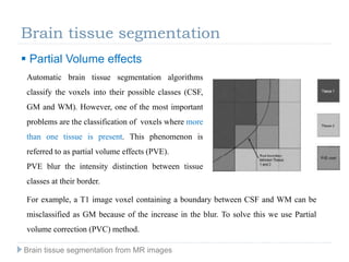
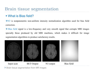
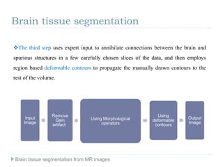
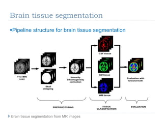
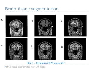
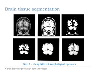
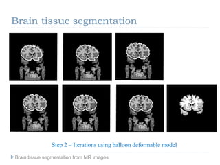
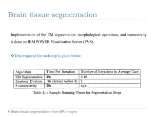
SlideShare a Scribd company logo
Contents
Brain tissue segmentation from MR images
1. Introduction
2. Magnetic Resonance Imaging
3. Brain tissue segmentation
Introduction
Manual segmentation of brain tissue is both challenging and time-consuming
due to of the large number of MRI slices for each patient which composes the
three-dimensional information and also due to intra/inter-observer variability
of manually segmented scans. Thus developing a robust automated brain tissue
segmentation method is an active research field.
However, automated segmentation of brain tissue is still a challenging
problem due to the complexity of the images, differences in tissue intensities,
noise, intensity non-uniformities, partial volume effects or absence of models
of the anatomy that fully capture the possible deformations in each structure.
Brain tissue segmentation from MR images
 Need for automatic segmentation
 Why to detect brain tissues?
Brain tissue is a particularly complex structure, and its segmentation is an
important step for tasks such as
Cortical labeling
Change detection
Visualization in surgical planning.
Introduction
Brain tissue segmentation from MR images
 Brain tissues
Introduction
Central nervous system (CNS) is the part of the human nervous network
which integrates the coordination and processing of receiving neural
information. CNS is contained by the
brain and the spinal cord and
constituted by two tissue
components: gray matter tissue
(GM), which is the main CNS
element and consists in neuronal
cell bodies; and white matter
tissue (WM), which is the second
CNS component and it is mainly
composed of militated axon
tracts.
Brain tissue segmentation from MR images
Introduction
 Modalities of Medical Image Acquisition
 MRI (Magnetic Resonance Imaging)
 PET (Positron Emission Tomography)
 SPECT (Single Photon Emission Computed Tomography)
 MRS (Magnetic Resonance Spectroscopy)
 Ultrasound
 X-ray imaging
 CT (Computed Tomography)
Brain tissue segmentation from MR images
Introduction
The modalities have different strengths and thus are used under different circumstances.
• CT captures bone with accuracy, and is used for dosage planning in radiation therapy.
• PET, SPECT, and MRS are typically used to provide functional information with MRI
also gaining usage in that domain.
• MRI produces high contrast between soft tissues, and is therefore useful for detecting
lesions in the brain.
MRI scan
CT scan
Brain tissue segmentation from MR images
Magnetic Resonance Imaging
 How MRI works?
 Patient is bathed in a magnetic field 5000 times stronger than the earths.
This field causes some of the body’s nuclei to behave like tiny compasses and line
up
 Then the nuclei are hit by pulsing radio
 Once the pulses stop the nuclei go back to their state induced by the magnet
 The energy now released by the nuclei acts like miniature radio stations giving out a
signal
 These radio waves are picked up by a computer where they are translated into an
image.
Brain tissue segmentation from MR images
Magnetic Resonance Imaging
Nuclear
Magnetic
Resonance in
MRI
Brain tissue segmentation from MR images
 Construction
Magnetic Resonance Imaging
Brain tissue segmentation from MR images
MRI is based on the magnetization properties of atomic nuclei.
A powerful, uniform, external magnetic field is employed to align the protons that are
normally randomly oriented within the water nuclei of the tissue being examined.
This alignment (or magnetization) is next disrupted by introduction of an external Radio
Frequency (RF) energy. The nuclei return to their resting alignment through various
relaxation processes and in so doing emit RF energy.
After a certain period following the initial RF, the emitted signals are measured. Fourier
transformation is used to convert the frequency information contained in the signal from
each location in the imaged plane to corresponding intensity levels, which are then displayed
as shades of gray in a matrix arrangement of pixels. By varying the sequence of RF pulses
applied & collected, different types of images are created.
Repetition Time (TR) is the amount of time between successive pulse sequences
applied to the same slice.
Time to Echo (TE) is the time between the delivery of the RF pulse and the receipt of
the echo signal.
Magnetic Resonance Imaging
Brain tissue segmentation from MR images
Tissue can be characterized by two different relaxation times – T1 and T2.
T1 (longitudinal relaxation time) is the time constant which determines the rate at
which excited protons return to equilibrium. It is a measure of the time taken for
spinning protons to realign with the external magnetic field.
 T2 (transverse relaxation time) is the time constant which determines the rate at
which excited protons reach equilibrium or go out of phase with each other. It is a
measure of the time taken for spinning protons to lose phase coherence among the
nuclei spinning perpendicular to the main field.
Magnetic Resonance Imaging
Brain tissue segmentation from MR images
Magnetic Resonance Imaging
 T1 and T2 weighted imaging
T1- and T2-weighted images can be easily differentiated by looking the CSF.
CSF is dark on T1-weighted imaging and bright on T2-weighted imaging
In T1, CSF tissue has the darkest
intensities while WM has the
brightest. On the contrary, in T2
CSF has the brightest intensities
while WM is the darkest. On
both sequences, GM has an
intermediate gray level.
Brain tissue segmentation from MR images
Magnetic Resonance Imaging
T1-weighted images are produced by using short TE and TR times. The contrast and
brightness of the image are predominately determined by T1 properties of tissue.
T2-weighted images are produced by using longer TE and TR times.
Fluid Attenuated Inversion Recovery (Flair)
The Flair sequence is similar to a T2-weighted image except
that the TE and TR times are very long. By doing so,
abnormalities remain bright but normal CSF fluid is attenuated
and made dark. This sequence is very sensitive to pathology
and makes the differentiation between CSF and an abnormality
much easier. In FLAIR , WM and GM have an intermediate
grey level and lesions seems brighter.
Brain tissue segmentation from MR images
Magnetic Resonance Imaging
Most common MRI Sequences and their Approximate TR and TE times.
 Approximate TR and TE time
Brain tissue segmentation from MR images
Brain tissue segmentation
Pipeline structure for brain tissue segmentation
Brain tissue segmentation from MR images
Brain tissue segmentation
Skull stripping
BET brain extraction tool
The brain surface extractor (BSE)
statistical parametric mapping (SPM2)
The above mentioned algorithms are all MATLAB based tools and use T1
weighted images as inputs to extract skull and remove it from the MR image
• BET (Brain Extraction Tool) deletes non-brain tissue from an image of the whole
head. It can also estimate the inner and outer skull surfaces, and outer scalp surface,
if you have good quality T1 and T2 input images.
Brain tissue segmentation from MR images
Automated brain segmentation pipelines usually incorporate a preprocessing step by
which image inhomogeneities are removed.
Sources of inhomogeneities have been studied extensively. The artifacts causes have been
divided into two main groups by classifying them as inherent to the same MRI device or
provoked by the same scanned object.
Main causes in first group are especially derived from radio frequency (RF) transmissions
and receptions but also differences in the magnetic field or eddy currents driven by field
gradients.
Cause derivations in the second group are related to the imaged object itself (position,
shape, and orientation of the object inside the magnet) or dielectric properties of the object.
Brain tissue segmentation
Intensity inhomogeneity
Brain tissue segmentation from MR images
Brain tissue segmentation
 Partial Volume effects
Automatic brain tissue segmentation algorithms
classify the voxels into their possible classes (CSF,
GM and WM). However, one of the most important
problems are the classification of voxels where more
than one tissue is present. This phenomenon is
referred to as partial volume effects (PVE).
PVE blur the intensity distinction between tissue
classes at their border.
For example, a T1 image voxel containing a boundary between CSF and WM can be
misclassified as GM because of the increase in the blur. To solve this we use Partial
volume correction (PVC) method.
Brain tissue segmentation from MR images
Brain tissue segmentation
 How to evaluate accuracy segmentation?
 Quantitative evaluations are commonly based on the comparison between the
segmentation results and a manually expert labeled volume or ground truth.
 Usually, intra-inter observer variability is avoided by the utilization of labeled volumes
from more than one expert. Still, this is not a sufficient condition and it is difficult to find a
consensus among experts.
 Thus we use an algorithm to find simultaneous truth and performance level
estimation.(STAPLE).
The method take a collection of segments of an image, and compute simultaneously a
probabilistic estimate of the true segmentation and a measure of the performance level
represented by each segmentation.
Brain tissue segmentation from MR images
Brain tissue segmentation
Generally we report evaluation based on statistical analysis measures derived from
classification rates with respect to the ground truth such as true positive (TPR), true negative
(TNR), false positive (FPR) and false negative (FNR) rates. In a single tissue classification,
these rates are defined as:
 TPR is the percentage of voxels classified as tissue by the method that are labeled as
tissue by the expert.
 TNR is the percentage of voxels classified as non-tissue by the method that are labeled
as non-tissue by the expert.
 FPR is the percentage of voxels classified as tissue by the method that are labeled as
non-tissue by the expert.
 FNR is the percentage of voxels classified as non-tissue by the method that are labeled
as tissue by the expert.
Brain tissue segmentation from MR images
Sensitivity or true positive fraction (TPF) is the classifier ability to correctly identify
tissue voxels . It can be defined as:
 Similarly, specificity is defined as the classifier ability to identify non-tissue:
The accuracy of the classifier is usually computed as the rate of correct predicted voxels
over all predicted voxels. Hence
Brain tissue segmentation
Brain tissue segmentation from MR images
Brain tissue segmentation
 Conversely, the error rate of the classifier is given by the misclassified voxels over all
predicted voxels as:
 Furthermore, similarity indexes can be used to compute the accuracy of the method.
Dice coefficient is defined as the set agreement between classification and ground truth :
 Analogously, the Jaccard similarity index, measures the overlap between the segmentation
results and the ground truth as:
Brain tissue segmentation from MR images
Brain tissue segmentation
 Other measures based on intensity, distance or connectivity can be are
As we know, Dice coefficient is the most broadly used measure to quantitatively evaluate
the accuracy of brain tissue segmentation. The Fractional Brain Tissue of a given class
returns the normalized fraction of the given tissue in the brain.
It is defined as the amount of voxels which are classified as the given class divided by all
brain voxels. Hence:
Brain tissue segmentation from MR images
We measure the atrophy in MS lesion tissues using the Brain parenchyma
factor coefficient which is defined as the number of GM,WM voxels and tissue
lesion voxels L divided by the all the brain voxels. A decrease in BPF over time
might give early diagnostic clues about the onset of MS disease:
Brain tissue segmentation
 Diagnosis of MS Lesion
 What is Multiple Sclerosis?
Brain tissue segmentation from MR images
Brain tissue segmentation
Image morphology provides a way to incorporate neighborhood and distance information.
The basic idea in morphology is to convolve an image with a given mask and to binarize
the result of the convolution using a given function. Choice of convolution mask and
binarization function depend on the particular morphological operator being used.
Binary morphology has been used in several segmentation systems, their functional
description is given on next slide.
 Mathematical Morphology
Brain tissue segmentation from MR images
Brain tissue segmentation
 Mathematical Morphology
Erosion: An erosion operation on an image I containing labels 0 and 1, with a structuring
element S, changes the value of pixel i in I from 1 to 0, if the result of convolving S with I,
centered at i, is less than some predetermined value
Dilation: Dual to erosion, a dilation operation on an image I containing labels 0 and 1, with a
structuring element S, changes the value of pixel i in I from 0 to 1, if the result of convolving S
with I, centered at i, is more than some predetermined value.
Brain tissue segmentation from MR images
Brain tissue segmentation
 Deformable Models
Deformable Models are used for object recognition.
There are two type of deformable models – Parametric deformable model and
Geometric deformable model
Snakes and Balloons are the two deformable models we will need for brain tissue
segmentation.
Snakes model is a 2 D model which maps the curves.
Balloons model is a 3 D model which is implicit.
Brain tissue segmentation from MR images
Brain tissue segmentation
Brain tissue segmentation from MR images
Brain tissue segmentation
Procedure of Segmenting Brain tissue
1. EM segmentation for correction of gain due to RF coil inhomogeneities in the data
2. binary morphology and connectivity to incorporate topological information, and
3. active contours to add spatial knowledge to the segmentation process.
Brain tissue segmentation from MR images
Brain tissue segmentation
 EM segmentation
• Expectation Maximization Segmentation (EM) is a Iterative method consisting of
two steps:
Expectation Step: Given the current bias field , E step computes likelihood of each
tissue class.
Maximization Step: Given the likelihood of each tissue class, the M step estimates
the image homogeneities
• EM segmentation does not incorporate any spatial information for the classification
of voxels.
Brain tissue segmentation from MR images
N3 is nonparametric non-uniform intensity normalization algorithm used for bias field
correction.
 Bias field signal is a low-frequency and very smooth signal that corrupts MRI images
specially those produced by old MRI machines, which makes it difficult for image
segmentation algorithms to produce satisfactory results.
 What is Bias field?
Input scan BET Output N3 output Bias field
Brain tissue segmentation
Brain tissue segmentation from MR images
Using the model described above, we divide the segmentation of the brain
into three steps.
The first step is to remove the gain artifact from the data and to classify the voxels
into four classes: white matter, grey matter, CSF, and skin (or fat), purely on the basis
of their signal intensities.
Due to natural overlap between intensity distributions of the various structures,
misclassifications are likely at this stage.
 In particular, muscle is likely to be classified as gray matter, fat classified as white
matter, and nerve fibers classified as white or gray matter.
Brain tissue segmentation
Brain tissue segmentation from MR images
The Second step aims to reduce some of the misclassification by using neighborhood
and connectivity information. It uses morphological operators to "shave off" the nerve
fibers and muscles connecting the brain tissue to the cranium, and then uses
connectivity to find the largest connected component of white and grey matter in the
image.
The strategy is that misclassified fat, muscle, and nerve fibers will get cut off from the
central largest component, which is the brain tissue.
Due to the variation in the size of the connectors from the brain tissue to the cranium,
often the brain tissue is not isolated at the end of this step, which is when we use the
third step.
Brain tissue segmentation
Brain tissue segmentation from MR images
The third step uses expert input to annihilate connections between the brain and
spurious structures in a few carefully chosen slices of the data, and then employs
region based deformable contours to propagate the manually drawn contours to the
rest of the volume.
Brain tissue segmentation
Brain tissue segmentation from MR images
Input
Image
Remove
Gain
artifact
Using Morphological
operators
Using
deformable
contours
Output
Image
1.
2. 3.
4. 5. 6.
Step 1 – Iterations of EM segmenter
Brain tissue segmentation
Brain tissue segmentation from MR images
Brain tissue segmentation
Step 2 – Using different morphological operators
Brain tissue segmentation from MR images
Step 2 – Iterations using balloon deformable model
Brain tissue segmentation
Brain tissue segmentation from MR images
Brain tissue segmentation
Implementation of the EM segmentation, morphological operations, and connectivity
is done on IBM POWER Visualization Server (PVS).
Time required for each step is given below.
Brain tissue segmentation from MR images
Thank You for your time !
All the questions are welcome and appreciated !
Brain tissue segmentation from MR images
S.M. Smith. Fast robust automated brain extraction. Human Brain Mapping,
17(3):143-155, November 2002.
Segmentation of brain tissue from magnetic resonance images by Tina Kapur,
Massachusetts Institute of Technology, February1995
MRI brain tissue segmentation by Sergi Valverde, university of Girona ,2012
http://www.brainvoyager.com/bvqx/doc/UsersGuide/Segmentation/IntensityInhomog
eneityCorrection.html
Brain tissue segmentation from MR images
References

More Related Content

What's hot

MR Neurography
MR NeurographyMR Neurography
MR Neurography
swalih121
 
Neural Network Based Brain Tumor Detection using MR Images
Neural Network Based Brain Tumor Detection using MR ImagesNeural Network Based Brain Tumor Detection using MR Images
Neural Network Based Brain Tumor Detection using MR Images
Aisha Kalsoom
 
Magnetic resonance spectroscopy
Magnetic resonance spectroscopyMagnetic resonance spectroscopy
Magnetic resonance spectroscopyAnurag Singh
 
Applications of Digital image processing in Medical Field
Applications of Digital image processing in Medical FieldApplications of Digital image processing in Medical Field
Applications of Digital image processing in Medical Field
Ashwani Srivastava
 
wavelet packets
wavelet packetswavelet packets
wavelet packets
ajayhakkumar
 
Brain tumor detection by scanning MRI images (using filtering techniques)
Brain tumor detection by scanning MRI images (using filtering techniques)Brain tumor detection by scanning MRI images (using filtering techniques)
Brain tumor detection by scanning MRI images (using filtering techniques)
Vivek reddy
 
Deep Learning in Bio-Medical Imaging
Deep Learning in Bio-Medical ImagingDeep Learning in Bio-Medical Imaging
Deep Learning in Bio-Medical Imaging
Joonhyung Lee
 
Brain Tumour Detection.pptx
Brain Tumour Detection.pptxBrain Tumour Detection.pptx
Brain Tumour Detection.pptx
RevolverRaja2
 
Image texture analysis techniques survey-1
Image texture analysis techniques  survey-1Image texture analysis techniques  survey-1
Image texture analysis techniques survey-1anitadixitjoshi
 
Brain tumor mri image segmentation and detection
Brain tumor mri image segmentation and detectionBrain tumor mri image segmentation and detection
Brain tumor mri image segmentation and detection
eSAT Publishing House
 
EEG analysis and Machine Learning
EEG  analysis and Machine LearningEEG  analysis and Machine Learning
EEG analysis and Machine Learning
Abbas Badran
 
Medical image processing studies
Medical image processing studiesMedical image processing studies
Medical image processing studies
Bằng Nguyễn Kim
 
Brain tumor detection using convolutional neural network
Brain tumor detection using convolutional neural network Brain tumor detection using convolutional neural network
Brain tumor detection using convolutional neural network
MD Abdullah Al Nasim
 
Neuro imaging in epilepsy
Neuro imaging in                                               epilepsyNeuro imaging in                                               epilepsy
Neuro imaging in epilepsyEhab Elftouh
 
A completed modeling of local binary pattern operator
A completed modeling of local binary pattern operatorA completed modeling of local binary pattern operator
A completed modeling of local binary pattern operatorWin Yu
 
Eyetracking
EyetrackingEyetracking
Eyetracking
MD Shafe
 
Magnetic resonance spectroscopy
Magnetic resonance spectroscopyMagnetic resonance spectroscopy
Magnetic resonance spectroscopy
Haidy Elbiady
 
BRAIN TUMOR MRI IMAGE SEGMENTATION AND DETECTION IN IMAGE PROCESSING
BRAIN TUMOR MRI IMAGE SEGMENTATION AND DETECTION IN IMAGE PROCESSINGBRAIN TUMOR MRI IMAGE SEGMENTATION AND DETECTION IN IMAGE PROCESSING
BRAIN TUMOR MRI IMAGE SEGMENTATION AND DETECTION IN IMAGE PROCESSING
Dharshika Shreeganesh
 
COM2304: Digital Image Fundamentals - I
COM2304: Digital Image Fundamentals - I COM2304: Digital Image Fundamentals - I
COM2304: Digital Image Fundamentals - I
Hemantha Kulathilake
 
Brain Tumor Detection using CNN
Brain Tumor Detection using CNNBrain Tumor Detection using CNN
Brain Tumor Detection using CNN
MohammadRakib8
 

What's hot (20)

MR Neurography
MR NeurographyMR Neurography
MR Neurography
 
Neural Network Based Brain Tumor Detection using MR Images
Neural Network Based Brain Tumor Detection using MR ImagesNeural Network Based Brain Tumor Detection using MR Images
Neural Network Based Brain Tumor Detection using MR Images
 
Magnetic resonance spectroscopy
Magnetic resonance spectroscopyMagnetic resonance spectroscopy
Magnetic resonance spectroscopy
 
Applications of Digital image processing in Medical Field
Applications of Digital image processing in Medical FieldApplications of Digital image processing in Medical Field
Applications of Digital image processing in Medical Field
 
wavelet packets
wavelet packetswavelet packets
wavelet packets
 
Brain tumor detection by scanning MRI images (using filtering techniques)
Brain tumor detection by scanning MRI images (using filtering techniques)Brain tumor detection by scanning MRI images (using filtering techniques)
Brain tumor detection by scanning MRI images (using filtering techniques)
 
Deep Learning in Bio-Medical Imaging
Deep Learning in Bio-Medical ImagingDeep Learning in Bio-Medical Imaging
Deep Learning in Bio-Medical Imaging
 
Brain Tumour Detection.pptx
Brain Tumour Detection.pptxBrain Tumour Detection.pptx
Brain Tumour Detection.pptx
 
Image texture analysis techniques survey-1
Image texture analysis techniques  survey-1Image texture analysis techniques  survey-1
Image texture analysis techniques survey-1
 
Brain tumor mri image segmentation and detection
Brain tumor mri image segmentation and detectionBrain tumor mri image segmentation and detection
Brain tumor mri image segmentation and detection
 
EEG analysis and Machine Learning
EEG  analysis and Machine LearningEEG  analysis and Machine Learning
EEG analysis and Machine Learning
 
Medical image processing studies
Medical image processing studiesMedical image processing studies
Medical image processing studies
 
Brain tumor detection using convolutional neural network
Brain tumor detection using convolutional neural network Brain tumor detection using convolutional neural network
Brain tumor detection using convolutional neural network
 
Neuro imaging in epilepsy
Neuro imaging in                                               epilepsyNeuro imaging in                                               epilepsy
Neuro imaging in epilepsy
 
A completed modeling of local binary pattern operator
A completed modeling of local binary pattern operatorA completed modeling of local binary pattern operator
A completed modeling of local binary pattern operator
 
Eyetracking
EyetrackingEyetracking
Eyetracking
 
Magnetic resonance spectroscopy
Magnetic resonance spectroscopyMagnetic resonance spectroscopy
Magnetic resonance spectroscopy
 
BRAIN TUMOR MRI IMAGE SEGMENTATION AND DETECTION IN IMAGE PROCESSING
BRAIN TUMOR MRI IMAGE SEGMENTATION AND DETECTION IN IMAGE PROCESSINGBRAIN TUMOR MRI IMAGE SEGMENTATION AND DETECTION IN IMAGE PROCESSING
BRAIN TUMOR MRI IMAGE SEGMENTATION AND DETECTION IN IMAGE PROCESSING
 
COM2304: Digital Image Fundamentals - I
COM2304: Digital Image Fundamentals - I COM2304: Digital Image Fundamentals - I
COM2304: Digital Image Fundamentals - I
 
Brain Tumor Detection using CNN
Brain Tumor Detection using CNNBrain Tumor Detection using CNN
Brain Tumor Detection using CNN
 

Similar to Brain tissue segmentation from MR images

Neuroimaging Lecture
Neuroimaging LectureNeuroimaging Lecture
Neuroimaging Lecture
test
 
fMRI Segmentation Using Echo State Neural Network
fMRI Segmentation Using Echo State Neural NetworkfMRI Segmentation Using Echo State Neural Network
fMRI Segmentation Using Echo State Neural Network
CSCJournals
 
Magnetic resonance imaging
Magnetic resonance imagingMagnetic resonance imaging
Magnetic resonance imaging
Albein Vivek
 
Image Processing Technique for Brain Abnormality Detection
Image Processing Technique for Brain Abnormality DetectionImage Processing Technique for Brain Abnormality Detection
Image Processing Technique for Brain Abnormality Detection
CSCJournals
 
Brain imaging in psychiatry
Brain imaging in psychiatryBrain imaging in psychiatry
Brain imaging in psychiatry
Dr. Subhendu Sekhar Dhar
 
An introduction to MRI
An introduction to MRIAn introduction to MRI
An introduction to MRI
ghazale boroumand
 
ANALYTICAL STUDY OF BRAIN MRI PROTOCOLS, SEQUENCES AND PARAMETERS FOR DETECTI...
ANALYTICAL STUDY OF BRAIN MRI PROTOCOLS, SEQUENCES AND PARAMETERS FOR DETECTI...ANALYTICAL STUDY OF BRAIN MRI PROTOCOLS, SEQUENCES AND PARAMETERS FOR DETECTI...
ANALYTICAL STUDY OF BRAIN MRI PROTOCOLS, SEQUENCES AND PARAMETERS FOR DETECTI...
ijistjournal
 
Neuroimaging in psychiatry
Neuroimaging in psychiatryNeuroimaging in psychiatry
Neuroimaging in psychiatrySantanu Ghosh
 
Ijeee 16-19-a novel approach to brain tumor classification using wavelet and ...
Ijeee 16-19-a novel approach to brain tumor classification using wavelet and ...Ijeee 16-19-a novel approach to brain tumor classification using wavelet and ...
Ijeee 16-19-a novel approach to brain tumor classification using wavelet and ...Kumar Goud
 
MRI Presentation Final
MRI Presentation FinalMRI Presentation Final
MRI Presentation Final
sganzeveld29
 
Computer Aided Diagnosis Sytem- A decision support system for clinical Diagno...
Computer Aided Diagnosis Sytem- A decision support system for clinical Diagno...Computer Aided Diagnosis Sytem- A decision support system for clinical Diagno...
Computer Aided Diagnosis Sytem- A decision support system for clinical Diagno...
PUNEET TIWARI
 
1. MRI interpretation.docx
1. MRI interpretation.docx1. MRI interpretation.docx
1. MRI interpretation.docx
ssuserc88386
 
International Journal of Image Processing (IJIP) Volume (2) Issue (1)
International Journal of Image Processing (IJIP) Volume (2) Issue (1)International Journal of Image Processing (IJIP) Volume (2) Issue (1)
International Journal of Image Processing (IJIP) Volume (2) Issue (1)CSCJournals
 
Fully Automatic Method for 3D T1-Weighted Brain Magnetic Resonance Images Seg...
Fully Automatic Method for 3D T1-Weighted Brain Magnetic Resonance Images Seg...Fully Automatic Method for 3D T1-Weighted Brain Magnetic Resonance Images Seg...
Fully Automatic Method for 3D T1-Weighted Brain Magnetic Resonance Images Seg...
CSCJournals
 
MRI Brain
MRI BrainMRI Brain
MRI Brain
Qamar Zaman
 
A Wavelet Based Automatic Segmentation of Brain Tumor in CT Images Using Opti...
A Wavelet Based Automatic Segmentation of Brain Tumor in CT Images Using Opti...A Wavelet Based Automatic Segmentation of Brain Tumor in CT Images Using Opti...
A Wavelet Based Automatic Segmentation of Brain Tumor in CT Images Using Opti...
CSCJournals
 
Icbme 2011
Icbme 2011Icbme 2011
Icbme 2011
Naresh Shah
 
Mripresenation
MripresenationMripresenation
Mripresenation
FLI
 
01_Overview.pptx
01_Overview.pptx01_Overview.pptx
01_Overview.pptx
OksanaPersaud1
 

Similar to Brain tissue segmentation from MR images (20)

Neuroimaging Lecture
Neuroimaging LectureNeuroimaging Lecture
Neuroimaging Lecture
 
fMRI Segmentation Using Echo State Neural Network
fMRI Segmentation Using Echo State Neural NetworkfMRI Segmentation Using Echo State Neural Network
fMRI Segmentation Using Echo State Neural Network
 
Magnetic resonance imaging
Magnetic resonance imagingMagnetic resonance imaging
Magnetic resonance imaging
 
Image Processing Technique for Brain Abnormality Detection
Image Processing Technique for Brain Abnormality DetectionImage Processing Technique for Brain Abnormality Detection
Image Processing Technique for Brain Abnormality Detection
 
Brain imaging in psychiatry
Brain imaging in psychiatryBrain imaging in psychiatry
Brain imaging in psychiatry
 
An introduction to MRI
An introduction to MRIAn introduction to MRI
An introduction to MRI
 
ANALYTICAL STUDY OF BRAIN MRI PROTOCOLS, SEQUENCES AND PARAMETERS FOR DETECTI...
ANALYTICAL STUDY OF BRAIN MRI PROTOCOLS, SEQUENCES AND PARAMETERS FOR DETECTI...ANALYTICAL STUDY OF BRAIN MRI PROTOCOLS, SEQUENCES AND PARAMETERS FOR DETECTI...
ANALYTICAL STUDY OF BRAIN MRI PROTOCOLS, SEQUENCES AND PARAMETERS FOR DETECTI...
 
Neuroimaging in psychiatry
Neuroimaging in psychiatryNeuroimaging in psychiatry
Neuroimaging in psychiatry
 
Ijeee 16-19-a novel approach to brain tumor classification using wavelet and ...
Ijeee 16-19-a novel approach to brain tumor classification using wavelet and ...Ijeee 16-19-a novel approach to brain tumor classification using wavelet and ...
Ijeee 16-19-a novel approach to brain tumor classification using wavelet and ...
 
MRI Presentation Final
MRI Presentation FinalMRI Presentation Final
MRI Presentation Final
 
Computer Aided Diagnosis Sytem- A decision support system for clinical Diagno...
Computer Aided Diagnosis Sytem- A decision support system for clinical Diagno...Computer Aided Diagnosis Sytem- A decision support system for clinical Diagno...
Computer Aided Diagnosis Sytem- A decision support system for clinical Diagno...
 
1. MRI interpretation.docx
1. MRI interpretation.docx1. MRI interpretation.docx
1. MRI interpretation.docx
 
International Journal of Image Processing (IJIP) Volume (2) Issue (1)
International Journal of Image Processing (IJIP) Volume (2) Issue (1)International Journal of Image Processing (IJIP) Volume (2) Issue (1)
International Journal of Image Processing (IJIP) Volume (2) Issue (1)
 
Fully Automatic Method for 3D T1-Weighted Brain Magnetic Resonance Images Seg...
Fully Automatic Method for 3D T1-Weighted Brain Magnetic Resonance Images Seg...Fully Automatic Method for 3D T1-Weighted Brain Magnetic Resonance Images Seg...
Fully Automatic Method for 3D T1-Weighted Brain Magnetic Resonance Images Seg...
 
MRI Brain
MRI BrainMRI Brain
MRI Brain
 
A Wavelet Based Automatic Segmentation of Brain Tumor in CT Images Using Opti...
A Wavelet Based Automatic Segmentation of Brain Tumor in CT Images Using Opti...A Wavelet Based Automatic Segmentation of Brain Tumor in CT Images Using Opti...
A Wavelet Based Automatic Segmentation of Brain Tumor in CT Images Using Opti...
 
Icbme 2011
Icbme 2011Icbme 2011
Icbme 2011
 
Imaging of brain tumours
Imaging of brain tumoursImaging of brain tumours
Imaging of brain tumours
 
Mripresenation
MripresenationMripresenation
Mripresenation
 
01_Overview.pptx
01_Overview.pptx01_Overview.pptx
01_Overview.pptx
 

Recently uploaded

Industrial Training at Shahjalal Fertilizer Company Limited (SFCL)
Industrial Training at Shahjalal Fertilizer Company Limited (SFCL)Industrial Training at Shahjalal Fertilizer Company Limited (SFCL)
Industrial Training at Shahjalal Fertilizer Company Limited (SFCL)
MdTanvirMahtab2
 
H.Seo, ICLR 2024, MLILAB, KAIST AI.pdf
H.Seo,  ICLR 2024, MLILAB,  KAIST AI.pdfH.Seo,  ICLR 2024, MLILAB,  KAIST AI.pdf
H.Seo, ICLR 2024, MLILAB, KAIST AI.pdf
MLILAB
 
MCQ Soil mechanics questions (Soil shear strength).pdf
MCQ Soil mechanics questions (Soil shear strength).pdfMCQ Soil mechanics questions (Soil shear strength).pdf
MCQ Soil mechanics questions (Soil shear strength).pdf
Osamah Alsalih
 
road safety engineering r s e unit 3.pdf
road safety engineering  r s e unit 3.pdfroad safety engineering  r s e unit 3.pdf
road safety engineering r s e unit 3.pdf
VENKATESHvenky89705
 
Investor-Presentation-Q1FY2024 investor presentation document.pptx
Investor-Presentation-Q1FY2024 investor presentation document.pptxInvestor-Presentation-Q1FY2024 investor presentation document.pptx
Investor-Presentation-Q1FY2024 investor presentation document.pptx
AmarGB2
 
Water Industry Process Automation and Control Monthly - May 2024.pdf
Water Industry Process Automation and Control Monthly - May 2024.pdfWater Industry Process Automation and Control Monthly - May 2024.pdf
Water Industry Process Automation and Control Monthly - May 2024.pdf
Water Industry Process Automation & Control
 
Railway Signalling Principles Edition 3.pdf
Railway Signalling Principles Edition 3.pdfRailway Signalling Principles Edition 3.pdf
Railway Signalling Principles Edition 3.pdf
TeeVichai
 
ASME IX(9) 2007 Full Version .pdf
ASME IX(9)  2007 Full Version       .pdfASME IX(9)  2007 Full Version       .pdf
ASME IX(9) 2007 Full Version .pdf
AhmedHussein950959
 
CFD Simulation of By-pass Flow in a HRSG module by R&R Consult.pptx
CFD Simulation of By-pass Flow in a HRSG module by R&R Consult.pptxCFD Simulation of By-pass Flow in a HRSG module by R&R Consult.pptx
CFD Simulation of By-pass Flow in a HRSG module by R&R Consult.pptx
R&R Consult
 
Governing Equations for Fundamental Aerodynamics_Anderson2010.pdf
Governing Equations for Fundamental Aerodynamics_Anderson2010.pdfGoverning Equations for Fundamental Aerodynamics_Anderson2010.pdf
Governing Equations for Fundamental Aerodynamics_Anderson2010.pdf
WENKENLI1
 
Final project report on grocery store management system..pdf
Final project report on grocery store management system..pdfFinal project report on grocery store management system..pdf
Final project report on grocery store management system..pdf
Kamal Acharya
 
一比一原版(IIT毕业证)伊利诺伊理工大学毕业证成绩单专业办理
一比一原版(IIT毕业证)伊利诺伊理工大学毕业证成绩单专业办理一比一原版(IIT毕业证)伊利诺伊理工大学毕业证成绩单专业办理
一比一原版(IIT毕业证)伊利诺伊理工大学毕业证成绩单专业办理
zwunae
 
Hierarchical Digital Twin of a Naval Power System
Hierarchical Digital Twin of a Naval Power SystemHierarchical Digital Twin of a Naval Power System
Hierarchical Digital Twin of a Naval Power System
Kerry Sado
 
NO1 Uk best vashikaran specialist in delhi vashikaran baba near me online vas...
NO1 Uk best vashikaran specialist in delhi vashikaran baba near me online vas...NO1 Uk best vashikaran specialist in delhi vashikaran baba near me online vas...
NO1 Uk best vashikaran specialist in delhi vashikaran baba near me online vas...
Amil Baba Dawood bangali
 
DESIGN A COTTON SEED SEPARATION MACHINE.docx
DESIGN A COTTON SEED SEPARATION MACHINE.docxDESIGN A COTTON SEED SEPARATION MACHINE.docx
DESIGN A COTTON SEED SEPARATION MACHINE.docx
FluxPrime1
 
一比一原版(UofT毕业证)多伦多大学毕业证成绩单如何办理
一比一原版(UofT毕业证)多伦多大学毕业证成绩单如何办理一比一原版(UofT毕业证)多伦多大学毕业证成绩单如何办理
一比一原版(UofT毕业证)多伦多大学毕业证成绩单如何办理
ydteq
 
Immunizing Image Classifiers Against Localized Adversary Attacks
Immunizing Image Classifiers Against Localized Adversary AttacksImmunizing Image Classifiers Against Localized Adversary Attacks
Immunizing Image Classifiers Against Localized Adversary Attacks
gerogepatton
 
Sachpazis:Terzaghi Bearing Capacity Estimation in simple terms with Calculati...
Sachpazis:Terzaghi Bearing Capacity Estimation in simple terms with Calculati...Sachpazis:Terzaghi Bearing Capacity Estimation in simple terms with Calculati...
Sachpazis:Terzaghi Bearing Capacity Estimation in simple terms with Calculati...
Dr.Costas Sachpazis
 
ethical hacking-mobile hacking methods.ppt
ethical hacking-mobile hacking methods.pptethical hacking-mobile hacking methods.ppt
ethical hacking-mobile hacking methods.ppt
Jayaprasanna4
 
English lab ppt no titlespecENG PPTt.pdf
English lab ppt no titlespecENG PPTt.pdfEnglish lab ppt no titlespecENG PPTt.pdf
English lab ppt no titlespecENG PPTt.pdf
BrazilAccount1
 

Recently uploaded (20)

Industrial Training at Shahjalal Fertilizer Company Limited (SFCL)
Industrial Training at Shahjalal Fertilizer Company Limited (SFCL)Industrial Training at Shahjalal Fertilizer Company Limited (SFCL)
Industrial Training at Shahjalal Fertilizer Company Limited (SFCL)
 
H.Seo, ICLR 2024, MLILAB, KAIST AI.pdf
H.Seo,  ICLR 2024, MLILAB,  KAIST AI.pdfH.Seo,  ICLR 2024, MLILAB,  KAIST AI.pdf
H.Seo, ICLR 2024, MLILAB, KAIST AI.pdf
 
MCQ Soil mechanics questions (Soil shear strength).pdf
MCQ Soil mechanics questions (Soil shear strength).pdfMCQ Soil mechanics questions (Soil shear strength).pdf
MCQ Soil mechanics questions (Soil shear strength).pdf
 
road safety engineering r s e unit 3.pdf
road safety engineering  r s e unit 3.pdfroad safety engineering  r s e unit 3.pdf
road safety engineering r s e unit 3.pdf
 
Investor-Presentation-Q1FY2024 investor presentation document.pptx
Investor-Presentation-Q1FY2024 investor presentation document.pptxInvestor-Presentation-Q1FY2024 investor presentation document.pptx
Investor-Presentation-Q1FY2024 investor presentation document.pptx
 
Water Industry Process Automation and Control Monthly - May 2024.pdf
Water Industry Process Automation and Control Monthly - May 2024.pdfWater Industry Process Automation and Control Monthly - May 2024.pdf
Water Industry Process Automation and Control Monthly - May 2024.pdf
 
Railway Signalling Principles Edition 3.pdf
Railway Signalling Principles Edition 3.pdfRailway Signalling Principles Edition 3.pdf
Railway Signalling Principles Edition 3.pdf
 
ASME IX(9) 2007 Full Version .pdf
ASME IX(9)  2007 Full Version       .pdfASME IX(9)  2007 Full Version       .pdf
ASME IX(9) 2007 Full Version .pdf
 
CFD Simulation of By-pass Flow in a HRSG module by R&R Consult.pptx
CFD Simulation of By-pass Flow in a HRSG module by R&R Consult.pptxCFD Simulation of By-pass Flow in a HRSG module by R&R Consult.pptx
CFD Simulation of By-pass Flow in a HRSG module by R&R Consult.pptx
 
Governing Equations for Fundamental Aerodynamics_Anderson2010.pdf
Governing Equations for Fundamental Aerodynamics_Anderson2010.pdfGoverning Equations for Fundamental Aerodynamics_Anderson2010.pdf
Governing Equations for Fundamental Aerodynamics_Anderson2010.pdf
 
Final project report on grocery store management system..pdf
Final project report on grocery store management system..pdfFinal project report on grocery store management system..pdf
Final project report on grocery store management system..pdf
 
一比一原版(IIT毕业证)伊利诺伊理工大学毕业证成绩单专业办理
一比一原版(IIT毕业证)伊利诺伊理工大学毕业证成绩单专业办理一比一原版(IIT毕业证)伊利诺伊理工大学毕业证成绩单专业办理
一比一原版(IIT毕业证)伊利诺伊理工大学毕业证成绩单专业办理
 
Hierarchical Digital Twin of a Naval Power System
Hierarchical Digital Twin of a Naval Power SystemHierarchical Digital Twin of a Naval Power System
Hierarchical Digital Twin of a Naval Power System
 
NO1 Uk best vashikaran specialist in delhi vashikaran baba near me online vas...
NO1 Uk best vashikaran specialist in delhi vashikaran baba near me online vas...NO1 Uk best vashikaran specialist in delhi vashikaran baba near me online vas...
NO1 Uk best vashikaran specialist in delhi vashikaran baba near me online vas...
 
DESIGN A COTTON SEED SEPARATION MACHINE.docx
DESIGN A COTTON SEED SEPARATION MACHINE.docxDESIGN A COTTON SEED SEPARATION MACHINE.docx
DESIGN A COTTON SEED SEPARATION MACHINE.docx
 
一比一原版(UofT毕业证)多伦多大学毕业证成绩单如何办理
一比一原版(UofT毕业证)多伦多大学毕业证成绩单如何办理一比一原版(UofT毕业证)多伦多大学毕业证成绩单如何办理
一比一原版(UofT毕业证)多伦多大学毕业证成绩单如何办理
 
Immunizing Image Classifiers Against Localized Adversary Attacks
Immunizing Image Classifiers Against Localized Adversary AttacksImmunizing Image Classifiers Against Localized Adversary Attacks
Immunizing Image Classifiers Against Localized Adversary Attacks
 
Sachpazis:Terzaghi Bearing Capacity Estimation in simple terms with Calculati...
Sachpazis:Terzaghi Bearing Capacity Estimation in simple terms with Calculati...Sachpazis:Terzaghi Bearing Capacity Estimation in simple terms with Calculati...
Sachpazis:Terzaghi Bearing Capacity Estimation in simple terms with Calculati...
 
ethical hacking-mobile hacking methods.ppt
ethical hacking-mobile hacking methods.pptethical hacking-mobile hacking methods.ppt
ethical hacking-mobile hacking methods.ppt
 
English lab ppt no titlespecENG PPTt.pdf
English lab ppt no titlespecENG PPTt.pdfEnglish lab ppt no titlespecENG PPTt.pdf
English lab ppt no titlespecENG PPTt.pdf
 

Brain tissue segmentation from MR images

  • 1.
  • 2. Contents Brain tissue segmentation from MR images 1. Introduction 2. Magnetic Resonance Imaging 3. Brain tissue segmentation
  • 3. Introduction Manual segmentation of brain tissue is both challenging and time-consuming due to of the large number of MRI slices for each patient which composes the three-dimensional information and also due to intra/inter-observer variability of manually segmented scans. Thus developing a robust automated brain tissue segmentation method is an active research field. However, automated segmentation of brain tissue is still a challenging problem due to the complexity of the images, differences in tissue intensities, noise, intensity non-uniformities, partial volume effects or absence of models of the anatomy that fully capture the possible deformations in each structure. Brain tissue segmentation from MR images  Need for automatic segmentation
  • 4.  Why to detect brain tissues? Brain tissue is a particularly complex structure, and its segmentation is an important step for tasks such as Cortical labeling Change detection Visualization in surgical planning. Introduction Brain tissue segmentation from MR images
  • 5.  Brain tissues Introduction Central nervous system (CNS) is the part of the human nervous network which integrates the coordination and processing of receiving neural information. CNS is contained by the brain and the spinal cord and constituted by two tissue components: gray matter tissue (GM), which is the main CNS element and consists in neuronal cell bodies; and white matter tissue (WM), which is the second CNS component and it is mainly composed of militated axon tracts. Brain tissue segmentation from MR images
  • 6. Introduction  Modalities of Medical Image Acquisition  MRI (Magnetic Resonance Imaging)  PET (Positron Emission Tomography)  SPECT (Single Photon Emission Computed Tomography)  MRS (Magnetic Resonance Spectroscopy)  Ultrasound  X-ray imaging  CT (Computed Tomography) Brain tissue segmentation from MR images
  • 7. Introduction The modalities have different strengths and thus are used under different circumstances. • CT captures bone with accuracy, and is used for dosage planning in radiation therapy. • PET, SPECT, and MRS are typically used to provide functional information with MRI also gaining usage in that domain. • MRI produces high contrast between soft tissues, and is therefore useful for detecting lesions in the brain. MRI scan CT scan Brain tissue segmentation from MR images
  • 8. Magnetic Resonance Imaging  How MRI works?  Patient is bathed in a magnetic field 5000 times stronger than the earths. This field causes some of the body’s nuclei to behave like tiny compasses and line up  Then the nuclei are hit by pulsing radio  Once the pulses stop the nuclei go back to their state induced by the magnet  The energy now released by the nuclei acts like miniature radio stations giving out a signal  These radio waves are picked up by a computer where they are translated into an image. Brain tissue segmentation from MR images
  • 9. Magnetic Resonance Imaging Nuclear Magnetic Resonance in MRI Brain tissue segmentation from MR images
  • 10.  Construction Magnetic Resonance Imaging Brain tissue segmentation from MR images
  • 11. MRI is based on the magnetization properties of atomic nuclei. A powerful, uniform, external magnetic field is employed to align the protons that are normally randomly oriented within the water nuclei of the tissue being examined. This alignment (or magnetization) is next disrupted by introduction of an external Radio Frequency (RF) energy. The nuclei return to their resting alignment through various relaxation processes and in so doing emit RF energy. After a certain period following the initial RF, the emitted signals are measured. Fourier transformation is used to convert the frequency information contained in the signal from each location in the imaged plane to corresponding intensity levels, which are then displayed as shades of gray in a matrix arrangement of pixels. By varying the sequence of RF pulses applied & collected, different types of images are created. Repetition Time (TR) is the amount of time between successive pulse sequences applied to the same slice. Time to Echo (TE) is the time between the delivery of the RF pulse and the receipt of the echo signal. Magnetic Resonance Imaging Brain tissue segmentation from MR images
  • 12. Tissue can be characterized by two different relaxation times – T1 and T2. T1 (longitudinal relaxation time) is the time constant which determines the rate at which excited protons return to equilibrium. It is a measure of the time taken for spinning protons to realign with the external magnetic field.  T2 (transverse relaxation time) is the time constant which determines the rate at which excited protons reach equilibrium or go out of phase with each other. It is a measure of the time taken for spinning protons to lose phase coherence among the nuclei spinning perpendicular to the main field. Magnetic Resonance Imaging Brain tissue segmentation from MR images
  • 13. Magnetic Resonance Imaging  T1 and T2 weighted imaging T1- and T2-weighted images can be easily differentiated by looking the CSF. CSF is dark on T1-weighted imaging and bright on T2-weighted imaging In T1, CSF tissue has the darkest intensities while WM has the brightest. On the contrary, in T2 CSF has the brightest intensities while WM is the darkest. On both sequences, GM has an intermediate gray level. Brain tissue segmentation from MR images
  • 14. Magnetic Resonance Imaging T1-weighted images are produced by using short TE and TR times. The contrast and brightness of the image are predominately determined by T1 properties of tissue. T2-weighted images are produced by using longer TE and TR times. Fluid Attenuated Inversion Recovery (Flair) The Flair sequence is similar to a T2-weighted image except that the TE and TR times are very long. By doing so, abnormalities remain bright but normal CSF fluid is attenuated and made dark. This sequence is very sensitive to pathology and makes the differentiation between CSF and an abnormality much easier. In FLAIR , WM and GM have an intermediate grey level and lesions seems brighter. Brain tissue segmentation from MR images
  • 15. Magnetic Resonance Imaging Most common MRI Sequences and their Approximate TR and TE times.  Approximate TR and TE time Brain tissue segmentation from MR images
  • 16. Brain tissue segmentation Pipeline structure for brain tissue segmentation Brain tissue segmentation from MR images
  • 17. Brain tissue segmentation Skull stripping BET brain extraction tool The brain surface extractor (BSE) statistical parametric mapping (SPM2) The above mentioned algorithms are all MATLAB based tools and use T1 weighted images as inputs to extract skull and remove it from the MR image • BET (Brain Extraction Tool) deletes non-brain tissue from an image of the whole head. It can also estimate the inner and outer skull surfaces, and outer scalp surface, if you have good quality T1 and T2 input images. Brain tissue segmentation from MR images
  • 18. Automated brain segmentation pipelines usually incorporate a preprocessing step by which image inhomogeneities are removed. Sources of inhomogeneities have been studied extensively. The artifacts causes have been divided into two main groups by classifying them as inherent to the same MRI device or provoked by the same scanned object. Main causes in first group are especially derived from radio frequency (RF) transmissions and receptions but also differences in the magnetic field or eddy currents driven by field gradients. Cause derivations in the second group are related to the imaged object itself (position, shape, and orientation of the object inside the magnet) or dielectric properties of the object. Brain tissue segmentation Intensity inhomogeneity Brain tissue segmentation from MR images
  • 19. Brain tissue segmentation  Partial Volume effects Automatic brain tissue segmentation algorithms classify the voxels into their possible classes (CSF, GM and WM). However, one of the most important problems are the classification of voxels where more than one tissue is present. This phenomenon is referred to as partial volume effects (PVE). PVE blur the intensity distinction between tissue classes at their border. For example, a T1 image voxel containing a boundary between CSF and WM can be misclassified as GM because of the increase in the blur. To solve this we use Partial volume correction (PVC) method. Brain tissue segmentation from MR images
  • 20. Brain tissue segmentation  How to evaluate accuracy segmentation?  Quantitative evaluations are commonly based on the comparison between the segmentation results and a manually expert labeled volume or ground truth.  Usually, intra-inter observer variability is avoided by the utilization of labeled volumes from more than one expert. Still, this is not a sufficient condition and it is difficult to find a consensus among experts.  Thus we use an algorithm to find simultaneous truth and performance level estimation.(STAPLE). The method take a collection of segments of an image, and compute simultaneously a probabilistic estimate of the true segmentation and a measure of the performance level represented by each segmentation. Brain tissue segmentation from MR images
  • 21. Brain tissue segmentation Generally we report evaluation based on statistical analysis measures derived from classification rates with respect to the ground truth such as true positive (TPR), true negative (TNR), false positive (FPR) and false negative (FNR) rates. In a single tissue classification, these rates are defined as:  TPR is the percentage of voxels classified as tissue by the method that are labeled as tissue by the expert.  TNR is the percentage of voxels classified as non-tissue by the method that are labeled as non-tissue by the expert.  FPR is the percentage of voxels classified as tissue by the method that are labeled as non-tissue by the expert.  FNR is the percentage of voxels classified as non-tissue by the method that are labeled as tissue by the expert. Brain tissue segmentation from MR images
  • 22. Sensitivity or true positive fraction (TPF) is the classifier ability to correctly identify tissue voxels . It can be defined as:  Similarly, specificity is defined as the classifier ability to identify non-tissue: The accuracy of the classifier is usually computed as the rate of correct predicted voxels over all predicted voxels. Hence Brain tissue segmentation Brain tissue segmentation from MR images
  • 23. Brain tissue segmentation  Conversely, the error rate of the classifier is given by the misclassified voxels over all predicted voxels as:  Furthermore, similarity indexes can be used to compute the accuracy of the method. Dice coefficient is defined as the set agreement between classification and ground truth :  Analogously, the Jaccard similarity index, measures the overlap between the segmentation results and the ground truth as: Brain tissue segmentation from MR images
  • 24. Brain tissue segmentation  Other measures based on intensity, distance or connectivity can be are As we know, Dice coefficient is the most broadly used measure to quantitatively evaluate the accuracy of brain tissue segmentation. The Fractional Brain Tissue of a given class returns the normalized fraction of the given tissue in the brain. It is defined as the amount of voxels which are classified as the given class divided by all brain voxels. Hence: Brain tissue segmentation from MR images
  • 25. We measure the atrophy in MS lesion tissues using the Brain parenchyma factor coefficient which is defined as the number of GM,WM voxels and tissue lesion voxels L divided by the all the brain voxels. A decrease in BPF over time might give early diagnostic clues about the onset of MS disease: Brain tissue segmentation  Diagnosis of MS Lesion  What is Multiple Sclerosis? Brain tissue segmentation from MR images
  • 26. Brain tissue segmentation Image morphology provides a way to incorporate neighborhood and distance information. The basic idea in morphology is to convolve an image with a given mask and to binarize the result of the convolution using a given function. Choice of convolution mask and binarization function depend on the particular morphological operator being used. Binary morphology has been used in several segmentation systems, their functional description is given on next slide.  Mathematical Morphology Brain tissue segmentation from MR images
  • 27. Brain tissue segmentation  Mathematical Morphology Erosion: An erosion operation on an image I containing labels 0 and 1, with a structuring element S, changes the value of pixel i in I from 1 to 0, if the result of convolving S with I, centered at i, is less than some predetermined value Dilation: Dual to erosion, a dilation operation on an image I containing labels 0 and 1, with a structuring element S, changes the value of pixel i in I from 0 to 1, if the result of convolving S with I, centered at i, is more than some predetermined value. Brain tissue segmentation from MR images
  • 28. Brain tissue segmentation  Deformable Models Deformable Models are used for object recognition. There are two type of deformable models – Parametric deformable model and Geometric deformable model Snakes and Balloons are the two deformable models we will need for brain tissue segmentation. Snakes model is a 2 D model which maps the curves. Balloons model is a 3 D model which is implicit. Brain tissue segmentation from MR images
  • 29. Brain tissue segmentation Brain tissue segmentation from MR images
  • 30. Brain tissue segmentation Procedure of Segmenting Brain tissue 1. EM segmentation for correction of gain due to RF coil inhomogeneities in the data 2. binary morphology and connectivity to incorporate topological information, and 3. active contours to add spatial knowledge to the segmentation process. Brain tissue segmentation from MR images
  • 31. Brain tissue segmentation  EM segmentation • Expectation Maximization Segmentation (EM) is a Iterative method consisting of two steps: Expectation Step: Given the current bias field , E step computes likelihood of each tissue class. Maximization Step: Given the likelihood of each tissue class, the M step estimates the image homogeneities • EM segmentation does not incorporate any spatial information for the classification of voxels. Brain tissue segmentation from MR images
  • 32. N3 is nonparametric non-uniform intensity normalization algorithm used for bias field correction.  Bias field signal is a low-frequency and very smooth signal that corrupts MRI images specially those produced by old MRI machines, which makes it difficult for image segmentation algorithms to produce satisfactory results.  What is Bias field? Input scan BET Output N3 output Bias field Brain tissue segmentation Brain tissue segmentation from MR images
  • 33. Using the model described above, we divide the segmentation of the brain into three steps. The first step is to remove the gain artifact from the data and to classify the voxels into four classes: white matter, grey matter, CSF, and skin (or fat), purely on the basis of their signal intensities. Due to natural overlap between intensity distributions of the various structures, misclassifications are likely at this stage.  In particular, muscle is likely to be classified as gray matter, fat classified as white matter, and nerve fibers classified as white or gray matter. Brain tissue segmentation Brain tissue segmentation from MR images
  • 34. The Second step aims to reduce some of the misclassification by using neighborhood and connectivity information. It uses morphological operators to "shave off" the nerve fibers and muscles connecting the brain tissue to the cranium, and then uses connectivity to find the largest connected component of white and grey matter in the image. The strategy is that misclassified fat, muscle, and nerve fibers will get cut off from the central largest component, which is the brain tissue. Due to the variation in the size of the connectors from the brain tissue to the cranium, often the brain tissue is not isolated at the end of this step, which is when we use the third step. Brain tissue segmentation Brain tissue segmentation from MR images
  • 35. The third step uses expert input to annihilate connections between the brain and spurious structures in a few carefully chosen slices of the data, and then employs region based deformable contours to propagate the manually drawn contours to the rest of the volume. Brain tissue segmentation Brain tissue segmentation from MR images Input Image Remove Gain artifact Using Morphological operators Using deformable contours Output Image
  • 36. 1. 2. 3. 4. 5. 6. Step 1 – Iterations of EM segmenter Brain tissue segmentation Brain tissue segmentation from MR images
  • 37. Brain tissue segmentation Step 2 – Using different morphological operators Brain tissue segmentation from MR images
  • 38. Step 2 – Iterations using balloon deformable model Brain tissue segmentation Brain tissue segmentation from MR images
  • 39. Brain tissue segmentation Implementation of the EM segmentation, morphological operations, and connectivity is done on IBM POWER Visualization Server (PVS). Time required for each step is given below. Brain tissue segmentation from MR images
  • 40. Thank You for your time ! All the questions are welcome and appreciated ! Brain tissue segmentation from MR images
  • 41. S.M. Smith. Fast robust automated brain extraction. Human Brain Mapping, 17(3):143-155, November 2002. Segmentation of brain tissue from magnetic resonance images by Tina Kapur, Massachusetts Institute of Technology, February1995 MRI brain tissue segmentation by Sergi Valverde, university of Girona ,2012 http://www.brainvoyager.com/bvqx/doc/UsersGuide/Segmentation/IntensityInhomog eneityCorrection.html Brain tissue segmentation from MR images References

Editor's Notes

  1. A typical image acquisition consists of exposing patients to the imaging equipment, sometimes with contrast enhancing agents or markers, and generating an image of their anatomy. This image can be a 2D projection of a 3D scene as is produced with X-ray or ultrasound, or it can be a full 3D image, as generated by CT or MRI.
  2. 60Mhz, 10^16 billion particles of hydrogen
  3. Give earth example
  4. Repetition time and time to echo
  5. The 3-dimensionsal image it provides is built up in units called voxels. Each one represents a tidy cube of brain tissue
  6. Parenchyma - the functional tissue of an organ as distinguished from the connective and supporting tissue Atrophy- (of body tissue or an organ) waste away, especially as a result of the degeneration of cells
  7. Homogeneity- the quality or state of being all the same or all of the same kind.
  8. Annihilate- destruction