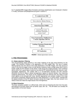
In the domain of medical imaging, accurate segmentation of brain MR images is of interest for many brain disorders. However, due to several factors such noise, imaging artefacts, intrinsic tissue variation and partial volume effects, tissue segmentation remains a challenging task. So, in this paper, a full automatic method for segmentation of brain MR images is presented. The method consists of four steps segmentation procedure. First, noise removing by median filtering is done; second segmentation of brain/non-brain tissue is performed by using a Threshold Morphologic Brain Extraction method (TMBE). Then initial centroids estimation by gray level histogram analysis based is executed. Finally, Fuzzy C-means Algorithm is used for MRI tissue segmentation. The efficiency of the proposed method is demonstrated by extensive segmentation experiments using simulated and real MR images.
Segmentation of Brain MR Images for Tumor Extraction by Combining Kmeans Clus...CSCJournals
Segmentation of images holds an important position in the area of image processing. It becomes more important while typically dealing with medical images where pre-surgery and post surgery decisions are required for the purpose of initiating and speeding up the recovery process [5] Computer aided detection of abnormal growth of tissues is primarily motivated by the necessity of achieving maximum possible accuracy. Manual segmentation of these abnormal tissues cannot be compared with modern day’s high speed computing machines which enable us to visually observe the volume and location of unwanted tissues. A well known segmentation problem within MRI is the task of labeling voxels according to their tissue type which include White Matter (WM), Grey Matter (GM) , Cerebrospinal Fluid (CSF) and sometimes pathological tissues like tumor etc. This paper describes an efficient method for automatic brain tumor segmentation for the extraction of tumor tissues from MR images. It combines Perona and Malik anisotropic diffusion model for image enhancement and Kmeans clustering technique for grouping tissues belonging to a specific group. The proposed method uses T1, T2 and PD weighted gray level intensity images. The proposed technique produced appreciative results
International Journal of Computational Engineering Research(IJCER) is an intentional online Journal in English monthly publishing journal. This Journal publish original research work that contributes significantly to further the scientific knowledge in engineering and Technology
Classification of Brain Cancer is implemented
by using Back Propagation Neural network and Principle
Component Analysis, Magnetic Resonance Imaging of brain
cancer affected patients are taken for classification of brain
cancer. Image processing techniques are used for processing
the MRI images which are image preprocessing, image
segmentation and feature extraction is used. We extract the
Texture feature of segmented image by using Gray Level Cooccurrence
Matrix (GLCM). Steps involve for brain cancer
classification are taking the MRI images, remove the noise by
using image pre-processing, applying the segmentation
method which isolate the tumor region from rest part of the
MRI image by setting the pixel value 1 to tumor region and 0
to rest of the region, after this feature extraction technique
has been applied for extracting texture feature and feature
are stored in knowledge based, this features are used for
classification of new MRI images taken for testing by
comparing the feature of new images with stored features. We
implemented three classifiers to classify the brain cancer, first
classifier is back propagation neural network which perform
classification in two phase which are training phase and
testing phase, second classifier is the combination of PCA and
BPNN means by using PCA to reduce the dimensionality of
feature matrix and by using BPNN to classify the brain
cancer, third classifier is Principle Component Analysis which
reduce the dimensionality of dataset and perform
classification. And finally compare the performance of that
classifiers.
Automatic Diagnosis of Abnormal Tumor Region from Brain Computed Tomography I...ijcseit
The research work presented in this paper is to achieve the tissue classification and automatically
diagnosis the abnormal tumor region present in Computed Tomography (CT) images using the wavelet
based statistical texture analysis method. Comparative studies of texture analysis method are performed
for the proposed wavelet based texture analysis method and Spatial Gray Level Dependence Method
(SGLDM). Our proposed system consists of four phases i) Discrete Wavelet Decomposition (ii)
Feature extraction (iii) Feature selection (iv) Analysis of extracted texture features by classifier. A
wavelet based statistical texture feature set is derived from normal and tumor regions. Genetic Algorithm
(GA) is used to select the optimal texture features from the set of extracted texture features. We construct
the Support Vector Machine (SVM) based classifier and evaluate the performance of classifier by
comparing the classification results of the SVM based classifier with the Back Propagation Neural network
classifier(BPN). The results of Support Vector Machine (SVM), BPN classifiers for the texture analysis
methods are evaluated using Receiver Operating Characteristic (ROC) analysis. Experimental results
show that the classification accuracy of SVM is 96% for 10 fold cross validation method. The system
has been tested with a number of real Computed Tomography brain images and has achieved satisfactory
results.
Efficient Brain Tumor Detection Using Wavelet TransformIJERA Editor
Brain tumor detection is a challenging task and its very important to analyze the structure of the tumor correctly so a automatic method is used now a days for the detection of the tumor. This method saves time as well as it reduces the error which occurs in the method of manual detection. In this paper the tumor is detected using wavelet transform. MRI is an important tool used in many fields of medicine and is capable of generating a detailed image of any part of the human body. The tumor is segmented from the MRI images, features are extracted and then the area of the tumor is determined. PNN can successfully handle the process of brain tumor classification
Brain Image Fusion using DWT and Laplacian Pyramid Approach and Tumor Detecti...INFOGAIN PUBLICATION
Image fusion is the process of combining important information from two or more images into a single image. The resulting image will be more enhanced than any of the input pictures. The idea of combining multiple image modalities to furnish a single, more enhanced image is well established, special fusion methods have been proposed in literature. This paper is based on image fusion using laplacian pyramid and Discreet Wavelet Transform (DWT) methods. This system uses an easy and effective algorithm for multi-focus image fusion which uses fusion rules to create fused image. Subsequently, the fused image is obtained by applying inverse discreet wavelet transform. After fused image is obtained, watershed segmentation algorithm is applied to detect the tumor part in fused image.
Brain tumor is a malformed growth of cells within brain which may be
cancerous or non-cancerous. The term ‘malformed’ indicates the existence of tumor. The
tumor may be benign or malignant and it needs medical support for further classification.
Brain tumor must be detected, diagnosed and evaluated in earliest stage. The medical
problems become grave if tumor is detected at the later stage. Out of various technologies
available for diagnosis of brain tumor, MRI is the preferred technology which enables the
diagnosis and evaluation of brain tumor. The current work presents various clustering
techniques that are employed to detect brain tumor. The classification involves classification
of images into normal and malformed (if detected the tumor). The algorithm deals with
steps such as preprocessing, segmentation, feature extraction and classification of MR brain
images. Finally, the confirmatory step is specifying the tumor area by technique called
region of interest.
International Journal of Engineering Research and Applications (IJERA) is an open access online peer reviewed international journal that publishes research and review articles in the fields of Computer Science, Neural Networks, Electrical Engineering, Software Engineering, Information Technology, Mechanical Engineering, Chemical Engineering, Plastic Engineering, Food Technology, Textile Engineering, Nano Technology & science, Power Electronics, Electronics & Communication Engineering, Computational mathematics, Image processing, Civil Engineering, Structural Engineering, Environmental Engineering, VLSI Testing & Low Power VLSI Design etc.
IOSR Journal of Electronics and Communication Engineering(IOSR-JECE) is an open access international journal that provides rapid publication (within a month) of articles in all areas of electronics and communication engineering and its applications. The journal welcomes publications of high quality papers on theoretical developments and practical applications in electronics and communication engineering. Original research papers, state-of-the-art reviews, and high quality technical notes are invited for publications.
Image Binarization for the uses of Preprocessing to Detect Brain Abnormality ...Journal For Research
Computerized MR of brain image binarization for the uses of preprocessing of features extraction and brain abnormality identification of brain has been described. Binarization is used as intermediate steps of many MR of brain normal and abnormal tissues detection. One of the main problems of MRI binarization is that many pixels of brain part cannot be correctly binarized due to the extensive black background or the large variation in contrast between background and foreground of MRI. Proposed binarization determines a threshold value using mean, variance, standard deviation and entropy followed by a non-gamut enhancement that can overcome the binarization problem. The proposed binarization technique is extensively tested with a variety of MRI and generates good binarization with improved accuracy and reduced error.
Survey on Brain MRI Segmentation TechniquesEditor IJMTER
Image segmentation is aimed at cutting out, a ROI (Region of Interest) from an image. For
medical images, segmentation is done for: studying the anatomical structure, identifying ROI ie tumor
or any other abnormalities, identifying the increase in tissue volume in a region, treatment planning.
Currently there are many different algorithms available for image segmentation. This paper lists and
compares some of them. Each has their own advantages and limitations.
Segmentation of Tumor Region in MRI Images of Brain using Mathematical Morpho...CSCJournals
This paper introduces an efficient detection of brain tumor from cerebral MRI images. The methodology consists of two steps: enhancement and segmentation. To improve the quality of images and limit the risk of distinct regions fusion in the segmentation phase an enhancement process is applied. We applied mathematical morphology to increase the contrast in MRI images and to segment MRI images. Some of experimental results on brain images show the feasibility and the performance of the proposed approach.
Literature survey for 3 d reconstruction of brain mri imageseSAT Journals
Abstract
Since Doctors had only the 2D Image Data to visualize the tumors in the MRI images, which never gave the actual feel of how the tumor would exactly look like . The doctors were deprived from the exact visualization of the tumor the amount of the tumor to be removed by operation was not known, which caused a lot of deformation in the faces and structure of the patients face or skull. The diversity and complexity of tumor cells makes it very challenging to visualize tumor present in magnetic resonance image (MRI) data. Hence to visualize the tumor properly 2D MRI image has to be converted to 3D image. With the development of computer image processing technology, three-dimensional (3D) visualization has become an important method of the medical diagnose, it offers abundant and accurate information for medical experts. Three-dimensional (3-D) reconstruction of medical images is widely applied to tumor localization; surgical planning and brain electromagnetic field computation etc. The brain MR images have unique characteristics, i.e., very complicated changes of the gray-scales and highly irregular boundaries. Traditional 3-D reconstruction algorithms are challenged in solving this problem. Many reconstruction algorithms, such as marching cubes and dividing cubes, need to establish the topological relationship between the slices of images. The results of these traditional approaches vary depending on the number of input sections, their positions, the shape of the original body and the applied interpolation technique. These make the task tedious and time-consuming. Moreover, satisfied reconstruction result may not even be obtained when the highly irregular objects such as the encephalic tissues are considered. Due to complexity and irregularity of each encephalic tissue boundary, three-dimensional (3D) reconstruction for MRI image is necessary. A Literature survey is done to study different methods of 3D reconstruction of brain images from MRI images. Keywords: 3-D reconstruction, region growing, segmentation method, immune algorithm (IA), one class support vector machine (OCSVM) and sphere shaped support vector machine (SSSVM).
IJRET : International Journal of Research in Engineering and Technology is an international peer reviewed, online journal published by eSAT Publishing House for the enhancement of research in various disciplines of Engineering and Technology. The aim and scope of the journal is to provide an academic medium and an important reference for the advancement and dissemination of research results that support high-level learning, teaching and research in the fields of Engineering and Technology. We bring together Scientists, Academician, Field Engineers, Scholars and Students of related fields of Engineering and Technology
Multistage Classification of Alzheimer’s DiseaseIJLT EMAS
Alzheimer’s disease is a type of dementia that destroys
memory and other mental functions. During the progression of
the disease certain proteins called plaques and tangles get
deposited in hippocampus which is located in the temporal lobe
of brain. The disease is not a normal part of aging and gets
worsen over time. Medical imaging techniques like Magnetic
Resonance Imaging (MRI), Computed Tomography (CT) and
Positron Emission Tomography (PET) play significant role in the
disease diagnosis. In this paper, we propose a method for
classifying MRI into Normal Control (NC), Mild Cognitive
Impairment (MCI) and Alzheimer’s Disease(AD). An overall
outline of the methodology includes textural feature extraction,
feature reduction process and classification of the images into
various stages. Classification has been performed with three
classifiers namely Support Vector Machine (SVM), Artificial
Neural Network (ANN) and k-Nearest Neighbours (k-NN)
Image Segmentation and Identification of Brain Tumor using FFT Techniques of ...IDES Editor
The image processing tools are extensively used on
the development of new algorithms and mathematical tools
for the advanced processing of medical and biological images.
Given an MRI scan, first segment the tumor region in the
MRI brain image and study the pixel intensity values. A
detailed procedure using Matlab script is written to extract
tumor region in CT scan Brain Image and MRI Scan Brain
Image. MRI Scan has higher resolution and easier
identification compare to CT scan Brain image. Fast Fourier
Transform is used here to study the tumor region of MRI
Brain Image in terms of its pixel intensity. Types of FFT like
Zero padded FFT, Windowed FFT are used to study the signal
converted from the MRI Brain Image. It is found that lesser
spectral leakage for Zero Padded Windowed FFT than other
Types of FFT and hence the tumor cell identification is easier
than other methods. Finally higher pixel intensity values of
the cells gives identification of presence and activeness of
tumor cells.
A Wavelet Based Automatic Segmentation of Brain Tumor in CT Images Using Opti...CSCJournals
This paper presents an automated segmentation of brain tumors in computed tomography images (CT) using combination of Wavelet Statistical Texture features (WST) obtained from 2-level Discrete Wavelet Transformed (DWT) low and high frequency sub bands and Wavelet Co-occurrence Texture features (WCT) obtained from two level Discrete Wavelet Transformed (DWT) high frequency sub bands. In the proposed method, the wavelet based optimal texture features that distinguish between the brain tissue, benign tumor and malignant tumor tissue is found. Comparative studies of texture analysis is performed for the proposed combined wavelet based texture analysis method and Spatial Gray Level Dependence Method (SGLDM). Our proposed system consists of four phases i) Discrete Wavelet Decomposition (ii) Feature extraction (iii) Feature selection (iv) Classification and evaluation. The combined Wavelet Statistical Texture feature set (WST) and Wavelet Co-occurrence Texture feature (WCT) sets are derived from normal and tumor regions. Feature selection is performed by Genetic Algorithm (GA). These optimal features are used to segment the tumor. An Probabilistic Neural Network (PNN) classifier is employed to evaluate the performance of these features and by comparing the classification results of the PNN classifier with the Feed Forward Neural Network classifier(FFNN).The results of the Probabilistic Neural Network, FFNN classifiers for the texture analysis methods are evaluated using Receiver Operating Characteristic (ROC) analysis. The performance of the algorithm is evaluated on a series of brain tumor images. The results illustrate that the proposed method outperforms the existing methods.
An Efficient Brain Tumor Detection Algorithm based on Segmentation for MRI Sy...ijtsrd
A collection, or mass, of abnormal cells in the brain is called as Brain Tumor . The skull, which encloses your brain, is very rigid. Growth inside such a restricted space can cause problems. Brain tumors can be malignant or benign. Segmentation in magnetic resonance imaging (MRI) was an emergent research area in the field of medical imaging system. In this an efficient algorithm is proposed for tumor detection based on segmentation and morphological operators. Quality of scanned image is enhanced and then morphological operators are applied to detect the tumor in the scanned image. Merlin Asha. M | G. Naveen Balaji | S. Mythili | A. Karthikeyan | N. Thillaiarasu"An Efficient Brain Tumor Detection Algorithm based on Segmentation for MRI System" Published in International Journal of Trend in Scientific Research and Development (ijtsrd), ISSN: 2456-6470, Volume-2 | Issue-2 , February 2018, URL: http://www.ijtsrd.com/papers/ijtsrd9667.pdf http://www.ijtsrd.com/engineering/electronics-and-communication-engineering/9667/an-efficient-brain-tumor-detection-algorithm-based-on-segmentation-for-mri-system/merlin-asha-m
Se support présente l'outil d'intégration Maven dans le processus d'industrialisation du génie logiciel. Tout ce qu'il faut savoir sur maven.
La deuxième partie de ce cours traite la mise en oeuvre de maven dans des projets basés sur JPA, Hibernate, Spring et Struts.
Bon apprentissage à tous
Un support de cours complet sur l'architecture JEE et l'industrialisation du génie logiciel. Ce support contient les parties suivantes :
- Tendances du génie logiciel
- Architecture JEE
- Services de l'infrastructure JEE (jdbc, jndi, rmi,servlet, jsp, jstl, jsf,EJB, JaxWS, JaxRS, JMS, JMX, ....)
- Maven : Outil d'industrialisation du génie logiciel
- Junit : Test Unitaires
- Hibernate
- Spring IOC et Spring MVC
- Struts 2
Bon apprentissage à tous
Maven
Exemple d'application qui montre comment utiliser les bonnes pratiques de JEE pour développer un site web de commerce électronique en utilisant les outils :
- Eclipse comme environnement de développement
- Maven comme outil d’intégration
- JUnit comme Framework des tsts unitaire
- Spring IOC pour l'inversion de contrôle
- Spring MVC pour la couche web
- Spring Security pour la sécurité
- JPA, Hibernate
- Web Service SOAP
- Web Service REST
- Service RMI
Ce Support de cours Spring contient :
- Architecture JEE (Over view)
- Spring Overview
- Spring IOC
- Spring MVC
- Spring Integration (RMI, JaxWS, JaxRS, JMS, JMX,...)
- Spring Security
Bon apprentissage à tous
Support Dot Net avec C#. Ce cours traite les points suivants :
- Architecture .Net
- Les bases de C#
- Objet et Classe
- Héritage
- Encapsulation
- Polymorphisme
- Les exceptions
- Les entrées sorties
- Les interfaces graphiques
- Le multi Threading
- Programmation réseaux (Sockets et DataGram)
- Accès aux bases de données
Support de cours EJB :
- Architecture JEE
- Séparer le code métier et le code technique
- Inversion de contrôle
- EJB session
- EJB Entity
- Mise en oeuvre des projets EJB
- EJB et Web services
- JMS
- MDB
- Gestion de transactions avec EJB
- Application
MRI Image Segmentation Using Gradient Based Watershed Transform In Level Set ...IJERA Editor
Brain image classification is one of the utmost imperative parts of clinical investigative tools. Brain images
typically comprise noise, inhomogeneity and sometimes deviation. Therefore, precise segmentation of brain
images is a very challenging task. Nevertheless, the process of perfect segmentation of these images is very
important and crucial for a spot-on diagnosis by clinical tools. Also, intensity inhomogeneity often arises in realworld
images, which presents a substantial challenge in image segmentation. The most extensively used image
segmentation algorithms are region-based and usually rely on the homogeneousness of the image intensities in
the sections of interest, which often fail to afford precise segmentation results due to the intensity
inhomogeneity. This Research presents a more accurate segmentation using Gradient Based watershed
transform in level set method for a medical diagnosis system. Experimental results proved that our method
validating a much better rate of segmentation accuracy as compare to the traditional approaches, results are also
validated in terms of certain Measure properties of image regions like eccentricity, perimeter etc.
Brain Tumor Segmentation and Volume Estimation from T1-Contrasted and T2 MRIsCSCJournals
Amid the variations of the cancer disease, brain tumors account for the majority deaths among young people. To diagnose and treat this deadly disease effectively, analysis of hundreds of medical images such as Magnetic Resonance Imaging (MRI) scans is usually performed. However, the analyses of these scans are still mainly performed manually, making the procedure not only very tedious and time-consuming for doctors, but also error prone and non-repeatable. Attempts have been made to automate this procedure by performing image processing techniques such as thresholding, region-growing, unsupervised learning (e.g. k-means, fuzzy c-means clustering), and supervised learning (e.g. support vector machines). Some require human interaction. The techniques may be applied on one or more MRI sequence scans. Unfortunately, these automated attempts still result in a high level of error, and more computationally complex algorithms do not guarantee an increase in accuracy. This paper presents a novel, fully automatic brain tumor segmentation and volume estimation method using simple techniques on T1-contrasted and T2 MRIs. This new approach implemented five main steps: preprocessing using anisotropic diffusion, segmentation of tumor regions using k-means clustering, region combination using logical and Morphological operations, error checking using temporal smoothing, and volumetric measurement. When compared with five state-of-the-art algorithms, the proposed algorithm outperformed those in past works. Advances were seen by its noise reduction, increase in accuracy and closeness to actual tumor volume.
An evaluation of automated tumor detection techniques of brain magnetic reson...Salam Shah
Image processing is a technique developed by computer and Information technology scientist and being used in all field of research including medical sciences. The focus of this paper is the use of image processing in tumor detection from the brain Magnetic Resonance Imaging (MRI). For the brain tumor detection, Computed Tomography (CT) and Magnetic Resonance Imaging (MRI) are the prominent imaging techniques, but most of the experts prefer MRI over CT. The traditional method of tumor detection in MRI images is a manual inspection which provides variations in the results when analyzed by different experts, therefore, in view of the limitations of the manual analysis of MRI, there is a need for an automated system that can produce globally acceptable and accurate results. There is enough amount of published literature available to replace the manual inspection process of MRI images with the digital computer system using image processing techniques. In this paper, we have provided a review of digital image processing techniques in the context of brain MRI processing and critically analyzed them for the identification of the gaps and limitations of the techniques so that the gaps can be filled and limitations of various techniques can be improved for precise and better results.
Classification of Abnormalities in Brain MRI Images Using PCA and SVMIJERA Editor
The impact of digital image processing is increasing by the day for its use in the medical and research areas. Medical image classification scheme has been on the increase in order to help physicians and medical practitioners in their evaluation and analysis of diseases. Several classification schemes such as Artificial Neural Network (ANN), Bayes Classification, Support Vector Machine (SVM) and K-Means Nearest Neighbor have been used. In this paper, we evaluate and compared the performance of SVM and PCA by analyzing diseased image of the brain (Alzheimer) and normal (MRI) brain. The results show that Principal Components Analysis outperforms the Support Vector Machine in terms of training time and recognition time.
International Refereed Journal of Engineering and Science (IRJES) is a peer reviewed online journal for professionals and researchers in the field of computer science. The main aim is to resolve emerging and outstanding problems revealed by recent social and technological change. IJRES provides the platform for the researchers to present and evaluate their work from both theoretical and technical aspects and to share their views.
PERFORMANCE ANALYSIS OF TEXTURE IMAGE RETRIEVAL FOR CURVELET, CONTOURLET TR...ijfcstjournal
Texture represents spatial or statistical repetition in pixel intensity and orientation. Brain tumor is an
abnormal cell or tissue forms within a brain. In this paper, a model based on texture feature is useful to
detect the MRI brain tumor images. There are two parts, namely; feature extraction process and
classification. First, the texture features are extracted using techniques like Curvelet transform, Contourlet
transform and Local ternary pattern (LTP).
Similar to Fully Automatic Method for 3D T1-Weighted Brain Magnetic Resonance Images Segmentation (20)
Honest Reviews of Tim Han LMA Course Program.pptxtimhan337
Personal development courses are widely available today, with each one promising life-changing outcomes. Tim Han’s Life Mastery Achievers (LMA) Course has drawn a lot of interest. In addition to offering my frank assessment of Success Insider’s LMA Course, this piece examines the course’s effects via a variety of Tim Han LMA course reviews and Success Insider comments.
2024.06.01 Introducing a competency framework for languag learning materials ...Sandy Millin
http://sandymillin.wordpress.com/iateflwebinar2024
Published classroom materials form the basis of syllabuses, drive teacher professional development, and have a potentially huge influence on learners, teachers and education systems. All teachers also create their own materials, whether a few sentences on a blackboard, a highly-structured fully-realised online course, or anything in between. Despite this, the knowledge and skills needed to create effective language learning materials are rarely part of teacher training, and are mostly learnt by trial and error.
Knowledge and skills frameworks, generally called competency frameworks, for ELT teachers, trainers and managers have existed for a few years now. However, until I created one for my MA dissertation, there wasn’t one drawing together what we need to know and do to be able to effectively produce language learning materials.
This webinar will introduce you to my framework, highlighting the key competencies I identified from my research. It will also show how anybody involved in language teaching (any language, not just English!), teacher training, managing schools or developing language learning materials can benefit from using the framework.
Unit 8 - Information and Communication Technology (Paper I).pdfThiyagu K
This slides describes the basic concepts of ICT, basics of Email, Emerging Technology and Digital Initiatives in Education. This presentations aligns with the UGC Paper I syllabus.
Biological screening of herbal drugs: Introduction and Need for
Phyto-Pharmacological Screening, New Strategies for evaluating
Natural Products, In vitro evaluation techniques for Antioxidants, Antimicrobial and Anticancer drugs. In vivo evaluation techniques
for Anti-inflammatory, Antiulcer, Anticancer, Wound healing, Antidiabetic, Hepatoprotective, Cardio protective, Diuretics and
Antifertility, Toxicity studies as per OECD guidelines
Synthetic Fiber Construction in lab .pptxPavel ( NSTU)
Synthetic fiber production is a fascinating and complex field that blends chemistry, engineering, and environmental science. By understanding these aspects, students can gain a comprehensive view of synthetic fiber production, its impact on society and the environment, and the potential for future innovations. Synthetic fibers play a crucial role in modern society, impacting various aspects of daily life, industry, and the environment. ynthetic fibers are integral to modern life, offering a range of benefits from cost-effectiveness and versatility to innovative applications and performance characteristics. While they pose environmental challenges, ongoing research and development aim to create more sustainable and eco-friendly alternatives. Understanding the importance of synthetic fibers helps in appreciating their role in the economy, industry, and daily life, while also emphasizing the need for sustainable practices and innovation.
Macroeconomics- Movie Location
This will be used as part of your Personal Professional Portfolio once graded.
Objective:
Prepare a presentation or a paper using research, basic comparative analysis, data organization and application of economic information. You will make an informed assessment of an economic climate outside of the United States to accomplish an entertainment industry objective.
Model Attribute Check Company Auto PropertyCeline George
In Odoo, the multi-company feature allows you to manage multiple companies within a single Odoo database instance. Each company can have its own configurations while still sharing common resources such as products, customers, and suppliers.
A Strategic Approach: GenAI in EducationPeter Windle
Artificial Intelligence (AI) technologies such as Generative AI, Image Generators and Large Language Models have had a dramatic impact on teaching, learning and assessment over the past 18 months. The most immediate threat AI posed was to Academic Integrity with Higher Education Institutes (HEIs) focusing their efforts on combating the use of GenAI in assessment. Guidelines were developed for staff and students, policies put in place too. Innovative educators have forged paths in the use of Generative AI for teaching, learning and assessments leading to pockets of transformation springing up across HEIs, often with little or no top-down guidance, support or direction.
This Gasta posits a strategic approach to integrating AI into HEIs to prepare staff, students and the curriculum for an evolving world and workplace. We will highlight the advantages of working with these technologies beyond the realm of teaching, learning and assessment by considering prompt engineering skills, industry impact, curriculum changes, and the need for staff upskilling. In contrast, not engaging strategically with Generative AI poses risks, including falling behind peers, missed opportunities and failing to ensure our graduates remain employable. The rapid evolution of AI technologies necessitates a proactive and strategic approach if we are to remain relevant.
Embracing GenAI - A Strategic ImperativePeter Windle
Artificial Intelligence (AI) technologies such as Generative AI, Image Generators and Large Language Models have had a dramatic impact on teaching, learning and assessment over the past 18 months. The most immediate threat AI posed was to Academic Integrity with Higher Education Institutes (HEIs) focusing their efforts on combating the use of GenAI in assessment. Guidelines were developed for staff and students, policies put in place too. Innovative educators have forged paths in the use of Generative AI for teaching, learning and assessments leading to pockets of transformation springing up across HEIs, often with little or no top-down guidance, support or direction.
This Gasta posits a strategic approach to integrating AI into HEIs to prepare staff, students and the curriculum for an evolving world and workplace. We will highlight the advantages of working with these technologies beyond the realm of teaching, learning and assessment by considering prompt engineering skills, industry impact, curriculum changes, and the need for staff upskilling. In contrast, not engaging strategically with Generative AI poses risks, including falling behind peers, missed opportunities and failing to ensure our graduates remain employable. The rapid evolution of AI technologies necessitates a proactive and strategic approach if we are to remain relevant.
Fully Automatic Method for 3D T1-Weighted Brain Magnetic Resonance Images Segmentation
1. Bouchaib CHERRADI, Omar BOUATTANE, Mohamed YOUSSFI & Abdelhadi RAIHANI
International Journal of Image Processing (IJIP), Volume (5) : Issue (2) : 2011 220
Fully Automatic Method for 3D T1-Weighted Brain Magnetic
Resonance Images Segmentation
Bouchaib CHERRADI cherradi1@hotmail.com
Ph. D student
UFR: MCM&SCP
F.S.T, BP 146, Mohammedia, Morocco.
Omar BOUATTANE bouattane@hotmail.com
Professor
E.N.S.E.T, Informatics Department
Hassan II St, Mohammedia, Morocco.
Mohamed YOUSSFI med@youssfi.net
Ph. D student
F.S, Information Processing Department
Mohamed V University, Rabat. Morocco.
Abdelhadi RAIHANI abraihani@yahoo.fr
Ph. D student
F.S, Information Processing Department
Hassan II University, Mohammedia. Morocco.
Abstract
Accurate segmentation of brain MR images is of interest for many brain disorders. However, due
to several factors such noise, imaging artefacts, intrinsic tissue variation and partial volume
effects, brain extraction and tissue segmentation remains a challenging task. So, in this paper, a
full automatic method for segmentation of anatomical 3D brain MR images is proposed. The
method consists of many steps. First, noise reduction by median filtering is done; second
segmentation of brain/non-brain tissue is performed by using a Threshold Morphologic Brain
Extraction method (TMBE). Then initial centroids estimation by gray level histogram analysis is
executed, this stage yield to a Modified version of Fuzzy C-means Algorithm (MFCM) that is used
for MRI tissue segmentation. Finally 3D visualisation of the three clusters (CSF, GM and WM) is
performed. The efficiency of the proposed method is demonstrated by extensive segmentation
experiments using simulated and real MR images. A confrontation of the method with similar
methods of the literature has been undertaken trough different performance measures. The
MFCM for tissue segmentation introduce a gain in rapidity of convergence of about 70%.
Keywords: Noise Reduction, Brain Extraction, Clustering, MRI Segmentation, Performance
Measures.
1. INTRODUCTION
Magnetic resonance (MR) imaging has been widely applied in biological research and
diagnostics, primarily because of its excellent soft tissue contrast, non-invasive character, high
spatial resolution and easy slice selection at any orientation. In many applications, its
segmentation plays an important role on the following sides: (a) identifying anatomical areas of
interest for diagnosis, treatment, or surgery planning paradigms; (b) pre-processing for
multimodality image registration; and (c) improved correlation of anatomical areas of interest with
localized functional metrics [1].
Intracranial segmentation commonly referred to as brain extraction, aims to segment the brain
tissue (cortex and cerebellum) from the skull and non-brain intracranial tissues in magnetic
2. Bouchaib CHERRADI, Omar BOUATTANE, Mohamed YOUSSFI & Abdelhadi RAIHANI
International Journal of Image Processing (IJIP), Volume (5) : Issue (2) : 2011 221
resonance (MR) images of the brain. Brain extraction is an important pre-processing step in
neuroimaging analysis because brain images must typically be skull stripped before other
processing algorithms such as registration, tissue classification or bias field correction can be
applied [2-6]. In practice, brain extraction is widely used in neuroimaging analyses such as multi-
modality image fusion and inter-subject image comparisons [2], [3]; examination of the
progression of brain disorders such as Alzheimer’s Disease [7, 8], multiple sclerosis [9-12] and
schizophrenia [13], [14]; monitoring the development or aging of the brain [15], [16]; and creating
probabilistic atlases from large groups of subjects [2]. Numerous automated brain extraction
methods have been proposed [17-24]. However, the performance of these methods, which rely
on signal intensity and signal contrast, may be influenced by numerous factors including MR
signal inhomogeneities, type of MR image set, stability of system electronics, and extent of
neurodegeneration in the subjects studied. In [25] we have proposed simple hybrid method,
based on optimal thresholding and mathematical morphology operators for extracting brain
tissues from 2D T1-weighted cerebral MRI images.
From the pattern recognition point of view, the tissue segmentation stage is to classify a set of
elements defined by a set of features among which a set of classes can be previously known. In
the MRI segmentation domain, the vector pattern X corresponds to the gray level of the studied
point (pixel or voxel). From these approaches, one distinguishes the supervised methods where
the class features are known a priori, and the unsupervised ones which use the features auto-
learning. From this point of view, several algorithms have been proposed such as: c-means [26],
fuzzy c-means (FCM) [27], adaptive fuzzy c-means [28], modified fuzzy c-means [29] using
illumination patterns and fuzzy c-means combined with neutrosophic set [30].
Segmentation is a very large problem; it requires several algorithmic techniques and different
computational models, which can be sequential or parallel using processor elements (PE),
cellular automata or neural networks. In [31], we have presented Parallel implementation of c-
means clustering algorithm to demonstrate the effectiveness and how the complexity of the
parallel algorithm can be reduced in the reconfigurable mesh computer (RMC) computational
model. In [32] the authors present the design, the modelling and the realisation of an emulator for
this massively parallel re-configurable mesh computer.
Fully automatic brain tissue segmentation of magnetic resonance images (MRI) is of great
importance for research and clinical study of much neurological pathology. The accurate
segmentation of MR images into different tissue classes, especially gray matter (GM), white
matter (WM) and cerebrospinal fluid (CSF), is an important task. Moreover, regional volume
calculations of these tissues may bring even more useful diagnostic information. Among them,
the quantization of gray and white matter volumes may be of major interest in neurodegenerative
disorders such as Alzheimer disease, in movements disorders such as Parkinson or Parkinson
related syndrome, in white matter metabolic or inflammatory disease, in congenital brain
malformations or prenatal brain damage, or in post traumatic syndrome. The automatic
segmentation of brain MR images, however, remains a persistent problem. Automated and
reliable tissue classification is further complicated by the overlap of MR intensities of different
tissue classes and by the presence of a spatially smoothly varying intensity inhomogeneity.
In this paper we present fully automatic method for brain MRI volume segmentation. The system
combines noise reduction by median filtering, the proposed TMBE method for non brain tissue
removal, initial centroids estimation by gray level histogram analysis, and Fuzzy C-means
Algorithm for tissue segmentation. Extensive experiments using simulated and real MR image
data show that the proposed method can produce good segmentation results. Quantitative
evaluation of the efficiency of the proposed method for brain extraction and tissue segmentation
is confronted to some well known methods trough standard performance measure in the
literature.
The reminder of this paper is organized as follows. Section 2 presents the pre-processing
procedure in witch we represent our proposed method for brain extraction (TMBE) and a
procedure for noise removing. Tissue classification method and performance measure are
presented in section 3. Simulation results for the two main stages in the fully automatic method
3. Bouchaib CHERRADI, Omar BOUATTANE, Mohamed YOUSSFI & Abdelhadi RAIHANI
International Journal of Image Processing (IJIP), Volume (5) : Issue (2) : 2011 222
for T1-weighted MRI images (Brain Extraction and tissue classification) are introduced in Section
4. Finally, conclusion and perspectives are given in section 5.
FIGURE 1: Global segmentation scheme.
2. PRE-PROCESSING
2.1. Noise reduction: Filtering
This pre-processing stage performs a non linear mapping of the grey level dynamics for the
image. This transform consists in the application of a 3x3 median filter. The use of median
filtering derives from the nature of the noise distribution in the MR images. The main source of
noise in this kind of images is due to small density variations inside a single tissue which tend to
locally modify the RF emission of the atomic nuclei during the imaging process. Such variations
derive either from casual tissue motions or by external RF interferences and they assume a salt-
and-pepper appearance. The median filter is a non-derivative low-pass one that removes
efficiently this kind of disturb, allows homogeneous regions to become denser, thus improving
clustering performance. The choice of the neighbourhood size derives from the need to avoid
small regions to be confused with noise. Several approaches use complex models of the noise
and perform anisotropic filtering because the noise distribution is, to some extent, oriented with
the spatial direction of the RF atomic emission across the image. Here, the main concern is to
reduce noise and make the single tissues more homogeneous.
2.2 Brain Extraction
The Brain extraction problem is a difficult task, which aims at extracting the brain from the skull,
eliminating all non-brain tissue such as bones, eyes, skin, fat… This will allow us to simplify the
segmentation of the brain tissues.
Brain Extraction (TMBE)
Noise reduction: Median Filtering
Tissue Segmentation: Modified Fuzzy
C-means Algorithm (MFCM)
Initial Cluster center
Estimation: Histogram
analysis based
3D Visualization
T1-weighted brain MRI
Labeling: Maximum Membership
Procedure
4. Bouchaib CHERRADI, Omar BOUATTANE, Mohamed YOUSSFI & Abdelhadi RAIHANI
International Journal of Image Processing (IJIP), Volume (5) : Issue (2) : 2011 223
2.2.1 Some Previous Brain Extraction Techniques
2.2.1.1 Brain Extraction Tool (BET)
BET [21] is developed by FMRIB (Oxford Centre for Functional Magnetic Resonance Imaging of
the Brain) and is available at http://www.fmrib.ox.ac.uk/fsl/ for research purposes. In BET, the
intensity histogram is processed to find “robust” lower and upper intensity values for the image,
and a rough brain/non-brain threshold is determined. The center-of-gravity of the head image is
found, along with the rough size of the head in the image. Next a triangular tessellation of a
sphere’s surface is initialized inside the brain, and allowed to slowly deform, one vertex at a time,
following forces that keep the surface well-spaced and smooth, whilst attempting to move towards
the brain’s edge. If a suitably clean solution is not arrived at then this process is re-run with a
higher smoothness constraint.
2.2.1.2 Brain Surface Extractor (BSE)
BSE [22] is developed by NeuroImaging Research Group, University of Southern California and
the executable is available from http://neuroimage.usc.edu/BSE/. BSE is an edge based
method that employs anisotropic diffusion filtering. Edge detection is implemented using a 2D
Marr-Hildreth operator, employing low-pass filtering with a Gaussian kernel and localization of
zero crossings in the Laplacian of the filtered image. The final step is morphological processing of
the edge map.
2.2.1.3 McStrip (Minneapolis Consensus Stripping)
McStrip [16], [17] is developed by International Neuroimaging Consortium (INC) and is available
for download at http://www.neurovia.umn.edu/incweb/McStrip_download.html. McStrip is
initialized with a warp mask using AIR (http://bishopw.loni.ucla.edu/AIR5/), and dilates the AIR
mask to form a Coarse Mask. It then estimates a brain/ non-brain threshold based on the intensity
histogram, and automatically adjusts this threshold to produce a Threshold Mask. The volume of
tissue within the Threshold Mask determines the choice of the BSE Mask from among a suite of
15 masks computed using parameter combinations spanning both smoothing and edge
parameters. The final, McStrip Mask is a union of the Threshold and BSE masks after void filling
and smoothing.
2.2.2 Threshold Morphologic Brain Extraction (TMBE)
Our simple and effective method is divided in five steps [25]
2.2.2.1 Binarisation by Thresholding
This step is based on global binary image thresholding using Otsu's method [33]. Figure 2-b
shows a result of this operation.
2.2.2.2 Greatest Connected Component Extraction
A survey based on a statistical analysis of the existing connected components on the binary
image, permits to extract the region whose area is the biggest. Figure 2-c shows a result of this
operation.
2.2.2.3 Filling the Holes
The remaining holes in the binary image obtained in step 2, containing the greatest connected
component, are filled using morphologic operation consisting of filling holes in the binary image.
A hole is a set of background voxels within connected component. The result of this operation is
shown in figure 2-d.
2.2.2.4 Dilatation
This morphologic operation consists of eliminating all remaining black spots on the white surface
of the image. These spots are covered by the dilatation of the white parts. This carried out by
moving a square structuring element of size (S*S) on binary image and applying logical OR
operator on each of the (S2
-1) neighbouring pixels (figure 2-e). Here we choose S=3.
5. Bouchaib CHERRADI, Omar BOUATTANE, Mohamed YOUSSFI & Abdelhadi RAIHANI
International Journal of Image Processing (IJIP), Volume (5) : Issue (2) : 2011 224
2.2.2.5 Brain Extracting
The region of interest is the brain. To extract this region we use the AND operator between the
original filtered image (figure 2-a) and the binary mask obtained in last step as is shown in figure
2-f. The non-brain tissues are obtained by applying AND operator between the image in figure 2a
and the logical complement of the mask in figure 2e, the result is in figure 2-g.
a) b) c) d)
e) f) g) h)
FIGURE 2: TMBE steps on sagitale slice number 120/181 from normal brain simulated phantom [40].
The figure 2-h shows the region of interest corresponding to the effective brain tissues in original
MRI delimited by contour.
2.3. Histogram Based Centroids Initialization
Clustering algorithms requires an initialisation of the centroids values. Usually, this is randomly
made. However, an adequate selection permits generally to improve the accuracy and reduces
considerably the number of required iterations to the convergence of these algorithms.
Among the methods used to estimate initial cluster values in the image, we used the histogram
information analysis [25], [34].
The choice of the class number is done according to the quantity of information that we want to
extract from the image. In our case this number is known in advance since we have extract three
clusters from normal images (CSF, GM and WM).
2. TISSUE CLASSIFICATION
3.1. Image Segmentation
The objective of image segmentation is to divide an image into meaningful regions. Errors made
at this stage would affect all higher level activities. In an ideally segmented image, each region
should be homogeneous with respect to some criteria such as gray level, color or texture, and
adjacent regions should have significantly different characteristics or features. More formally,
segmentation is the process of partitioning the entire image into C regions {Ri} such that each Ri
is homogeneous with respect to some criteria. In many situations, it is not easy to determine if a
voxel should belong to a region or not. This is because the features used to determine
homogeneity may not have sharp transitions at region boundaries. To alleviate this situation, we
inset fuzzy set concepts into the segmentation process. In fuzzy segmentation, each voxel is
assigned a membership value in each of the C regions. If the memberships are taken into
6. Bouchaib CHERRADI, Omar BOUATTANE, Mohamed YOUSSFI & Abdelhadi RAIHANI
International Journal of Image Processing (IJIP), Volume (5) : Issue (2) : 2011 225
account while computing properties of regions, we obtain more accurate estimates of region
properties. One of the known techniques to obtain such a classification is the FCM algorithm.
3.2. Clustering: Modified FCM Algorithm
The fuzzy c-means (FCM) clustering algorithm was first introduced by DUNN [35] and later was
extended by BEZDEK [36]. Fuzzy C-means (FCM) is a clustering technique that employs fuzzy
partitioning such that a data point can belong to all classes with different membership grades
between 0 and 1.
The aim of FCM is to find C cluster centers (centroids) in the data set { } pRxxxX N
⊆= ,..., 21
that
minimize the following dissimilarity function:
)(∑ ∑∑ = ==
==
C
i
n
j
xV
m
ij
C
i
iFCM jiduJJ
1 1
,
2
1
(1)
uij: Membership of data xj in the cluster Vi;
Vi : Centroid of cluster i;
d(Vi,xj) : Euclidian distance between ith
centroid (Vi) and jth
data point xj;
m є [1,∞[ : Fuzzy weighting exponent (generally equals 2).
N: Number of data.
C: Number of clusters, 2≤C≤N.
p: Number of features in each data xj.
With the constraints:
[ ] jiuij ,,1,0 ∀∈ (2a)
∑=
=∀=
C
i
ij Nju
1
,...,1,1 (2b)
CiNu
N
j
ij ,...,1,0
1
=∀<< ∑=
(2c)
To reach a minimum of dissimilarity function there are two conditions.
∑
∑
=
=
= N
j
m
ij
N
j j
m
ij
i
u
xu
V
1
1
(3)
∑ =
−
=
C
k
m
kj
ij
ij
d
d
u
1
)1/(2
1
(4)
We have modified this iterative algorithm to include the proposed procedure for estimating
initial centroids with histogram analysis; this algorithm is in the following steps.
Step 0. Estimate the number of clusters C according to the procedure in section 2-3, choose the
correspondent’s gray level values as initial values of cluster centres
)0(
V , Choose
fuzzification parameter m ( ∞<< m1 ) m=2, and choose threshold ε>0. Initialize the
membership matrix (U) according to the constraints of equations 2a, 2b and 2c.
At iteration Ni
{ Step 1. Calculate centroids V(Ni)
using Equation (3).
Step 2. Compute dissimilarity function JNi using equation (1). If its improvement over previous
iteration (JNi –JNi-1) is below a threshold ε>0, Go to Step 4.
Step 3. Compute a new membership matrix (UNi) using Equation (4). Go to Step 1.
Step 4. Stop. }
7. Bouchaib CHERRADI, Omar BOUATTANE, Mohamed YOUSSFI & Abdelhadi RAIHANI
International Journal of Image Processing (IJIP), Volume (5) : Issue (2) : 2011 226
3.3. Performance Measures
To compare the performance of various segmentation techniques, we compute different
coefficients reflecting how well two segmented regions match. The manually segmented regions
are used as a gold standard (Truth Verity), and the automatically segmented ones are compared
to them. To provide comparison between methods, we use a different performance measure:
3.3.1. Jaccard Similarity Coefficient
According to [37] the Jaccard similarity coefficient JSC is formulated as:
)(/)( 2121 RRCardRRCardJSC ∪∩= (5)
Where R1 is the automatically segmented region, R2 is the correspondent region of the manually
segmented image, and Card(X) denotes the number of voxels in the region X. A JSC of 1.0
represents perfect overlap, whereas an index of 0.0 represents no overlap. JSC values of 1.0 are
desired.
3.3.2. Dice Similarity Coefficient [38]
Dice Similarity Coefficient is used to show the similarity level of automatically segmented region
to manual segmented one. The Dice coefficient is defined as:
)(/)(*2 2121 RRCardRRCardDSC +∩= (6)
Where R1 is the automatically segmented region, R2 is the region of the manually segmented
image, and Card(X) denotes the number of voxels in the region X. A DSC of 1.0 represents
perfect overlap, whereas an index of 0.0 represents no overlap. DSC values of 1.0 are desired.
3.3.3. Sensitivity and Specificity [39]
We also compute the sensitivity and specificity coefficient of the automated segmentation result
using the manually segmented mask. The Sensitivity is the percentage of voxels recognized by
the algorithm (Equation 7). The Specificity is the percentage of non recognized voxels by the
algorithm (Equation 8).
FNTP
TP
ySensitivit
+
= (7)
FPTN
TN
ySpecificit
+
= (8)
Where TP and FP stand for true positive and false positive, which were defined as the number of
voxels in R1 correctly and incorrectly classified as R2 by the automated algorithm. TN and FN
stand for true negative and false negative, which were defined as the number of voxels in R1
correctly and incorrectly classified as non R2 by the automated algorithm.
4. RESULTS AND DISCUSSION
4.1. Brain Extraction
To prove the effectiveness of the proposed method for the skull stripping problem we have
massively experiment TMBE using simulated and real MR image data in different modalities of
acquisition. The figure 3 shows some samples of pre-processed images.
To evaluate the TMBE method we used a set of simulated and real volumes given from reference
sites, they are presented as follows:
8. Bouchaib CHERRADI, Omar BOUATTANE, Mohamed YOUSSFI & Abdelhadi RAIHANI
International Journal of Image Processing (IJIP), Volume (5) : Issue (2) : 2011 227
a) b) c)
d) e) f)
g) h) k)
FIGURE 3: Some examples of pre-processed images by the proposed TMBE method (for Qualitative
evaluation). a)-c) Simulated T1-weighted image number 127/217 in coronal direction, d)-f) T2-weighted
image with tumor, g)-k) PD-weighted image with Multiple Sclerosis (MS) lesion.
- 20 simulated volumes of size 181x217x181 voxels given from the Brainweb simulated brain
database [40] and their given manual segmentation (determined by union of the three tissues to
form the correspondent region of interest (Brain) for each volume). This T1-weighted data are
provided with 1mm×1mm×1mm in spacing.
- 18 real T1-weighted volumes which were acquired coronally with size 256×256×128 voxels and
0.94mm×0.94mm×1.5mm as spatial resolution from the International Brain Segmentation
Repository IBSR V2.0 [41]. The MR brain data sets and their manual segmentations in three
tissues by expert radiologists were provided by the Center for Morphometric Analysis at
Massachusetts General Hospital (The image data sets used were named IBSR_01 through
IBSR_18).
T1-weighted modality, that belong to the fastest MRI modalities available, are often preferred,
since they offer a good contrast between gray (GM) and white cerebral matter (WM) as well as
between GM and cerebrospinal fluid (CSF).
To compare the performance of TMBE with three well known brain extraction techniques BET
[21], BSE [22] and McStrip [23, 24] we compute the different coefficients described in section 3.3.
The manually segmented brains are used as a Truth Verity (TV), and the automatically extracted
brains by the proposed method TBME are compared to them.
9. Bouchaib CHERRADI, Omar BOUATTANE, Mohamed YOUSSFI & Abdelhadi RAIHANI
International Journal of Image Processing (IJIP), Volume (5) : Issue (2) : 2011 228
Quantitative comparison of the proposed Brain Extraction method TMBE to these brain extraction
methods for two real datasets (IBSR_07 and IBSR_12) is summarised in Table.1 and Table.2.
The values indicated in tables are average values for multiple essays.
Notice that, we have implemented the method in MATLAB 7.8 and have been used on a Pentium
IV personal computer (Intel) with 2.6 GHz, 1024 MB of main memory, and an NVIDIA Geforce
7900 graphics card with 256 MB of graphics memory.
JSC DSC Sensitivity Specificity time
BET [21] 0.81 0.76 0.603 0.912 3 min
BSE [22] 0.82 0.88 0.607 0.973 2 min
McStrip [23,24] 0.80 0.84 0.600 0.903 6 min
TMBE 0.80 0.87 0.599 0.901 4 min
TABLE 1: Different similarity index calculated for brain extraction of ISBR_7.
JSC DSC Sensitivity Specificity time
BET [21] 0.85 0.78 0.600 0.902 3 min
BSE [22] 0.86 0.89 0.622 0.923 2 min
McStrip [23,24] 0.82 0.82 0.610 0.913 6 min
TMBE 0.84 0.86 0.591 0.923 4 min
TABLE 2: Different similarity index calculated for brain extraction of ISBR_12.
The comparison of TMBE with the three brain extraction techniques against expertly hand
stripped T1-weighted MRI volumes revealed that TMBE method gives comparable results to BSE
and BET in term of accuracy but with lowest time processing (creating mask in about 4 min). But
when compared with McStrip technique it is faster.
4.2 Tissues Classification
The tissues classification aims to divide the extracted volume by TMBE in three clusters:
Cerebrospinal fluid (CSF), gray matter (GM), and white matter (WM). The background voxels are
removed by simple thresholding before the clustering starts.
4.2.1 Classification Results
10. Bouchaib CHERRADI, Omar BOUATTANE, Mohamed YOUSSFI & Abdelhadi RAIHANI
International Journal of Image Processing (IJIP), Volume (5) : Issue (2) : 2011 229
a) b) c) d)
e) f) g) h)
FIGURE 4: Example of segmentation results comparison. a) Segmented image by the proposed method, b)
Cerebrospinal fluid (CSF) cluster, c) Gray matter (GM) cluster and d) The white matter (WM) cluster. e)
Truth Verity image. f), g) and h) Manual segmentation of the same brain tissues (Brainweb).
For qualitative evaluation, Figure.4 shows segmentation results of axial T1-weighted slice of
number 84 in axial direction obtained from the web site Brainweb [40] its about
t1_icbm_normal_1mm_pn0_rf0 volume file which we call Dataset1, the image was segmented in
three clusters (Truth Verity). It is very clear from this figure that the separation of the three
clusters is very effective in comparison with the correspondent’s results (TV).
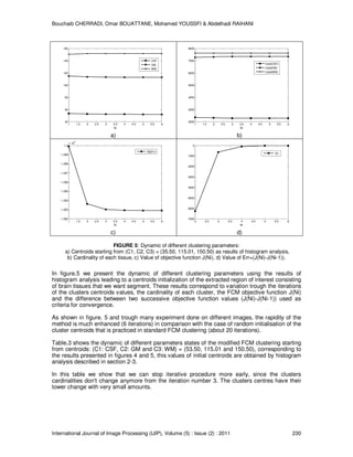
4.2.2 Parameters Dynamic of the MFCM.
Table.3 shows the Different parameters states of the Modified FCM clustering by starting from
centroids: (C1: CSF, C2: GM and C3: WM) = (53.50, 115.01 and 150.50), corresponding to the
results in figure 4 and figure 5.
Value of each Cluster Number of voxels in each Cluster ObjFcn value
Ni CSF GM WM Card(CSF) Card(GM) Card(WM) J(Ni)
1 53.50 115.01 150.50 2364 7270 7617 1299906.70
2 52.93 113.42 151.71 2322 7312 7617 1293634.77
3 52.51 113.05 151.78 2288 7346 7617 1293020.71
4 52.31 112.92 151.76 2288 7346 7617 1292923.43
5 52.23 112.87 151.74 2288 7346 7617 1292907.25
6 52.20 112.86 151.74 2288 7346 7617 1292904.56
TABLE 3: Different parameters states of the clustering method starting from centroids:
(C1: CSF, C2: GM and C3: WM) = (53.50, 115.01and 150.50).
11. Bouchaib CHERRADI, Omar BOUATTANE, Mohamed YOUSSFI & Abdelhadi RAIHANI
International Journal of Image Processing (IJIP), Volume (5) : Issue (2) : 2011 230
1 1.5 2 2.5 3 3.5 4 4.5 5 5.5 6
40
60
80
100
120
140
160
Ni
CSF
GM
WM
a)
1 1.5 2 2.5 3 3.5 4 4.5 5 5.5 6
2000
3000
4000
5000
6000
7000
8000
Ni
Card(CSF)
Card(GM)
Card(WM)
b)
1 1.5 2 2.5 3 3.5 4 4.5 5 5.5 6
1.292
1.293
1.294
1.295
1.296
1.297
1.298
1.299
1.3
x 10
6
Ni
ObjFcn
c)
2 2.5 3 3.5 4 4.5 5 5.5 6
-7000
-6000
-5000
-4000
-3000
-2000
-1000
0
Ni
Err
d)
FIGURE 5: Dynamic of different clustering parameters:
a) Centroids starting from (C1, C2, C3) = (35.50, 115.01, 150.50) as results of histogram analysis,
b) Cardinality of each tissue, c) Value of objective function J(Ni), d) Value of Err=(J(Ni)-J(Ni-1)).
In figure.5 we present the dynamic of different clustering parameters using the results of
histogram analysis leading to a centroids initialization of the extracted region of interest consisting
of brain tissues that we want segment. These results correspond to variation trough the iterations
of the clusters centroids values, the cardinality of each cluster, the FCM objective function J(Ni)
and the difference between two successive objective function values (J(Ni)-J(Ni-1)) used as
criteria for convergence.
As shown in figure. 5 and trough many experiment done on different images, the rapidity of the
method is much enhanced (6 iterations) in comparison with the case of random initialisation of the
cluster centroids that is practiced in standard FCM clustering (about 20 iterations).
Table.3 shows the dynamic of different parameters states of the modified FCM clustering starting
from centroids: (C1: CSF, C2: GM and C3: WM) = (53.50, 115.01 and 150.50), corresponding to
the results presented in figures 4 and 5, this values of initial centroids are obtained by histogram
analysis described in section 2-3.
In this table we show that we can stop iterative procedure more early, since the clusters
cardinalities don't change anymore from the iteration number 3. The clusters centres have their
tower change with very small amounts.
12. Bouchaib CHERRADI, Omar BOUATTANE, Mohamed YOUSSFI & Abdelhadi RAIHANI
International Journal of Image Processing (IJIP), Volume (5) : Issue (2) : 2011 231
4.2.3 Noise Robustness of the Proposed Method
To evaluate the robustness of the proposed method for tissue classification to the presence of
noise, we used Dataset1 with additional noise levels: 0%, 1%, 3%, 5%, 7% and 9%.
Dataset1: t1_icbm_normal_1mm_pn0_rf0 [40], is simulated normal brain phantom of
181x217x181 voxels with 1mm
3
for each voxel without any noise or intensities inhomogeneity.
TABLE 4: Performance measures for Modified FCM Clustering results of WM tissue (Dataset1).
In table.4 and table.5 we summarise the performance measure results calculated for segmented
GM and WM of different variants of Dataset1 obtained with additional noise of different amounts.
Noise JSC DSC Sensitivity Specificity
0% 0.952 0.975 0.657 0.897
1% 0.948 0.973 0.653 0.887
3% 0.929 0.963 0.642 0.880
5% 0.903 0.949 0.625 0.870
7% 0.874 0.932 0.614 0.850
9% 0.849 0.918 0.598 0.840
TABLE 5: Performance measures for Modified FCM Clustering results of GM tissue (Dataset1).
4.2.4 Visualisation of 3D Rendered Surface.
To appreciate the segmentation results obtained slice by slice, in 3D space, we export our
segmentation results to ANALYZE 10.0 that is a comprehensive and interactive package for
multidimensional image visualization, processing and analysis developed by The Biomedical
Imaging Resource at Mayo Clinic, Rochester, MN [42].
The figure.6 shows 3D rendered surface of the segmentation results for the three tissues
extracted from Dataset1. For the CSF we have limited the visualisation to the lateral ventricles.
a) b) c)
FIGURE 6: 3D visualization of rendered surface for segmented volume (Dataset1). a) Lateral Ventricular
CSF, b) GM, c) WM.
Noise JSC DSC Sensitivity Specificity
0% 0.942 0.955 0.757 0.895
1% 0.948 0.953 0.743 0.895
3% 0.919 0.943 0.732 0.899
5% 0.893 0.929 0.715 0.899
7% 0.854 0.922 0.704 0.897
9% 0.849 0.908 0.688 0.897
13. Bouchaib CHERRADI, Omar BOUATTANE, Mohamed YOUSSFI & Abdelhadi RAIHANI
International Journal of Image Processing (IJIP), Volume (5) : Issue (2) : 2011 232
5. CONCLUSION AND PERSPECTIVES
In this paper, we have presented a complete MRI images segmentation method. Unlike other
brain segmentation methods described in the literature, the one described in this paper is truly
automatic because it does not require a user to determine image-specific parameters, thresholds,
or regions of interest.
The automatic proposed method for extracting the brain from the T1-weighted MRI head scans is
based on a hybrid processing techniques including global optimal thresholding and mathematical
morphology operators. Our quantitative results show that the proposed method achieves
comparable performance with synthetic BrainWeb data, and real IBSR V2.0 data against
standard techniques such as BSE and BET, and McStrip. Our results are also more consistent
across the datasets, making the proposed method suited for measuring brain volumes in a clinical
setting.
Concerning the tissues classification we used modified FCM that is a clustering technique that
utilizes the distance between voxels and cluster centres in the spatial domain to compute the
membership function. The modification consists of using the histogram analysis for the
determination of initial cluster centroids instead of a random initialization. The segmentation
process is achieved in 6 iterations instead of about 20 iterations when we used standard FCM
with random initial centroids. This is important improvement (about 70%) especially in our case
where we manipulate big quantity of data.
The accuracy and the effectiveness of the fully automatic proposed method for 3D brain MR
images segmentation has been evaluated qualitatively and quantitatively, but more work can be
done to improve the method that need to be tested on many more data sets to expose
unexpected segmentation errors that might occur infrequently. The method should also be tested
with more recent images database.
More comprehensive comparison of MFCM with other clustering models will be addressed.
Future work will focus on developing an automatic image based classification system for brain
tumor using MRI data of different modalities and taking into account the intensity nonuniformity
artefact.
Another improvement in time processing can be gained while modifying the convergence criteria
for FCM clustering by considering a threshold on the change of clusters centres instead of
calculating the objective function.
An implementation of the proposed method as well as other algorithms for MRI segmentation on
massively parallel reconfigurable mesh computer emulator [32] is being finalised.
REFERENCES.
[1] L.P Clarke, R.P Velthijizen, M.A Camacho, J. J Heine, “MRI segmentation methods and
applications”, Magnetic Resonance Imaging, Vol. 13, No. 3, pp. 343-368, 1995.
[2] R. P. Woods, S. T. Grafton, J. D. G. Watson, N. L.Sicotte, and J. C. Mazziotta, “Automated
image registration: II. Intersubject validation of linear and nonlinear models”, J. of Computer
Assisted Tomography, vol. 22, pp: 139-152, 1998.
[3] R. P. Woods, M. Dapretto, N. L. Sicotte, A. W. Toga, and J. C. Mazziotta, “Creation and use of
a Talairach-Compatible atlas for accurate, automated, nonlinear intersubject registration, and
analysis of functional imaging data”, Human Brain Mapping, vol. 8, pp: 73-79, 1999.
[4] J. Van Horn, T. M. Ellmore, G. Esposito, K. F. and Berman, “Mapping Voxel-based Statistical
Power on Parametric Imaging”, NeuroImage, vol. 7, pp: 97-107, 1998.
14. Bouchaib CHERRADI, Omar BOUATTANE, Mohamed YOUSSFI & Abdelhadi RAIHANI
International Journal of Image Processing (IJIP), Volume (5) : Issue (2) : 2011 233
[5] D. W. Shattuck, S.R. Sandor-Leahy, K. A. Schaper, D. A. Rottenberg, and R. M. Leahy,
“Magnetic Resonance Image Tissue Classification Using a Partial Volume Model”,
NeuroImage, vol. 13, pp: 856-876, 2001.
[6] S. Strother, S. La Conte, L. Kai Hansen, J. Anderson, J. Zhang, S. Pulapura, and D.
Rottenberg, “Optimizing the fMRI data-processing pipeline using prediction and
reproducibility performance metrics: I. A preliminary group analysis”, NeuroImage, vol. 23,
pp:196-207, 2004.
[7] H. Rusinek, M. J. de Leon, A. E. George, L. A. Stylopoulos, R. Chandra, G. Smith, T. Rand, M.
Mourino, and H. Kowalski, “Alzheimer disease: measuring loss of cerebral gray matter with
MR imaging”, Radiology, vol. 178, pp: 109-114, 1991.
[8] P. M Thompson, M. S. Mega, R. P. Woods, C. I. Zoumalan, C. J. Lindshield, R. E. Blanton, J.
Moussai, C. J. Holmes, J. L. Cummings, and A. W. Toga, “Cortical change in Alzheimer’s
disease in detected with a disease specific population-based brain atlas”, Cerebral Cortex
vol. 11, pp: 1-16, 2001.
[9] R. A. Bermel, J. Sharma, C. W. Tjoa, S. R. Puli, and R. Bakshi, “A semiautomated measure of
whole- brain atrophy in multiple sclerosis”, Neurological Sciences, vol. 208, pp: 57-65, 2003.
[10] M. A. Horsfield, M. Rovaris, M. A. Rocca, P. Rossi, R. H. B. Benedict, M. Filippi, and R.
Bakshi, “Whole-brain atrophy in multiple sclerosis measured by two segmentation processes
from various MRI sequences”, Neurological Sciences, vol. 216, pp: 169-177, 2003.
[11] R. Zivadinov, F. Bagnato, D. Nasuelli, S. Bastianello, A. Bratina, L. Locatelli, K. Watts, L.
Finamore, A. Grop, M. Dwyer, M. Catalan, A. Clemenzi, E. Millefiorini, R. Bakshi, and M.
Zorzon, M., “ Short-term brain atrophy changes in relapsing-remitting multiple sclerosis”,
Neurological Sciences, vol. 223, pp: 185-193, 2004.
[12] J. Sharma, M. P. Sanfilipo, R. H. B. Benedict, B. Weinstock-Guttman, F. E. Munschauer, and
R. Bakshi, “Whole-brain atrophy in multiple sclerosis measured by automated versus semi-
automated MR imaging segmentation”, Neuroradiology, vol. 25, pp: 985-996, 2004.
[13] K. L. Narr, P. M. Thompson, P. Szeszko, D. Robinson, S. Jang, R. P. Woods, S. Kim, K. M.
Hayashi, D. Asunction, A. W. Toga, and R. M. Bilder, “Regional specificity of hippocampal
volume reductions in first episode schizophrenia”, NeuroImage, vol. 21, pp: 1563- 1575,
2004.
[14] P. Tanskanen, J. M. Veijola, U. K. Piippo, M. Haapea, J. A. Miettunen, J. Pyhtinen, E. T.
Bullmore, P. B. Jones, and M. K. Isohanni, “Hippocampus and amygdale volumes in
schizophrenia and other psychoses in the Northern Finland 1966 birth cohort”. Schizophrenia
Research, vol. 75, pp: 283-294, 2005.
[15] T. L. Jernigan, S. L. Archibald, C. Fennema-Notestine, A. C. Gamst, J. C. Stout, J. Bonner,
and J. R. Hesselink, “Effects of age on tissues and regions of the cerebrum and cerebellum”,
Neurobiology of Aging, vol. 22, pp: 581-594, 2001.
[16] R. E. Blanton, J. G. Levitt, J. R. Peterson, D. Fadale, M.L. Sporty, M. Lee, D. To, E. C.
Mormino, P. M.Thompson, J. T. McCracken, and A. W. Toga, “Gender differences in the left
inferior frontal gyrus in normal children”, NeuroImage, vol. 22, pp: 626-636, 2004.
[17] A.M. Dale AM, B. Fischl , and M. Sereno, “Cortical surface-based analysis. Segmentation
and surface reconstruction”, Neuroimage, vol. 9, pp: 179-94, 1999.
15. Bouchaib CHERRADI, Omar BOUATTANE, Mohamed YOUSSFI & Abdelhadi RAIHANI
International Journal of Image Processing (IJIP), Volume (5) : Issue (2) : 2011 234
[18] H. Hahn, and H-O. Peitgen, “The skull stripping problem in MRI solved by a single 3D
watershed transform”, MICCAI, LNCS 1935, pp: 134-143, 2000.
[19] S. Sandor, and R. Leahy, “Surface-based labeling of cortical anatomy using a deformable
database”, IEEE Transactions on Medical Imaging, vol. 16, pp: 41-54, 1997.
[20] F. Segonne, A. M. Dale, E. Busa, M. Glessner, D. Salat, H. K. Hahn, and B. Fischl, “A hybrid
approach to the skull stripping problem in MRI”, Neuroimage, vol. 22, pp :1060-75, 2004.
[21] S. M. Smith, “Fast robust automated brain extraction”, Human Brain Mapping, vol. 17, pp:
143-55, 2002.
[22] Shattuck, D.W., Sandor-Leahy, S.R., Shaper, K.A., Rottenberg, D.A., Leahy, R.M., Magnetic
resonance image tissue classification using a partial volume model. NeuroImage. 13 (5),
856–876. 2001.
[23] Rehm K, Shattuck D, Leahy R, Schaper K, Rottenberg DA. “Semi-automated stripping of T1
MRI volumes: I. Consensus of intensity- and edge-based methods”. NeuroImage. 9(6) S86,
1999.
[24] Rehm K, Schaper K, Anderson J, Woods R, Stoltzner S, Rottenberg D. “Putting our heads
together: a consensus approach to brain/non-brain segmentation in T1-weighted MR
volumes”. NeuroImage, November, 2003.
[25] B. Cherradi O. Bouattane, M. Youssfi and A. Raihani,” Brain Extraction and Fuzzy Tissue
Segmentation in Cerebral 2D T1-Weigthed Magnetic Resonance Images”. International
Journal of Computer Science Issues, Vol. 8, Issue 3, May 2011, In Press
[26] J. B. MacQueen, “Some Methods for classification and Analysis of Multivariate
Observations”, Proceedings of 5th Berkeley Symposium on Mathematical Statistics and
Probability, Berkeley, University of California Press, 1:281-297,1967.
[27] J.S.R. Jang, C. Sun, T.E. Mizutani, Neuro-Fuzzy and Soft Computing, Prentice Hall, pp. 426–
427. 1997.
[28] L. Dzung Pham, L.P. Jerry, “An adaptative fuzzy c-means algorithm for image segmentation
in the presence of intensity inhomogeneities”, Pattern Recognition Letters 20 57–68,1999.
[29] L. Ma, R.C. Staunton, “A modified fuzzy c-means image segmentation algorithm for use with
uneven illumination patterns”, Pattern Recognition 40 3005–3011, 2007.
[30] Y. Guo, H.D. Cheng, W. Zaho, Y. Zhang, “A novel image segmentation algorithm based on
fuzzy c-means algorithm and neutrosophic set”, in: Proceeding of the 11th Joint Conference
on Information Sciences, Atlantis Press, 2008.
[31] O. Bouattane, B. Cherradi, M. Youssfi and M.O. Bensalah. “Parallel c-means algorithm for
image segmentation on a reconfigurable mesh computer”. Parallel Computing, Volume 37,
Issues 4-5, Pages 230-243, April-May 2011.
[32] M. Youssfi, O. Bouattane, M.O. Bensalah, “A massively parallel re-configurable mesh
computer emulator: design, modeling and realization”, Journal of Software Engineering and
Applications vol 3, pp 11–26, 2010.
[33] N. Otsu, “A Threshold Selection Method from Gray-Level Histograms”. IEEE Transactions on
Systems, Man and Cybernetics, Vol. 9, No. 1, pp. 62-661979.
[34] B. Cherradi, O.Bouattane, M. Youssfi and A. Raihani, “Fuzzy segmentation method for MRI
images with spatial information”, in the proceeding of Mediterranien Congres of
Telecommunications (CMT2010). Casablanca, Morocco. pp 296-299, 2010.
16. Bouchaib CHERRADI, Omar BOUATTANE, Mohamed YOUSSFI & Abdelhadi RAIHANI
International Journal of Image Processing (IJIP), Volume (5) : Issue (2) : 2011 235
[35] J.C. Dunn, “A fuzzy relative of the ISODATA process and its use in detecting compact well-
separated clusters”, Journal of Cybernetics 3(3), pp. 32–57, 1973.
[36] J.C. Bezdek, “Pattern Recognition with Fuzzy Objective Function Algorithms”, Plenum
Press, New York 1981.
[37] P. Jaccard, “The distribution of the flora in the alpine zone”. New Phytol. 11 (2), 37–50, 1912.
[38] L. Dice, “Measures of the amount of ecologic association between species”, Ecology, vol. 26,
pp: 297- 302, 1945.
[39] MJ. Gardner and DG. Altman, “Calculating confidence intervals for proportions and their
differences”. BMJ Publishing Group, pp 28-33, 1989.
[40] http://www.bic.mni.mcgill.ca/brainweb/.
[41] http://www.cma.mgh.harvard.edu/ibsr/.
[42] http://www.mayo.edu/bir/Software/Analyze/Analyze.html.