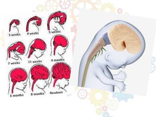
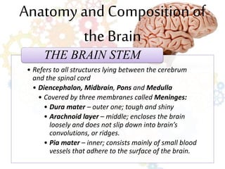
BRAIN DEVELOPMENT
THE BRAIN
 Three-pound organ of pinkish-gray tissue.
 10 billion nerve cells.
 Responsible for all mental functions.
 Control center for movement, sleep, hunger,
thirst and virtually other vital activity
necessary to survival.
 All human emotions, including love, hate,
fear, anger, elation and sadness.
 Receives and interprets the countless signals
sent from our body and environment.
The Brain Before and at Birth
4th month
• Medulla, Cerebellum, and Cerebrum
• All clearly separated
6th month (middle)
• A dentlike fissure appears on the surface of
the cerebrum
At birth
• Baby‘s brain contain 100 billion neurons
Anatomy and Composition of
the Brain
• Refers to all structures lying between the cerebrum
and the spinal cord
• Diencephalon, Midbrain, Pons and Medulla
• Covered by three membranes called Meninges:
• Dura mater – outer one; tough and shiny
• Arachnoid layer – middle; encloses the brain
loosely and does not slip down into brain’s
convolutions, or ridges.
• Pia mater – inner; consists mainly of small blood
vessels that adhere to the surface of the brain.
THE BRAIN STEM
THE CEREBRUM
The
largest
part of
the
human
brain.
85% of
the
brain’s
weight.
Cortex
- Large
surface area
- Intricate
development
account for
superior
intelligence.
Corpus
Callosum
- Slab of white
nerve fibers
that connects
the two
cerebral
hemispheres.
Cerebral
Cortex
- Outer layer
of gray matter
- 3 to 4 mm
thick
Five lobes
- Occipital,
frontal,
parietal, and
temporal
lobes.
- Insular lobe,
located
internally, not
visible at the
outside brain.
THE CEREBELLUM
- Lies in the
posterior or
hind part of
the cranium,
underneath
the cerebral
hemispheres,
Vermins
- White fibers
that connects
the two
multiridged
hemispheres.
Cerebral peduncles
- Three band of fibers
that connects
cerebellum to the other
parts of the brain.
- Superior (top) –
midbrain
- Middle – pons
- Inferior (bottom) -
medulla
- Essential to
the control
movement of
the body.
- Reflex
center for the
coordination
and precise
maintenance
of
equilibrium.
THALAMUS
- Part of
diencephalon
- Consist of two
rounded mass of
gray tissue. (very
middle of the brain)
- Crucial relay station
for incoming sensory
signals and outgoing
motor signals (going
to and from cerebral
cortex)
HYPOTHALAMUS
- Lies just below
the thalamus on
the midline base
of the brain.
Vital activities
necessary for
survival
- eating, drinking,
temperature
regulation, sleep,
emotional
behavior, and
sexual activity.
- Appears to act
as biological
timer.
Pons
- Located
between
medulla and
midbrain,
directly in
front of
cerebellum.
- Consist
mainly of
transverse
and
longitudinal
white nerve
fibers.
- A transverse
bridge of
fibers arises
from the
peduncle of
the
cerebellum
and join its
two halves.
- An intricate
longitudinal
fiber system
connects the
medulla with
the cerebral
hemispheres.
Limbic System
Makes up o
portions of the
thalamus,
hypothalamus,
hippocampsal
formation,
amygdala, caudate
nucleus, septum,
and
mesencephalon.
Linked together in
a unique way by
fiberpaths.
Control
multifaceted
behavior, including
emotional
expression,
seizure activity,
and memory
storage and recall.
CEREBRAL CORTEX
Brain’s outer layer
of gray matter
3 to 4 mm. thick
It is composed of
layers of
unmyelinated
(unsheated) cells
which in turn cover
an inner mass of
myelinated (white
sheathed) fibers
called white
matter.
• Somatomotor area – in front of central
fissure; responsible for nearly all
voluntary movements of the body
muscles.
• Somatosensory area – behind the central
fissure; receives impulses from the skin
surface as well as from structures
beneath the skin; sensation such as
touch and taste are processed here.
• Frontal lobe
– Personality, behavior, emotions
– Judgment, planning, problem solving
– Speech: speaking and writing (Broca’s area)
– Body movement (motor strip)
– Intelligence, concentration, self awareness
• Parietal lobe
– Interprets language, words
– Sense of touch, pain, temperature (sensory strip)
– Interprets signals from vision, hearing, motor, sensory and memory
– Spatial and visual perception
• Occipital lobe
– Interprets vision (color, light, movement)
• Temporal lobe
– Understanding language (Wernicke’s area)
– Memory
– Hearing
– Sequencing and organization
• Theories of neuroscientists on cellular
mechanisms by which nerve cells store memories
(Nash, 1998) :
– A change occurs in the ribonucleic acid (RNA) in the
cells of the cortex to code the memory trace in the
brain’s protein material.
– Peptides – (hormonlike substances) in the brain are
activated as an event is being stored as a memory.
– Neurotransmitters (chemicals that relay nerve
impulses between two or more neurons) are altered
as impulses stored.
THANK

Brain development

  • 1.
  • 2.
    THE BRAIN  Three-poundorgan of pinkish-gray tissue.  10 billion nerve cells.  Responsible for all mental functions.  Control center for movement, sleep, hunger, thirst and virtually other vital activity necessary to survival.  All human emotions, including love, hate, fear, anger, elation and sadness.  Receives and interprets the countless signals sent from our body and environment.
  • 3.
    The Brain Beforeand at Birth 4th month • Medulla, Cerebellum, and Cerebrum • All clearly separated 6th month (middle) • A dentlike fissure appears on the surface of the cerebrum At birth • Baby‘s brain contain 100 billion neurons
  • 5.
    Anatomy and Compositionof the Brain • Refers to all structures lying between the cerebrum and the spinal cord • Diencephalon, Midbrain, Pons and Medulla • Covered by three membranes called Meninges: • Dura mater – outer one; tough and shiny • Arachnoid layer – middle; encloses the brain loosely and does not slip down into brain’s convolutions, or ridges. • Pia mater – inner; consists mainly of small blood vessels that adhere to the surface of the brain. THE BRAIN STEM
  • 7.
    THE CEREBRUM The largest part of the human brain. 85%of the brain’s weight. Cortex - Large surface area - Intricate development account for superior intelligence. Corpus Callosum - Slab of white nerve fibers that connects the two cerebral hemispheres. Cerebral Cortex - Outer layer of gray matter - 3 to 4 mm thick Five lobes - Occipital, frontal, parietal, and temporal lobes. - Insular lobe, located internally, not visible at the outside brain.
  • 9.
    THE CEREBELLUM - Liesin the posterior or hind part of the cranium, underneath the cerebral hemispheres, Vermins - White fibers that connects the two multiridged hemispheres. Cerebral peduncles - Three band of fibers that connects cerebellum to the other parts of the brain. - Superior (top) – midbrain - Middle – pons - Inferior (bottom) - medulla - Essential to the control movement of the body. - Reflex center for the coordination and precise maintenance of equilibrium.
  • 10.
    THALAMUS - Part of diencephalon -Consist of two rounded mass of gray tissue. (very middle of the brain) - Crucial relay station for incoming sensory signals and outgoing motor signals (going to and from cerebral cortex)
  • 11.
    HYPOTHALAMUS - Lies justbelow the thalamus on the midline base of the brain. Vital activities necessary for survival - eating, drinking, temperature regulation, sleep, emotional behavior, and sexual activity. - Appears to act as biological timer.
  • 12.
    Pons - Located between medulla and midbrain, directlyin front of cerebellum. - Consist mainly of transverse and longitudinal white nerve fibers. - A transverse bridge of fibers arises from the peduncle of the cerebellum and join its two halves. - An intricate longitudinal fiber system connects the medulla with the cerebral hemispheres.
  • 13.
    Limbic System Makes upo portions of the thalamus, hypothalamus, hippocampsal formation, amygdala, caudate nucleus, septum, and mesencephalon. Linked together in a unique way by fiberpaths. Control multifaceted behavior, including emotional expression, seizure activity, and memory storage and recall.
  • 14.
    CEREBRAL CORTEX Brain’s outerlayer of gray matter 3 to 4 mm. thick It is composed of layers of unmyelinated (unsheated) cells which in turn cover an inner mass of myelinated (white sheathed) fibers called white matter.
  • 15.
    • Somatomotor area– in front of central fissure; responsible for nearly all voluntary movements of the body muscles. • Somatosensory area – behind the central fissure; receives impulses from the skin surface as well as from structures beneath the skin; sensation such as touch and taste are processed here.
  • 16.
    • Frontal lobe –Personality, behavior, emotions – Judgment, planning, problem solving – Speech: speaking and writing (Broca’s area) – Body movement (motor strip) – Intelligence, concentration, self awareness • Parietal lobe – Interprets language, words – Sense of touch, pain, temperature (sensory strip) – Interprets signals from vision, hearing, motor, sensory and memory – Spatial and visual perception • Occipital lobe – Interprets vision (color, light, movement) • Temporal lobe – Understanding language (Wernicke’s area) – Memory – Hearing – Sequencing and organization
  • 17.
    • Theories ofneuroscientists on cellular mechanisms by which nerve cells store memories (Nash, 1998) : – A change occurs in the ribonucleic acid (RNA) in the cells of the cortex to code the memory trace in the brain’s protein material. – Peptides – (hormonlike substances) in the brain are activated as an event is being stored as a memory. – Neurotransmitters (chemicals that relay nerve impulses between two or more neurons) are altered as impulses stored.
  • 18.