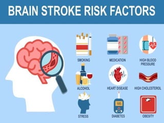
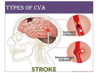
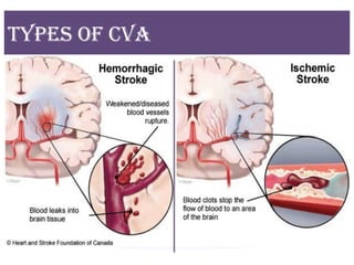
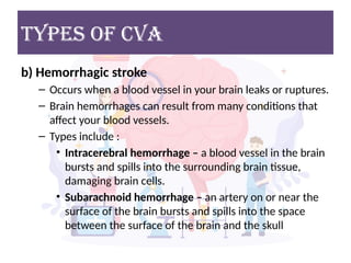
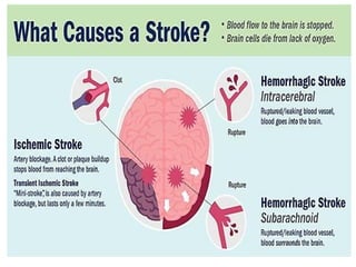
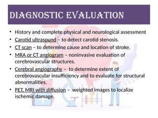
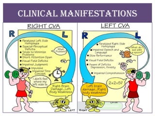
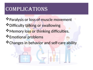
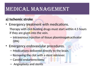
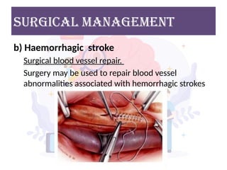
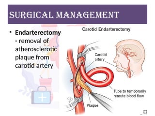
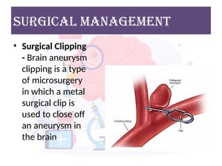
‘STROKE’
CVA / stroke
• An interruption of blood supply to a vital
center in the brain
• Also called “brain attack”, cerebral infarction,
cerebral hemorrhage, ischemic stroke or
stroke
DEFINITION
• “Cerebrovascular Accident (CVA) or Stroke” is
a term used to describe neurological changes
lasting more than 24 hours caused by an
interruption in the blood supply to a part of
the brain.
• If the blood flow ceases for an extended
period of time, the cerebral tissues involved
die causing permanent neurological deficits.
EPIDEMIOLOGY
• STROKE is one of the leading causes of death
and disability in INDIA.
• The estimated adjusted prevalence rate of
stroke range, 84-262/100,000 in rural and 334-
424/100,000 in urban area.
TYPES OF CVA
TYPES OF CVA
TYPES OF CVA
a) Ischemic stroke
– About 80 percent of strokes are ischemic strokes.
– Occur when the arteries to your brain become
narrowed or blocked, causing severely reduced
blood flow (ischemia).
– The most common ischemic strokes include:
• Thrombotic stroke – occurs when a blood clot (thrombus)
forms in one of the arteries that supply blood to the brain.
• Embolic stroke – occurs when a blood clot or other debris
forms away from the brain.
TYPES OF CVA
b) Hemorrhagic stroke
– Occurs when a blood vessel in your brain leaks or ruptures.
– Brain hemorrhages can result from many conditions that
affect your blood vessels.
– Types include :
• Intracerebral hemorrhage – a blood vessel in the brain
bursts and spills into the surrounding brain tissue,
damaging brain cells.
• Subarachnoid hemorrhage – an artery on or near the
surface of the brain bursts and spills into the space
between the surface of the brain and the skull
TYPES OF CVA
• c) Transient ischemic attack (TIA)
– A temporary decrease in blood supply to part of
the brain causes TIAs, which may last as little as
five minutes.
– Sometimes known as a ministroke.
Diagnostic evaluation
• History and complete physical and neurological assessment
• Carotid ultrasound – to detect carotid stenosis.
• CT scan – to determine cause and location of stroke.
• MRA or CT angiogram – noninvasive evaluation of
cerebrovascular structures.
• Cerebral angiography – to determine extent of
cerebrovascular insufficiency and to evaluate for structural
abnormalities.
• PET, MRI with diffusion – weighted images to localize
ischemic damage.
Brain functions
CLINICAL MANIFESTATIONS
Stroke recognition
complications
Paralysis or loss of muscle movement
Difficulty talking or swallowing
Memory loss or thinking difficulties.
Emotional problems
Changes in behavior and self-care ability
MEDICAL MANAGEMENT
a) Ischemic stroke
• Emergency treatment with medications.
Therapy with clot-busting drugs must start within 4.5 hours
if they are given into the vein.
– Intravenous injection of tissue plasminogen activator
(tPA)
• Emergency endovascular procedures.
– Medications delivered directly to the brain.
– Removing the clot with a stent retriever.
– Carotid endarterectomy.
– Angioplasty and stents
surgICAL MANAGEMENT
b) Haemorrhagic stroke
Surgical blood vessel repair.
Surgery may be used to repair blood vessel
abnormalities associated with hemorrhagic strokes
Surgical MANAGEMENT
• Endarterectomy
- removal of
atherosclerotic
plaque from
carotid artery
Surgical MANAGEMENT
• Surgical Clipping
- Brain aneurysm
clipping is a type
of microsurgery
in which a metal
surgical clip is
used to close off
an aneurysm in
the brain
• Anticoagulation after hemorrhage is ruled out.
• Antiplatelet agents
• Antispasmodic agents for spastic paralysis.
• A rehabilitation program, including physical
therapy, occupational therapy, speech therapy
(as soon as stable), and counseling as needed.
• Treatment of post-stroke depression with
antidepressants such as selective serotonin
reuptake inhibitors.
Subsequent Treatment
NURSING DIAGNOSIS
Ineffective tissue perfusion (cerebral)
Disturbed sensory perception
Impaired physical mobility
Risk for injury
Self-care deficit
Impaired verbal communication
Impaired swallowing
NURSING considerations
Maintain patent airway.
Monitor for changes in the client’s level of
consciousness (increased intracranial pressure
sign).
o Elevate the client’s head to reduce ICP and to
promote venous drainage. Avoid extreme flexion or
extension, maintain the head in a midline neutral
position and elevate the head of bed to 30 degrees.
o Institute seizure precautions
NURSING considerations
 Maintain a non-stimulating environment.
 Assist with communication skills if the client’s speech is
impaired.
 Assist with safe feeding
 Maintain skin integrity.
o Encourage PROM every 2 hr to the affected extremities and
AROM every 2 hr to the unaffected extremities.
 Elevate the affected extremities to promote venous
return and to reduce swelling.
 Maintain a safe environment to reduce the risks of falls.
THANK
YOU

CVA - Cerebrovascular Accident / Stroke

  • 1.
  • 2.
    CVA / stroke •An interruption of blood supply to a vital center in the brain • Also called “brain attack”, cerebral infarction, cerebral hemorrhage, ischemic stroke or stroke
  • 3.
    DEFINITION • “Cerebrovascular Accident(CVA) or Stroke” is a term used to describe neurological changes lasting more than 24 hours caused by an interruption in the blood supply to a part of the brain. • If the blood flow ceases for an extended period of time, the cerebral tissues involved die causing permanent neurological deficits.
  • 4.
    EPIDEMIOLOGY • STROKE isone of the leading causes of death and disability in INDIA. • The estimated adjusted prevalence rate of stroke range, 84-262/100,000 in rural and 334- 424/100,000 in urban area.
  • 6.
  • 7.
  • 8.
    TYPES OF CVA a)Ischemic stroke – About 80 percent of strokes are ischemic strokes. – Occur when the arteries to your brain become narrowed or blocked, causing severely reduced blood flow (ischemia). – The most common ischemic strokes include: • Thrombotic stroke – occurs when a blood clot (thrombus) forms in one of the arteries that supply blood to the brain. • Embolic stroke – occurs when a blood clot or other debris forms away from the brain.
  • 9.
    TYPES OF CVA b)Hemorrhagic stroke – Occurs when a blood vessel in your brain leaks or ruptures. – Brain hemorrhages can result from many conditions that affect your blood vessels. – Types include : • Intracerebral hemorrhage – a blood vessel in the brain bursts and spills into the surrounding brain tissue, damaging brain cells. • Subarachnoid hemorrhage – an artery on or near the surface of the brain bursts and spills into the space between the surface of the brain and the skull
  • 10.
    TYPES OF CVA •c) Transient ischemic attack (TIA) – A temporary decrease in blood supply to part of the brain causes TIAs, which may last as little as five minutes. – Sometimes known as a ministroke.
  • 12.
    Diagnostic evaluation • Historyand complete physical and neurological assessment • Carotid ultrasound – to detect carotid stenosis. • CT scan – to determine cause and location of stroke. • MRA or CT angiogram – noninvasive evaluation of cerebrovascular structures. • Cerebral angiography – to determine extent of cerebrovascular insufficiency and to evaluate for structural abnormalities. • PET, MRI with diffusion – weighted images to localize ischemic damage.
  • 13.
  • 14.
  • 15.
  • 16.
  • 17.
    complications Paralysis or lossof muscle movement Difficulty talking or swallowing Memory loss or thinking difficulties. Emotional problems Changes in behavior and self-care ability
  • 18.
    MEDICAL MANAGEMENT a) Ischemicstroke • Emergency treatment with medications. Therapy with clot-busting drugs must start within 4.5 hours if they are given into the vein. – Intravenous injection of tissue plasminogen activator (tPA) • Emergency endovascular procedures. – Medications delivered directly to the brain. – Removing the clot with a stent retriever. – Carotid endarterectomy. – Angioplasty and stents
  • 19.
    surgICAL MANAGEMENT b) Haemorrhagicstroke Surgical blood vessel repair. Surgery may be used to repair blood vessel abnormalities associated with hemorrhagic strokes
  • 20.
    Surgical MANAGEMENT • Endarterectomy -removal of atherosclerotic plaque from carotid artery
  • 21.
    Surgical MANAGEMENT • SurgicalClipping - Brain aneurysm clipping is a type of microsurgery in which a metal surgical clip is used to close off an aneurysm in the brain
  • 22.
    • Anticoagulation afterhemorrhage is ruled out. • Antiplatelet agents • Antispasmodic agents for spastic paralysis. • A rehabilitation program, including physical therapy, occupational therapy, speech therapy (as soon as stable), and counseling as needed. • Treatment of post-stroke depression with antidepressants such as selective serotonin reuptake inhibitors. Subsequent Treatment
  • 23.
    NURSING DIAGNOSIS Ineffective tissueperfusion (cerebral) Disturbed sensory perception Impaired physical mobility Risk for injury Self-care deficit Impaired verbal communication Impaired swallowing
  • 24.
    NURSING considerations Maintain patentairway. Monitor for changes in the client’s level of consciousness (increased intracranial pressure sign). o Elevate the client’s head to reduce ICP and to promote venous drainage. Avoid extreme flexion or extension, maintain the head in a midline neutral position and elevate the head of bed to 30 degrees. o Institute seizure precautions
  • 25.
    NURSING considerations  Maintaina non-stimulating environment.  Assist with communication skills if the client’s speech is impaired.  Assist with safe feeding  Maintain skin integrity. o Encourage PROM every 2 hr to the affected extremities and AROM every 2 hr to the unaffected extremities.  Elevate the affected extremities to promote venous return and to reduce swelling.  Maintain a safe environment to reduce the risks of falls.
  • 27.