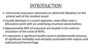
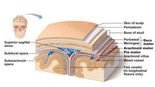
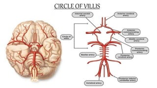
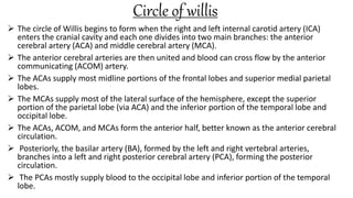
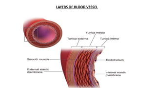
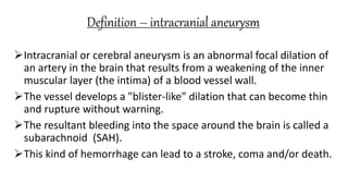
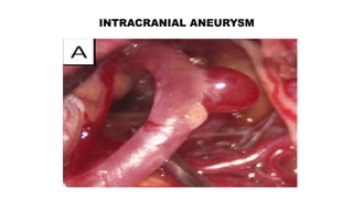
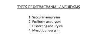
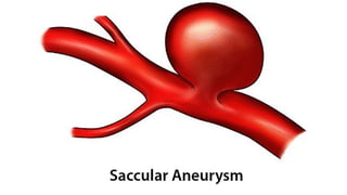
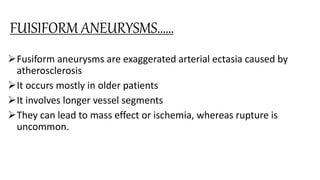
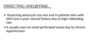
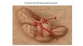
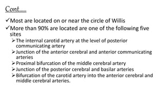
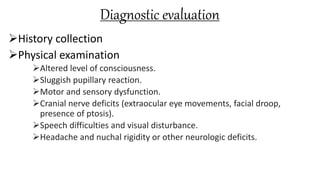
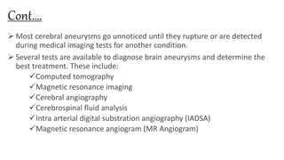
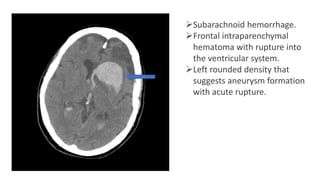
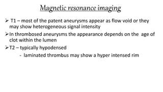
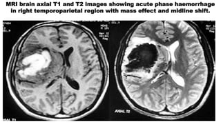
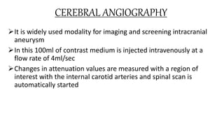
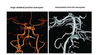
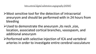
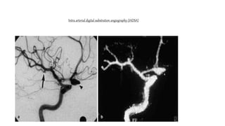
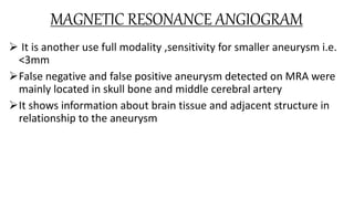
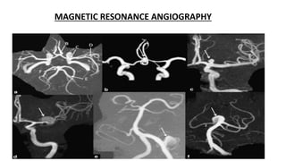
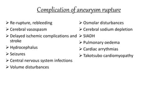
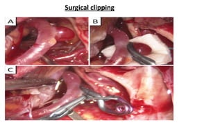
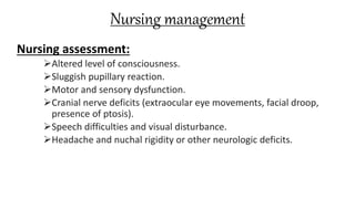
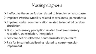
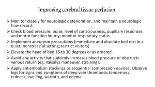
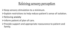
College Of Nursing
Madras Medical College
Chennai-03
INTRACRANIAL ANEURYSM
PRESENTED BY
EDWIN JOSE.L
MSc(nursing) I YEAR
College of nursing
Madras medical college
Chennai-03
INTRODUCTION
 Intracranial aneurysm represents an abnormal dilatation on the
arterial wall of the cerebral vessel.
Usually develops in a vessel segments ,most often near a
bifurcation point with an underlying structural abnormalities.
Approximately 85% of aneurysm are located in the anterior
circulation of the circle of Willis.
It represents a significant health concern predominantly because
of significant morbidity and mortality associated with rupture and
subarachnoid hemorrhage
CIRCLE OF VILLIS
Circle of willis
 The circle of Willis begins to form when the right and left internal carotid artery (ICA)
enters the cranial cavity and each one divides into two main branches: the anterior
cerebral artery (ACA) and middle cerebral artery (MCA).
 The anterior cerebral arteries are then united and blood can cross flow by the anterior
communicating (ACOM) artery.
 The ACAs supply most midline portions of the frontal lobes and superior medial parietal
lobes.
 The MCAs supply most of the lateral surface of the hemisphere, except the superior
portion of the parietal lobe (via ACA) and the inferior portion of the temporal lobe and
occipital lobe.
 The ACAs, ACOM, and MCAs form the anterior half, better known as the anterior cerebral
circulation.
 Posteriorly, the basilar artery (BA), formed by the left and right vertebral arteries,
branches into a left and right posterior cerebral artery (PCA), forming the posterior
circulation.
 The PCAs mostly supply blood to the occipital lobe and inferior portion of the temporal
lobe.
LAYERS OF BLOOD VESSEL
Definition – intracranial aneurysm
Intracranial or cerebral aneurysm is an abnormal focal dilation of
an artery in the brain that results from a weakening of the inner
muscular layer (the intima) of a blood vessel wall.
The vessel develops a "blister-like" dilation that can become thin
and rupture without warning.
The resultant bleeding into the space around the brain is called a
subarachnoid (SAH).
This kind of hemorrhage can lead to a stroke, coma and/or death.
INTRACRANIAL ANEURYSM
INCIDENCE
 Aneurysm are most prevalent between the ages of 35-60years
 The female to male ratio is 3:2 , but before 40 male and female s
are equally affected
 About 10-30%of patients can have multiple aneurysms
 About 3-5% of new strokes are due to aneurysm rupture related
to subarachnoid hemorrhage
RISK FACTORS
Alcohol abuse
Cigarette smoking
Female sex
Genetic condition
Hormonal therapy-estrogen therapy
Older age
Positive family history
Uncontrolled hypertension
GENETIC AND OTHEr MEDICAL CONDITIONS ASSOCIATED WITH
Aortic aneurysm
Bicuspid aortic valve defects
Coartcation of aorta
Ehrler's-Danlos syndrome
Fibromuscular dysplasia
Hereditary hemorrhagic telangiectasis
Intracranial arteriovenous malformation
Klinefelter syndrome
CONT……
Marfan’s syndrome
Microcephalic osteo dysplastic primordial dwarfism
Neuro fibromatosis
Noonan syndrome
Pheochromocytoma
Polycystic kidney disease
Pseudo xanthoma elasticum
Tuberous sclerosis
Alpha –antitrypsin deficiency
Alpha – glucosidase deficiency
TYPES OF INTRACRANIAL ANEURYSMS
1. Saccular aneurysm
2. Fusiform aneurysm
3. Dissecting aneurysm
4. Mycotic aneurysm
SACCULAR ANEURYSMS……
90% of intracranial aneurysm are saccular or “berry shaped” and
develop as a thin walled sac protruding from the arteries of the
circle of willis or its main branch
It consists of an out pouching of deficient collagenized tunica
muscularis that protrudes through a localized defect in the internal
elastic lamina
Tunica muscularis and the elastic lamina terminate at the
aneurysm neck and the aneurysm wall is very thin ,consisting of
only intima and adventitia.
85% of saccular aneurysm are found at the polygon of Willis.
Cont…..
 Anterior communicating artery – 30-35%
 Internal carotid artery – 30% (posterior communicating artery, carotid
bifurcation, ophthalmic artery)
 Middle cerebral artery – 22%
 Posterior circulation -8-10%
 According to size it is grouped into
 Small < 10mm
 Large 10-25mm
 Gaint >25mm
 According to the neck width
 Small neck < 4mm
 Large neck > 4mm
FUSIFORM ANEURYSM
FUISIFORM ANEURYSMS……
Fusiform aneurysms are exaggerated arterial ectasia caused by
atherosclerosis
It occurs mostly in older patients
It involves longer vessel segments
They can lead to mass effect or ischemia, whereas rupture is
uncommon.
DISSECTING ANEURYSMS….
 Dissecting aneurysm are rare and in patients who with
SAH have a poor natural history due to high rebleeding
rate
It usually seen on small perforated vessel due to chronic
hypertension
MYCOTIC ANEURYSM
It is very rare and develop from infection in the arterial wall often
deriving from bacterial endocarditis
Only distal branches of the cerebral arteries are commonly
involved and the aneurysm are fusiform and eccentric without the
neck that characterize saccular aneurysm.
Signs and symptoms
Unruptured aneurysm:
Most cerebral aneurysms do not show symptoms until they either become
very large or rupture. Small unchanging aneurysms generally will not
produce symptoms.
 A larger aneurysm that is steadily growing may press on tissues and nerves
causing:
 pain above and behind the eye
 numbness
 weakness
 paralysis on one side of the face
 a dilated pupil in the eye
 vision changes or double vision.
Cont…
 Ruptured aneurysm:
When an aneurysm ruptures (bursts), one always experiences a sudden and
extremely severe headache (e.g., the worst headache of one’s life) and may
also develop:
 double vision
 nausea
 vomiting
 Nuchal rigidity
 Photosensitivity- sensitivity to light
 Seizures
 loss of consciousness (this may happen briefly or may be prolonged)
 cardiac arrest.
Specific signs and symptoms
Anterior communicating artery:
 This is the most common site of aneurysmal SAH (34%).
 Usually, ACoA aneurysms are silent until they rupture.
 Suprachiasmatic pressure may cause visual field deficits, abulia or akinetic
mutism, amnestic syndromes, or hypothalamic dysfunction.
 Neurological deficits in aneurysmal rupture may reflect intraventricular
haemorrhage (79%), intraparenchymal haemorrhage (63%), acute
hydrocephalus (25%), or frontal lobe strokes (20%).
Anterior cerebral artery:
Aneurysms of this vessel, excluding ACoA, account for about 5% of
all cerebral aneurysms.
Most are asymptomatic until they rupture, although frontal lobe
syndromes, anosmia, or motor deficits may be noted.
Middle cerebral artery:
Aneurysms of the middle cerebral artery,account for about 20% of
aneurysms, typically at first or second division in the sylvian
fissure.
Aphasia, hemiparesis, hemisensory loss, anosognosia, or visual
field defects may be noted.
Internal carotid artery:
 Besides PCoA aneurysms, aneurysms of the ICA, account for about 4% of all
cerebral aneurysms.
 Supraglenoid aneurysms may cause ophthalmoplegia due to compression of
cranial nerve (CN) III or variable visual defects and optic atrophy due to
compression of the optic nerve.
 Chiasmal compression may produce bilateral temporal hemianopsia.
 Hypopituitarism or anosmia may be seen with giant aneurysms.
 Cavernous-carotid aneurysms exert mass effects within the cavernous sinus,
producing ophthalmoplegia and facial sensory loss.
 Rupture of these aneurysms typically produces a carotid-cavernous fistula,
SAH, or epistaxis.
Posterior communicating artery:
 Aneurysms present at the junction of the termination of the ICA and PCoA
account for 23% of cerebral aneurysms they are directed laterally,
posteriorly, and inferiorly.
 Pupillary dilatation, ophthalmoplegia, ptosis, mydriasis, and hemiparesis may
result.
Basilar artery:
 Basilar tip aneurysms,are the most common in the posterior circulation,
accounting for 5% of all aneurysms.
 Clinical findings usually are those associated with SAH, although bitemporal
hemianopsia or an oculomotor palsy may occur.
 Dolichoectatic aneurysms may cause bulbar dysfunction, respiratory
difficulties, or neurogenic pulmonary edema.
Leaking aneurysm
Sometimes an aneurysm may leak a small amount of blood into
the brain (called a sentinel bleed).
 Sentinel or warning headaches may result from an aneurysm that
suffers a tiny leak, days or weeks prior to a significant rupture.
However, only a minority of individuals have a sentinel headache
prior to rupture.
Common site of intracranial aneurysm
Cont…..
Most are located on or near the circle of Willis
More than 90% are located are one of the following five
sites
The internal carotid artery at the level of posterior
communicating artery
Junction of the anterior cerebral and anterior communicating
arteries
Proximal bifurcation of the middle cerebral artery
Junction of the posterior cerebral and basilar arteries
Bifurcation of the carotid artery into the anterior cerebral and
middle cerebral arteries.
Pathophysiology of intracranial aneurysm
Diagnostic evaluation
History collection
Physical examination
Altered level of consciousness.
Sluggish pupillary reaction.
Motor and sensory dysfunction.
Cranial nerve deficits (extraocular eye movements, facial droop,
presence of ptosis).
Speech difficulties and visual disturbance.
Headache and nuchal rigidity or other neurologic deficits.
Cont….
 Most cerebral aneurysms go unnoticed until they rupture or are detected
during medical imaging tests for another condition.
 Several tests are available to diagnose brain aneurysms and determine the
best treatment. These include:
Computed tomography
Magnetic resonance imaging
Cerebral angiography
Cerebrospinal fluid analysis
Intra arterial digital substration angiography (IADSA)
Magnetic resonance angiogram (MR Angiogram)
Computed tomography
 Reliable and simple diagnostic test for ruptured aneurysm
 Positivity rate is 98-100% of cases for up to 12 hours after onset
and 93% in the first 24 hours
 Positive results decreases with time
 Findings:
 Appears as a well defined round hyperattenuating lesions ,most
apparent on maximum intensity projection images
Subarachnoid hemorrhage.
Frontal intraparenchymal
hematoma with rupture into
the ventricular system.
Left rounded density that
suggests aneurysm formation
with acute rupture.
Fisher’s grade -sah
Magnetic resonance imaging
 T1 – most of the patent aneurysms appear as flow void or they
may show heterogeneous signal intensity
In thrombosed aneurysms the appearance depends on the age of
clot within the lumen
T2 – typically hypodensed
- laminated thrombus may show a hyper intensed rim
MRI brain axial T1 and T2 images showing acute phase haemorrhage
in right temporoparietal region with mass effect and midline shift.
CEREBRAL ANGIOGRAPHY
It is widely used modality for imaging and screening intracranial
aneurysm
In this 100ml of contrast medium is injected intravenously at a
flow rate of 4ml/sec
Changes in attenuation values are measured with a region of
interest with the internal carotid arteries and spinal scan is
automatically started
large vertebral junction aneurysm extravasation from the aneurysms
Cerebrospinal fluid analysis
Done in patient in whom CT reveals no abnormalities but hav
strong clinical history of SAH
Bloody CSF that fails to clear with continued egress of CSF
suggests SAH
Presence of xanthochromia ,a yellowish discoloration of the
cerebrospinal fluid representing bilirubin from the breakdown of
haemoglobin
Blood from SAH that occurred more than 12 hours before the
spinal tap will results in xanthochromic CSF where fresh blood
occurs with trauma
Intra arterial digital substration angiography (IADSA)
Most sensitive tool for the detection of intracranial
aneurysm and should be performed with in 24 hours from
bleeding
Used to demonstrate the aneurysm ,its neck ,size,
location, associated cortical branches, vasospasm, and
additional aneurysm
Performed with selective injection of ICA and vertebral
arteries in order to investigate entire cerebral vasculature
Intra arterial digital substration angiography (IADSA)
MAGNETIC RESONANCE ANGIOGRAM
 It is another use full modality ,sensitivity for smaller aneurysm i.e.
<3mm
False negative and false positive aneurysm detected on MRA were
mainly located in skull bone and middle cerebral artery
It shows information about brain tissue and adjacent structure in
relationship to the aneurysm
MAGNETIC RESONANCE ANGIOGRAPHY
Complication of aneurysm rupture
 Re-rupture, rebleeding
 Cerebral vasospasm
 Delayed ischemic complications and
stroke
 Hydrocephalus
 Seizures
 Central nervous system infections
 Volume disturbances
 Osmolar disturbances
 Cerebral sodium depletion
 SIADH
 Pulmonary oedema
 Cardiac arrythmias
 Takotsubo cardiomyopathy
management
Medical management
Unruptured aneurysm:
 Studies shows that there is correlation between modifiable risk factors such
as hypertension and smoking and aneurysm rupture
 Bed rest
 FFP
 Vitamin K
 Antiseizure drugs
 Analgesic agents
 Aspirin was successful in reducing aneurysm wall inflammation
Surgical management
Patients with a ruptured intracranial aneurysm should be treated
as soon as possible after the haemorrhage to prevent rebleeding
and to provide adequate medical treatment of vasospasm
Surgical clipping
Detachable coils- endovascular coiling
Surgical clipping
In 1937 Walter Dandy, an American neurosurgeon introduced
method of “clipping” ,who applied V shaped silver clip to the neck
of an internal carotid artery aneurysm.
Since then variety of aneurysm clips have been evolved
Aneurysm is clipped through a craniotomy ,a small metal clip
made up of titanium is then applied to neck of the aneurysm
Aneurysm clips comes in all different shapes and sizes and the
choice is based on the size and location of the aneurysm
Clip has a spring mechanism which allows the two jaws of the clip
to close around either side of the aneurysm thus occluding the
aneurysm
Surgical clipping
Endovascular coiling
 During 1980’s ,endovascular treatment of aneurysm with balloon was done
which has high rate of rupture
 In 1991, Guido Guglielmi ,an American neuroradiologist invented platinum
detachable micro-coil which was approved by FDA in 1995.
 Guglielmi detachable coil are soft wire spiral originally made out of
platinum
 These coils are deployed into the aneurysms via a microcatheter inserted
through the femoral artery and advanced into the brain
 Once the coil are released into the aneurysm the blood flow pattern within
the aneurysm is altered and the slow and sluggish remaining blood flow
leads to thrombosis
GUGLIELMI DETACHABLE COIL
Flow diversion
Flow diversion bridge the aneurysm neck and divert the blood
flow away from the aneurysm sac ,due to impedance created by
the mesh of the implant
Reduction of the blood flow into the aneurysmal sac causes stasis
of blood flow into the aneurysm which leads to thrombosis and
healing of the aneurysm
FLOW DIVERSION
Nursing management
Nursing assessment:
Altered level of consciousness.
Sluggish pupillary reaction.
Motor and sensory dysfunction.
Cranial nerve deficits (extraocular eye movements, facial droop,
presence of ptosis).
Speech difficulties and visual disturbance.
Headache and nuchal rigidity or other neurologic deficits.
Nursing diagnosis
Ineffective tissue perfusion related to bleeding or vasospasm.
Impaired Physical Mobility related to weakness ,paraesthesia
Impaired verbal communication related to impaired cerebral
circulation
Disturbed sensory perception related to altered sensory
reception, transmission, integration
Self care deficit related to neuromuscular impairment
Risk for impaired swallowing related to neuromuscular
impairment
Improving cerebral tissue perfusion
 Monitor closely for neurologic deterioration, and maintain a neurologic
flow record.
 Check blood pressure, pulse, level of consciousness, pupillary responses,
and motor function hourly; monitor respiratory status
 Implement aneurysm precautions (immediate and absolute bed rest in a
quiet, nonstressful setting; restrict visitors)
 Elevate the head of bed 15 to 30 degrees or as ordered.
 Avoid any activity that suddenly increases blood pressure or obstructs
venous return (eg, Valsalva maneuver, straining),
 Apply antiembolism stockings or sequential compression devices. Observe
legs for signs and symptoms of deep vein thrombosis tenderness,
redness, swelling, warmth, and edema.
Releiving sensory perception
Keep sensory stimulation to a minimum.
Explain restrictions to help reduce patient’s sense of isolation.
Relieving anxiety
Inform patient of plan of care.
Provide support and appropriate reassurance to patient and
family.
Monitoring and managing complications
 Assess for and immediately report signs of possible vasospasm, and administer
calcium channel blockers or fluid volume expanders as prescribed.
 Maintain seizure precautions and maintain airway and prevent injury if
a seizure occurs. Administer antiseizure medications as prescribed, phenytoin is
medication of choice.
 Monitor for onset of symptoms of hydrocephalus
 Monitor for and report symptoms of aneurysm rebleeding. Rebleeding occurs
most often in the first 2 weeks.
 Symptoms include sudden severe headache, nausea, vomiting, decreased level
of consciousness, and neurologic deficit.
 Administer medications as ordered.
 Hyponatremia: monitor sodium level.
Discharge and home care guidelines
Teach patients and family members about disease
condition
Medical treatments
Assistive devices
Follow-up environment
Thank you

Intracranial anurysm

  • 1.
    College Of Nursing MadrasMedical College Chennai-03 INTRACRANIAL ANEURYSM PRESENTED BY EDWIN JOSE.L MSc(nursing) I YEAR College of nursing Madras medical college Chennai-03
  • 2.
    INTRODUCTION  Intracranial aneurysmrepresents an abnormal dilatation on the arterial wall of the cerebral vessel. Usually develops in a vessel segments ,most often near a bifurcation point with an underlying structural abnormalities. Approximately 85% of aneurysm are located in the anterior circulation of the circle of Willis. It represents a significant health concern predominantly because of significant morbidity and mortality associated with rupture and subarachnoid hemorrhage
  • 6.
  • 7.
    Circle of willis The circle of Willis begins to form when the right and left internal carotid artery (ICA) enters the cranial cavity and each one divides into two main branches: the anterior cerebral artery (ACA) and middle cerebral artery (MCA).  The anterior cerebral arteries are then united and blood can cross flow by the anterior communicating (ACOM) artery.  The ACAs supply most midline portions of the frontal lobes and superior medial parietal lobes.  The MCAs supply most of the lateral surface of the hemisphere, except the superior portion of the parietal lobe (via ACA) and the inferior portion of the temporal lobe and occipital lobe.  The ACAs, ACOM, and MCAs form the anterior half, better known as the anterior cerebral circulation.  Posteriorly, the basilar artery (BA), formed by the left and right vertebral arteries, branches into a left and right posterior cerebral artery (PCA), forming the posterior circulation.  The PCAs mostly supply blood to the occipital lobe and inferior portion of the temporal lobe.
  • 8.
  • 9.
    Definition – intracranialaneurysm Intracranial or cerebral aneurysm is an abnormal focal dilation of an artery in the brain that results from a weakening of the inner muscular layer (the intima) of a blood vessel wall. The vessel develops a "blister-like" dilation that can become thin and rupture without warning. The resultant bleeding into the space around the brain is called a subarachnoid (SAH). This kind of hemorrhage can lead to a stroke, coma and/or death.
  • 10.
  • 11.
    INCIDENCE  Aneurysm aremost prevalent between the ages of 35-60years  The female to male ratio is 3:2 , but before 40 male and female s are equally affected  About 10-30%of patients can have multiple aneurysms  About 3-5% of new strokes are due to aneurysm rupture related to subarachnoid hemorrhage
  • 12.
    RISK FACTORS Alcohol abuse Cigarettesmoking Female sex Genetic condition Hormonal therapy-estrogen therapy Older age Positive family history Uncontrolled hypertension
  • 13.
    GENETIC AND OTHErMEDICAL CONDITIONS ASSOCIATED WITH Aortic aneurysm Bicuspid aortic valve defects Coartcation of aorta Ehrler's-Danlos syndrome Fibromuscular dysplasia Hereditary hemorrhagic telangiectasis Intracranial arteriovenous malformation Klinefelter syndrome
  • 14.
    CONT…… Marfan’s syndrome Microcephalic osteodysplastic primordial dwarfism Neuro fibromatosis Noonan syndrome Pheochromocytoma Polycystic kidney disease Pseudo xanthoma elasticum Tuberous sclerosis Alpha –antitrypsin deficiency Alpha – glucosidase deficiency
  • 15.
    TYPES OF INTRACRANIALANEURYSMS 1. Saccular aneurysm 2. Fusiform aneurysm 3. Dissecting aneurysm 4. Mycotic aneurysm
  • 17.
    SACCULAR ANEURYSMS…… 90% ofintracranial aneurysm are saccular or “berry shaped” and develop as a thin walled sac protruding from the arteries of the circle of willis or its main branch It consists of an out pouching of deficient collagenized tunica muscularis that protrudes through a localized defect in the internal elastic lamina Tunica muscularis and the elastic lamina terminate at the aneurysm neck and the aneurysm wall is very thin ,consisting of only intima and adventitia. 85% of saccular aneurysm are found at the polygon of Willis.
  • 18.
    Cont…..  Anterior communicatingartery – 30-35%  Internal carotid artery – 30% (posterior communicating artery, carotid bifurcation, ophthalmic artery)  Middle cerebral artery – 22%  Posterior circulation -8-10%  According to size it is grouped into  Small < 10mm  Large 10-25mm  Gaint >25mm  According to the neck width  Small neck < 4mm  Large neck > 4mm
  • 19.
  • 20.
    FUISIFORM ANEURYSMS…… Fusiform aneurysmsare exaggerated arterial ectasia caused by atherosclerosis It occurs mostly in older patients It involves longer vessel segments They can lead to mass effect or ischemia, whereas rupture is uncommon.
  • 22.
    DISSECTING ANEURYSMS….  Dissectinganeurysm are rare and in patients who with SAH have a poor natural history due to high rebleeding rate It usually seen on small perforated vessel due to chronic hypertension
  • 24.
    MYCOTIC ANEURYSM It isvery rare and develop from infection in the arterial wall often deriving from bacterial endocarditis Only distal branches of the cerebral arteries are commonly involved and the aneurysm are fusiform and eccentric without the neck that characterize saccular aneurysm.
  • 25.
    Signs and symptoms Unrupturedaneurysm: Most cerebral aneurysms do not show symptoms until they either become very large or rupture. Small unchanging aneurysms generally will not produce symptoms.  A larger aneurysm that is steadily growing may press on tissues and nerves causing:  pain above and behind the eye  numbness  weakness  paralysis on one side of the face  a dilated pupil in the eye  vision changes or double vision.
  • 26.
    Cont…  Ruptured aneurysm: Whenan aneurysm ruptures (bursts), one always experiences a sudden and extremely severe headache (e.g., the worst headache of one’s life) and may also develop:  double vision  nausea  vomiting  Nuchal rigidity  Photosensitivity- sensitivity to light  Seizures  loss of consciousness (this may happen briefly or may be prolonged)  cardiac arrest.
  • 27.
    Specific signs andsymptoms Anterior communicating artery:  This is the most common site of aneurysmal SAH (34%).  Usually, ACoA aneurysms are silent until they rupture.  Suprachiasmatic pressure may cause visual field deficits, abulia or akinetic mutism, amnestic syndromes, or hypothalamic dysfunction.  Neurological deficits in aneurysmal rupture may reflect intraventricular haemorrhage (79%), intraparenchymal haemorrhage (63%), acute hydrocephalus (25%), or frontal lobe strokes (20%).
  • 28.
    Anterior cerebral artery: Aneurysmsof this vessel, excluding ACoA, account for about 5% of all cerebral aneurysms. Most are asymptomatic until they rupture, although frontal lobe syndromes, anosmia, or motor deficits may be noted. Middle cerebral artery: Aneurysms of the middle cerebral artery,account for about 20% of aneurysms, typically at first or second division in the sylvian fissure. Aphasia, hemiparesis, hemisensory loss, anosognosia, or visual field defects may be noted.
  • 29.
    Internal carotid artery: Besides PCoA aneurysms, aneurysms of the ICA, account for about 4% of all cerebral aneurysms.  Supraglenoid aneurysms may cause ophthalmoplegia due to compression of cranial nerve (CN) III or variable visual defects and optic atrophy due to compression of the optic nerve.  Chiasmal compression may produce bilateral temporal hemianopsia.  Hypopituitarism or anosmia may be seen with giant aneurysms.  Cavernous-carotid aneurysms exert mass effects within the cavernous sinus, producing ophthalmoplegia and facial sensory loss.  Rupture of these aneurysms typically produces a carotid-cavernous fistula, SAH, or epistaxis.
  • 30.
    Posterior communicating artery: Aneurysms present at the junction of the termination of the ICA and PCoA account for 23% of cerebral aneurysms they are directed laterally, posteriorly, and inferiorly.  Pupillary dilatation, ophthalmoplegia, ptosis, mydriasis, and hemiparesis may result. Basilar artery:  Basilar tip aneurysms,are the most common in the posterior circulation, accounting for 5% of all aneurysms.  Clinical findings usually are those associated with SAH, although bitemporal hemianopsia or an oculomotor palsy may occur.  Dolichoectatic aneurysms may cause bulbar dysfunction, respiratory difficulties, or neurogenic pulmonary edema.
  • 31.
    Leaking aneurysm Sometimes ananeurysm may leak a small amount of blood into the brain (called a sentinel bleed).  Sentinel or warning headaches may result from an aneurysm that suffers a tiny leak, days or weeks prior to a significant rupture. However, only a minority of individuals have a sentinel headache prior to rupture.
  • 32.
    Common site ofintracranial aneurysm
  • 33.
    Cont….. Most are locatedon or near the circle of Willis More than 90% are located are one of the following five sites The internal carotid artery at the level of posterior communicating artery Junction of the anterior cerebral and anterior communicating arteries Proximal bifurcation of the middle cerebral artery Junction of the posterior cerebral and basilar arteries Bifurcation of the carotid artery into the anterior cerebral and middle cerebral arteries.
  • 34.
  • 35.
    Diagnostic evaluation History collection Physicalexamination Altered level of consciousness. Sluggish pupillary reaction. Motor and sensory dysfunction. Cranial nerve deficits (extraocular eye movements, facial droop, presence of ptosis). Speech difficulties and visual disturbance. Headache and nuchal rigidity or other neurologic deficits.
  • 36.
    Cont….  Most cerebralaneurysms go unnoticed until they rupture or are detected during medical imaging tests for another condition.  Several tests are available to diagnose brain aneurysms and determine the best treatment. These include: Computed tomography Magnetic resonance imaging Cerebral angiography Cerebrospinal fluid analysis Intra arterial digital substration angiography (IADSA) Magnetic resonance angiogram (MR Angiogram)
  • 37.
    Computed tomography  Reliableand simple diagnostic test for ruptured aneurysm  Positivity rate is 98-100% of cases for up to 12 hours after onset and 93% in the first 24 hours  Positive results decreases with time  Findings:  Appears as a well defined round hyperattenuating lesions ,most apparent on maximum intensity projection images
  • 38.
    Subarachnoid hemorrhage. Frontal intraparenchymal hematomawith rupture into the ventricular system. Left rounded density that suggests aneurysm formation with acute rupture.
  • 39.
  • 40.
    Magnetic resonance imaging T1 – most of the patent aneurysms appear as flow void or they may show heterogeneous signal intensity In thrombosed aneurysms the appearance depends on the age of clot within the lumen T2 – typically hypodensed - laminated thrombus may show a hyper intensed rim
  • 41.
    MRI brain axialT1 and T2 images showing acute phase haemorrhage in right temporoparietal region with mass effect and midline shift.
  • 42.
    CEREBRAL ANGIOGRAPHY It iswidely used modality for imaging and screening intracranial aneurysm In this 100ml of contrast medium is injected intravenously at a flow rate of 4ml/sec Changes in attenuation values are measured with a region of interest with the internal carotid arteries and spinal scan is automatically started
  • 43.
    large vertebral junctionaneurysm extravasation from the aneurysms
  • 44.
    Cerebrospinal fluid analysis Donein patient in whom CT reveals no abnormalities but hav strong clinical history of SAH Bloody CSF that fails to clear with continued egress of CSF suggests SAH Presence of xanthochromia ,a yellowish discoloration of the cerebrospinal fluid representing bilirubin from the breakdown of haemoglobin Blood from SAH that occurred more than 12 hours before the spinal tap will results in xanthochromic CSF where fresh blood occurs with trauma
  • 45.
    Intra arterial digitalsubstration angiography (IADSA) Most sensitive tool for the detection of intracranial aneurysm and should be performed with in 24 hours from bleeding Used to demonstrate the aneurysm ,its neck ,size, location, associated cortical branches, vasospasm, and additional aneurysm Performed with selective injection of ICA and vertebral arteries in order to investigate entire cerebral vasculature
  • 46.
    Intra arterial digitalsubstration angiography (IADSA)
  • 47.
    MAGNETIC RESONANCE ANGIOGRAM It is another use full modality ,sensitivity for smaller aneurysm i.e. <3mm False negative and false positive aneurysm detected on MRA were mainly located in skull bone and middle cerebral artery It shows information about brain tissue and adjacent structure in relationship to the aneurysm
  • 48.
  • 49.
    Complication of aneurysmrupture  Re-rupture, rebleeding  Cerebral vasospasm  Delayed ischemic complications and stroke  Hydrocephalus  Seizures  Central nervous system infections  Volume disturbances  Osmolar disturbances  Cerebral sodium depletion  SIADH  Pulmonary oedema  Cardiac arrythmias  Takotsubo cardiomyopathy
  • 50.
    management Medical management Unruptured aneurysm: Studies shows that there is correlation between modifiable risk factors such as hypertension and smoking and aneurysm rupture  Bed rest  FFP  Vitamin K  Antiseizure drugs  Analgesic agents  Aspirin was successful in reducing aneurysm wall inflammation
  • 51.
    Surgical management Patients witha ruptured intracranial aneurysm should be treated as soon as possible after the haemorrhage to prevent rebleeding and to provide adequate medical treatment of vasospasm Surgical clipping Detachable coils- endovascular coiling
  • 52.
    Surgical clipping In 1937Walter Dandy, an American neurosurgeon introduced method of “clipping” ,who applied V shaped silver clip to the neck of an internal carotid artery aneurysm. Since then variety of aneurysm clips have been evolved Aneurysm is clipped through a craniotomy ,a small metal clip made up of titanium is then applied to neck of the aneurysm Aneurysm clips comes in all different shapes and sizes and the choice is based on the size and location of the aneurysm Clip has a spring mechanism which allows the two jaws of the clip to close around either side of the aneurysm thus occluding the aneurysm
  • 53.
  • 54.
    Endovascular coiling  During1980’s ,endovascular treatment of aneurysm with balloon was done which has high rate of rupture  In 1991, Guido Guglielmi ,an American neuroradiologist invented platinum detachable micro-coil which was approved by FDA in 1995.  Guglielmi detachable coil are soft wire spiral originally made out of platinum  These coils are deployed into the aneurysms via a microcatheter inserted through the femoral artery and advanced into the brain  Once the coil are released into the aneurysm the blood flow pattern within the aneurysm is altered and the slow and sluggish remaining blood flow leads to thrombosis
  • 55.
  • 56.
    Flow diversion Flow diversionbridge the aneurysm neck and divert the blood flow away from the aneurysm sac ,due to impedance created by the mesh of the implant Reduction of the blood flow into the aneurysmal sac causes stasis of blood flow into the aneurysm which leads to thrombosis and healing of the aneurysm
  • 57.
  • 58.
    Nursing management Nursing assessment: Alteredlevel of consciousness. Sluggish pupillary reaction. Motor and sensory dysfunction. Cranial nerve deficits (extraocular eye movements, facial droop, presence of ptosis). Speech difficulties and visual disturbance. Headache and nuchal rigidity or other neurologic deficits.
  • 59.
    Nursing diagnosis Ineffective tissueperfusion related to bleeding or vasospasm. Impaired Physical Mobility related to weakness ,paraesthesia Impaired verbal communication related to impaired cerebral circulation Disturbed sensory perception related to altered sensory reception, transmission, integration Self care deficit related to neuromuscular impairment Risk for impaired swallowing related to neuromuscular impairment
  • 60.
    Improving cerebral tissueperfusion  Monitor closely for neurologic deterioration, and maintain a neurologic flow record.  Check blood pressure, pulse, level of consciousness, pupillary responses, and motor function hourly; monitor respiratory status  Implement aneurysm precautions (immediate and absolute bed rest in a quiet, nonstressful setting; restrict visitors)  Elevate the head of bed 15 to 30 degrees or as ordered.  Avoid any activity that suddenly increases blood pressure or obstructs venous return (eg, Valsalva maneuver, straining),  Apply antiembolism stockings or sequential compression devices. Observe legs for signs and symptoms of deep vein thrombosis tenderness, redness, swelling, warmth, and edema.
  • 61.
    Releiving sensory perception Keepsensory stimulation to a minimum. Explain restrictions to help reduce patient’s sense of isolation. Relieving anxiety Inform patient of plan of care. Provide support and appropriate reassurance to patient and family.
  • 62.
    Monitoring and managingcomplications  Assess for and immediately report signs of possible vasospasm, and administer calcium channel blockers or fluid volume expanders as prescribed.  Maintain seizure precautions and maintain airway and prevent injury if a seizure occurs. Administer antiseizure medications as prescribed, phenytoin is medication of choice.  Monitor for onset of symptoms of hydrocephalus  Monitor for and report symptoms of aneurysm rebleeding. Rebleeding occurs most often in the first 2 weeks.  Symptoms include sudden severe headache, nausea, vomiting, decreased level of consciousness, and neurologic deficit.  Administer medications as ordered.  Hyponatremia: monitor sodium level.
  • 63.
    Discharge and homecare guidelines Teach patients and family members about disease condition Medical treatments Assistive devices Follow-up environment
  • 65.