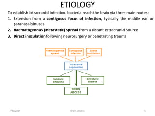
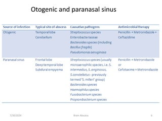
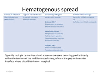
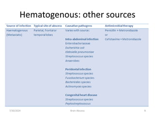
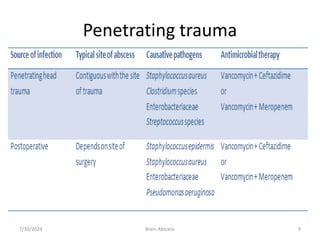
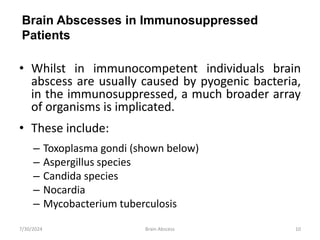
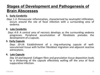
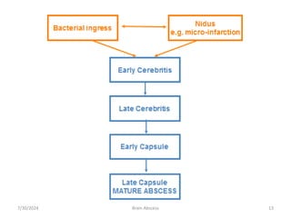
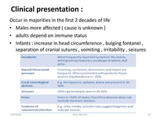
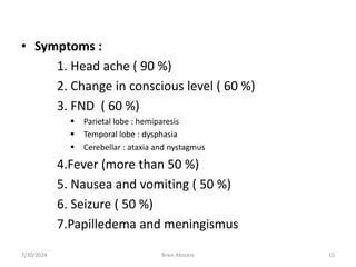
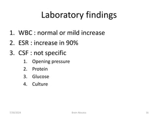
BENJAMIN KIZITO BIRUNGI
7/30/2024 1
Brain Abscess
BRAIN ABSCESS
By the end of this session you will be able to:
• Describe the stages of development and pathogenesis of brain
abscesses
• Identify the typical spectrum of pathogens implicated in the
aetiology of brain abscess
• Define the nonspecific nature of the clinical features of brain
abscess
• List the appropriate investigation of possible brain abscess and
to identify the important radiological features on CT and MR
scanning
• Develop a management plan for patients with brain abscess,
including antimicrobial treatment, surgical interventions and
adjunctive therapies
7/30/2024 Brain Abscess 2
• INCIDENCE:
• ETIOLOGY
• MICROBIOLOGY
• PATHOGENESIS
• CLINICAL PRESENTATION
• DIAGNOSIS
• MANAGEMENT
• OUTCOME
7/30/2024 3
Brain Abscess
INCIDENCE
• Is 1-2% of SOL in brain (USA)
• Is 8% (INDIA)
• Decreased incidence (because of antibiotic
and improved life)
• Lastly increased incidence because of
opportunistic infection in immune
compromised patient .
7/30/2024 4
Brain Abscess
ETIOLOGY
To establish intracranial infection, bacteria reach the brain via three main routes:
1. Extension from a contiguous focus of infection, typically the middle ear or
paranasal sinuses
2. Haematogenous (metastatic) spread from a distant extracranial source
3. Direct inoculation following neurosurgery or penetrating trauma
7/30/2024 5
Brain Abscess
7/30/2024 6
Brain Abscess
Otogenic and paranasal sinus
Hematogenous spread
7/30/2024 Brain Abscess 7
Typically, multiple or multi-loculated abscesses are seen, occurring predominantly
within the territory of the middle cerebral artery, often at the grey white matter
interface where blood flow is most marginal
Hematogenous: other sources
7/30/2024 Brain Abscess 8
Penetrating trauma
7/30/2024 Brain Abscess 9
• Whilst in immunocompetent individuals brain
abscess are usually caused by pyogenic bacteria,
in the immunosuppressed, a much broader array
of organisms is implicated.
• These include:
– Toxoplasma gondi (shown below)
– Aspergillus species
– Candida species
– Nocardia
– Mycobacterium tuberculosis
7/30/2024 10
Brain Abscess
Brain Abscesses in Immunosuppressed
Patients
PATHOGENESIS AND HISTOPATHOLOGY OF
BRAIN ABSCESS
• Brain abscesses occur at focal points of bacterial
multiplication within the brain parenchyma; they begin as a
localised area of cerebritis and later progress into a
collection of pus surrounded by a vascularised capsule.
• By virtue of the impermeability of the blood-brain barrier,
the brain parenchyma is relatively resistant to the
establishment of focal bacterial infection.
• An area of necrosis caused by for example micro-infarction
or hypoxaemia is necessary to act as a nidus for bacterial
multiplication.
• Several stages of development en route to the formation of
a mature encapsulated brain abscess following bacterial
ingress have been defined by neuroimaging studies
7/30/2024 11
Brain Abscess
1. Early Cerebritis
Days 1-3: Perivascular inflammation, characterised by neutrophil infiltration,
occurs around the site of focal infection with a surrounding area of
oedema.
2. Late Cerebritis
Days 4-9: A central area of necrosis develops as the surrounding oedema
progresses. Peripheral accumulation of fibroblasts preludes the
development of a capsule.
3. Early Capsule
Days 10-14: Establishment of a ring-enhancing capsule of well-
vascularised tissue with further fibroblast migration and adjacent reactive
astrocytosis.
4. Late Capsule
Day 14 and beyond: Collagen fibre and granulation tissue deposition leads
to a thickening of the capsule effectively walling off the area of focal
suppurative infection.
7/30/2024 12
Brain Abscess
Stages of Development and Pathogenesis of
Brain Abscesses
7/30/2024 Brain Abscess 13
Occur in majorities in the first 2 decades of life
• Males more affected ( cause is unknown )
• adults depend on immune status
• Infants : increase in head circumference , bulging fontanel ,
separation of cranial sutures , vomiting , irritability , seizures
7/30/2024 14
Brain Abscess
Clinical presentation :
• Symptoms :
1. Head ache ( 90 %)
2. Change in conscious level ( 60 %)
3. FND ( 60 %)
 Parietal lobe : hemiparesis
 Temporal lobe : dysphasia
 Cerebellar : ataxia and nystagmus
4.Fever (more than 50 %)
5. Nausea and vomiting ( 50 %)
6. Seizure ( 50 %)
7.Papilledema and meningismus
7/30/2024 Brain Abscess 15
Laboratory findings
1. WBC : normal or mild increase
2. ESR : increase in 90%
3. CSF : not specific
1. Opening pressure
2. Protein
3. Glucose
4. Culture
7/30/2024 Brain Abscess 16
4. radiological characteristic of brain abscess
1. Brain CTS with contrast
• ring enhancement
• Multi loculation
• Multiplicity
• Finding of gas
7/30/2024 Brain Abscess 17
• MRI :
• T1 :
• necrotic center ( hypointense)
• Capsule ( hyperintence)
• Edema ( hypointence)
• T2 :
• necrotic center ( hyperintence)
• Capsule ( hypointence)
• Edema ( hyperintence
7/30/2024 Brain Abscess 18
7/30/2024 Brain Abscess 19
7/30/2024 Brain Abscess 20
7/30/2024 Brain Abscess 21
7/30/2024 Brain Abscess 22
7/30/2024 Brain Abscess 23
management
The main treatment strategies are:
• Antimicrobial agents
• Needle aspiration - often using stereotactic guidance
• Complete surgical excision
In addition, adjunctive therapies such as corticosteroids and anticonvulsant agents are variably used.
A combination of antimicrobial therapy and aspiration is now used for the majority of cases, with
medical therapy alone and complete surgical excision reserved for particular circumstances
7/30/2024 Brain Abscess 24
Management
1. Antibiotic therapy :
• Antibiotic is mandatory and should given
• Antibiotics depends on C/S
• Imperial treatment depend on the etiology
– Sinusitis : ( penicillin + metronidazole )
– Otitis : ( penicillin + metronidazole + 3rd generation cephalosporin)
– Metastatic abscess :(metronidazole + 3rd generation cephalosporin)
– Post traumatic abscess ( vancomycin)
7/30/2024 Brain Abscess 25
It may also be considered for…
• Multiple small abscesses
• Abscesses located in surgically unaccessible or eloquent areas
It is most likely to be successful if…
• Abscesses are small (i.e. <1.5cm)
• In cerebritis stage
• Located in a well vascularised cortical area
Frequent interval scans should be performed to assess therapeutic response and to
identify complications requiring definitive surgical management.
7/30/2024 Brain Abscess 26
antibiotic therapy
Corticosteroids
– Adjuvant corticosteroids are often used to reduce vasogenic oedema
associated with brain abscesses.
There are important concerns regarding steroid use…
• Effectiveness in reducing oedema and mass effect not established in human
clinical trials
• May retard abscess capsule development
• May reduce antimicrobial penetration
• Give false impression of a therapeutic response by reducing ring-
enhancement on follow-up scans
Most authors recommend that corticosteroids are reserved for situations of
raised intracranial pressure resulting in a clear risk of brainstem herniation or
other significant neurological deficit.
7/30/2024 Brain Abscess 27
Anticonvulsants
– Seizures are a frequent complication of brain abscess both in the acute setting
and for a prolonged period after the resolution of the acute infection.
– Some advocate the use of seizure prophylaxis for extended periods in every
case of brain abscess
– If commenced, anticonvulsants should probably be continued for 6-12 months
and then only withdrawn if the patient is seizure-free, the EEG normal and no
signs of on going inflammation on neuroimaging.
7/30/2024 Brain Abscess 28
Aspiration
7/30/2024 Brain Abscess 29
Excision of brain abscess
• Advantages
1. Traumatic abscess ( contain foreign body and bone fragment )
2. Fungal abscess
3. Gas containing abscess
• Disadvantages
7/30/2024 Brain Abscess 30
Follow up
• CT weekly during antibiotic therapy
• And then monthly CT
• 2-3 week decrease size of abscess
• 3-4 months complete resolution of abscess
• 6-9 months no residual contrast
enhancement
7/30/2024 Brain Abscess 31
Outcome of abscess :
Mortality influenced by ( herniation , rupture of
abscess to the ventricle , clinical course of the
patient, type of abscess, neurological state of
patient at time of diagnosis)
7/30/2024 Brain Abscess 32
1. Long term morbidity : ( seizure , FND,
Cognitive dysfunction)
2. Recurrence: ( 5-10%) causes ( inadequate
antibiotic therapy, incorrect choice of AB,
presence of foreign body , failure to eradicate
source of the abscess)
7/30/2024 Brain Abscess 33
SUBDURAL AND EPIDURAL
EMPYEMA
7/30/2024 Brain Abscess 34
Subdural empyema
• This is an intracranial focal collection of pus
between the dura mater and the arachnoid
• Subdural empyema is usually unilateral
• There is tendecy to spread rapidly in the sub
dural space until limited by specific
boundaries( falx cerebr, tentorium cerebelli,
foramen magnum
• Causative bacteria same as those in prev slides
of brain abscess
7/30/2024 Brain Abscess 35
Pathophysiology
• Subdural empyema has a tendecy to behave like an
expanding mass
• This causes increased intracranial pressure and
cerebral intraparenchymal penetration
• Cerebral edema and hydrocephalus maybe develop
secondary to disruption in blood flow or CSF flow
• Cerebral infarction may develop due to thrombosis of
the the cortical veins or the carvenous sinus or from
septic venous thrombosis
7/30/2024 Brain Abscess 36
Pathophysisology
• In children, its often a complication of
meningitis. Therefore its important to
differentiate it from reactive subdural effusion.
• In older children and adults, it occurs as a
complication of paranasal sinusitis, otitis media
or mastoiditis
7/30/2024 Brain Abscess 37
Clinical presentation
• From history
– Fever above 38
– Headache: initially focal and later generalised
– Recent hx of sinusitis, otitis media, mastoiditis, meningitis,
cranial surgery, trauma, pulmonary infection,
– Confusion, drowsiness, stupor or coma
– Hemiparesis or hemiplegia
– Seizures: focal or generalized
– Nausea and vomiting
– Blurred vision: ambylopia
– Speech difficulty (dysphasia)
– Hx of intracranial abscess
7/30/2024 Brain Abscess 38
Physical examination
• Altered mental state
• Signs of meningeal irritation
• Focal neurologic deficits
• Aphasia or dysarthria
• Seizure
• Features of sinusitis
• Features of increased ICP
• Palsies of CN 3, 5, 6
7/30/2024 Brain Abscess 39
Investigations
• Labaratory studies
– CBC : leukocytosis
– Elevated ESR
– Blood culture
– Preoperative test: BUN, LFTs, electrolyts
Imaging studies
Cranial MRI is the choice( it outlines the extent of
subdyral empyema and greater morphological
details than CT scan
7/30/2024 Brain Abscess 40
• CT scan : shows hypodense area over the
hemisphere or along the falx
• Cranial ultrasound: important in
differentiating subdural empyema from
anechoic reactive subdural effusion in infants
with meningitis
• Other tests: EEG, chest radiograph,
7/30/2024 Brain Abscess 41
Treatment
• Maintain adequate airway and ensure
breathing and circulation
• Antibiotic therapy alone adequate in small
subdural empyema <1.5cm diameter
• Prophylactic anticonvulsants
• Treatment for increased ICP
7/30/2024 Brain Abscess 42
• Immediate surgical drainage should be
considered
– Craniotomy: best option
– Stereotatic burr hole placement with drainage and
irrigation
Grainage and debreidement of the primary source
of infection maybe necessary
7/30/2024 Brain Abscess 43
complications
• Seizures
• Cavernous sinus thrombosis from septic
thrombosis
• Increased ICP
• Hydrocephalous
• Cerebral infarction
• Cranial osteomyelitis
• Focal neurological deficits
7/30/2024 Brain Abscess 44
Epidural empyemaabscess
• Rare but pontentially life threatening
• Occus between the dura dnthe skull
• There are two types
– Spinal epidural abscess
– Intracranial epidural abscess
• The difference is where they develop and
some variationsi in risk factors
7/30/2024 Brain Abscess 45
Spinal epidural empyema
• Read more about it
7/30/2024 Brain Abscess 46
Intracranial epidural abscess
• Usually associated with subdural empyema
because the pus can cross the brain dura
along emissary veins
• Risk factirs include prior craniotomy, head
injury, sinusitis, otits media and mastoiditis.
• Common in males in the 2nd and 3rd decades
7/30/2024 Brain Abscess 47
Clinical presentation
• Fever
• Headache
• Malaise
• Lethargy
• Nausea and vomiting
• Focal neurological deficits
• Altered mental state
• evidence of infection
• seizure
7/30/2024 Brain Abscess 48
Management
• Craniotomy
• Antibiotic therapy
• Anticonvulsants
• corticosteroids
7/30/2024 Brain Abscess 49
7/30/2024 Brain Abscess 50

brain abscess explained in details, management

  • 1.
  • 2.
    BRAIN ABSCESS By theend of this session you will be able to: • Describe the stages of development and pathogenesis of brain abscesses • Identify the typical spectrum of pathogens implicated in the aetiology of brain abscess • Define the nonspecific nature of the clinical features of brain abscess • List the appropriate investigation of possible brain abscess and to identify the important radiological features on CT and MR scanning • Develop a management plan for patients with brain abscess, including antimicrobial treatment, surgical interventions and adjunctive therapies 7/30/2024 Brain Abscess 2
  • 3.
    • INCIDENCE: • ETIOLOGY •MICROBIOLOGY • PATHOGENESIS • CLINICAL PRESENTATION • DIAGNOSIS • MANAGEMENT • OUTCOME 7/30/2024 3 Brain Abscess
  • 4.
    INCIDENCE • Is 1-2%of SOL in brain (USA) • Is 8% (INDIA) • Decreased incidence (because of antibiotic and improved life) • Lastly increased incidence because of opportunistic infection in immune compromised patient . 7/30/2024 4 Brain Abscess
  • 5.
    ETIOLOGY To establish intracranialinfection, bacteria reach the brain via three main routes: 1. Extension from a contiguous focus of infection, typically the middle ear or paranasal sinuses 2. Haematogenous (metastatic) spread from a distant extracranial source 3. Direct inoculation following neurosurgery or penetrating trauma 7/30/2024 5 Brain Abscess
  • 6.
  • 7.
    Hematogenous spread 7/30/2024 BrainAbscess 7 Typically, multiple or multi-loculated abscesses are seen, occurring predominantly within the territory of the middle cerebral artery, often at the grey white matter interface where blood flow is most marginal
  • 8.
  • 9.
  • 10.
    • Whilst inimmunocompetent individuals brain abscess are usually caused by pyogenic bacteria, in the immunosuppressed, a much broader array of organisms is implicated. • These include: – Toxoplasma gondi (shown below) – Aspergillus species – Candida species – Nocardia – Mycobacterium tuberculosis 7/30/2024 10 Brain Abscess Brain Abscesses in Immunosuppressed Patients
  • 11.
    PATHOGENESIS AND HISTOPATHOLOGYOF BRAIN ABSCESS • Brain abscesses occur at focal points of bacterial multiplication within the brain parenchyma; they begin as a localised area of cerebritis and later progress into a collection of pus surrounded by a vascularised capsule. • By virtue of the impermeability of the blood-brain barrier, the brain parenchyma is relatively resistant to the establishment of focal bacterial infection. • An area of necrosis caused by for example micro-infarction or hypoxaemia is necessary to act as a nidus for bacterial multiplication. • Several stages of development en route to the formation of a mature encapsulated brain abscess following bacterial ingress have been defined by neuroimaging studies 7/30/2024 11 Brain Abscess
  • 12.
    1. Early Cerebritis Days1-3: Perivascular inflammation, characterised by neutrophil infiltration, occurs around the site of focal infection with a surrounding area of oedema. 2. Late Cerebritis Days 4-9: A central area of necrosis develops as the surrounding oedema progresses. Peripheral accumulation of fibroblasts preludes the development of a capsule. 3. Early Capsule Days 10-14: Establishment of a ring-enhancing capsule of well- vascularised tissue with further fibroblast migration and adjacent reactive astrocytosis. 4. Late Capsule Day 14 and beyond: Collagen fibre and granulation tissue deposition leads to a thickening of the capsule effectively walling off the area of focal suppurative infection. 7/30/2024 12 Brain Abscess Stages of Development and Pathogenesis of Brain Abscesses
  • 13.
  • 14.
    Occur in majoritiesin the first 2 decades of life • Males more affected ( cause is unknown ) • adults depend on immune status • Infants : increase in head circumference , bulging fontanel , separation of cranial sutures , vomiting , irritability , seizures 7/30/2024 14 Brain Abscess Clinical presentation :
  • 15.
    • Symptoms : 1.Head ache ( 90 %) 2. Change in conscious level ( 60 %) 3. FND ( 60 %)  Parietal lobe : hemiparesis  Temporal lobe : dysphasia  Cerebellar : ataxia and nystagmus 4.Fever (more than 50 %) 5. Nausea and vomiting ( 50 %) 6. Seizure ( 50 %) 7.Papilledema and meningismus 7/30/2024 Brain Abscess 15
  • 16.
    Laboratory findings 1. WBC: normal or mild increase 2. ESR : increase in 90% 3. CSF : not specific 1. Opening pressure 2. Protein 3. Glucose 4. Culture 7/30/2024 Brain Abscess 16
  • 17.
    4. radiological characteristicof brain abscess 1. Brain CTS with contrast • ring enhancement • Multi loculation • Multiplicity • Finding of gas 7/30/2024 Brain Abscess 17
  • 18.
    • MRI : •T1 : • necrotic center ( hypointense) • Capsule ( hyperintence) • Edema ( hypointence) • T2 : • necrotic center ( hyperintence) • Capsule ( hypointence) • Edema ( hyperintence 7/30/2024 Brain Abscess 18
  • 19.
  • 20.
  • 21.
  • 22.
  • 23.
  • 24.
    management The main treatmentstrategies are: • Antimicrobial agents • Needle aspiration - often using stereotactic guidance • Complete surgical excision In addition, adjunctive therapies such as corticosteroids and anticonvulsant agents are variably used. A combination of antimicrobial therapy and aspiration is now used for the majority of cases, with medical therapy alone and complete surgical excision reserved for particular circumstances 7/30/2024 Brain Abscess 24
  • 25.
    Management 1. Antibiotic therapy: • Antibiotic is mandatory and should given • Antibiotics depends on C/S • Imperial treatment depend on the etiology – Sinusitis : ( penicillin + metronidazole ) – Otitis : ( penicillin + metronidazole + 3rd generation cephalosporin) – Metastatic abscess :(metronidazole + 3rd generation cephalosporin) – Post traumatic abscess ( vancomycin) 7/30/2024 Brain Abscess 25
  • 26.
    It may alsobe considered for… • Multiple small abscesses • Abscesses located in surgically unaccessible or eloquent areas It is most likely to be successful if… • Abscesses are small (i.e. <1.5cm) • In cerebritis stage • Located in a well vascularised cortical area Frequent interval scans should be performed to assess therapeutic response and to identify complications requiring definitive surgical management. 7/30/2024 Brain Abscess 26 antibiotic therapy
  • 27.
    Corticosteroids – Adjuvant corticosteroidsare often used to reduce vasogenic oedema associated with brain abscesses. There are important concerns regarding steroid use… • Effectiveness in reducing oedema and mass effect not established in human clinical trials • May retard abscess capsule development • May reduce antimicrobial penetration • Give false impression of a therapeutic response by reducing ring- enhancement on follow-up scans Most authors recommend that corticosteroids are reserved for situations of raised intracranial pressure resulting in a clear risk of brainstem herniation or other significant neurological deficit. 7/30/2024 Brain Abscess 27
  • 28.
    Anticonvulsants – Seizures area frequent complication of brain abscess both in the acute setting and for a prolonged period after the resolution of the acute infection. – Some advocate the use of seizure prophylaxis for extended periods in every case of brain abscess – If commenced, anticonvulsants should probably be continued for 6-12 months and then only withdrawn if the patient is seizure-free, the EEG normal and no signs of on going inflammation on neuroimaging. 7/30/2024 Brain Abscess 28
  • 29.
  • 30.
    Excision of brainabscess • Advantages 1. Traumatic abscess ( contain foreign body and bone fragment ) 2. Fungal abscess 3. Gas containing abscess • Disadvantages 7/30/2024 Brain Abscess 30
  • 31.
    Follow up • CTweekly during antibiotic therapy • And then monthly CT • 2-3 week decrease size of abscess • 3-4 months complete resolution of abscess • 6-9 months no residual contrast enhancement 7/30/2024 Brain Abscess 31
  • 32.
    Outcome of abscess: Mortality influenced by ( herniation , rupture of abscess to the ventricle , clinical course of the patient, type of abscess, neurological state of patient at time of diagnosis) 7/30/2024 Brain Abscess 32
  • 33.
    1. Long termmorbidity : ( seizure , FND, Cognitive dysfunction) 2. Recurrence: ( 5-10%) causes ( inadequate antibiotic therapy, incorrect choice of AB, presence of foreign body , failure to eradicate source of the abscess) 7/30/2024 Brain Abscess 33
  • 34.
  • 35.
    Subdural empyema • Thisis an intracranial focal collection of pus between the dura mater and the arachnoid • Subdural empyema is usually unilateral • There is tendecy to spread rapidly in the sub dural space until limited by specific boundaries( falx cerebr, tentorium cerebelli, foramen magnum • Causative bacteria same as those in prev slides of brain abscess 7/30/2024 Brain Abscess 35
  • 36.
    Pathophysiology • Subdural empyemahas a tendecy to behave like an expanding mass • This causes increased intracranial pressure and cerebral intraparenchymal penetration • Cerebral edema and hydrocephalus maybe develop secondary to disruption in blood flow or CSF flow • Cerebral infarction may develop due to thrombosis of the the cortical veins or the carvenous sinus or from septic venous thrombosis 7/30/2024 Brain Abscess 36
  • 37.
    Pathophysisology • In children,its often a complication of meningitis. Therefore its important to differentiate it from reactive subdural effusion. • In older children and adults, it occurs as a complication of paranasal sinusitis, otitis media or mastoiditis 7/30/2024 Brain Abscess 37
  • 38.
    Clinical presentation • Fromhistory – Fever above 38 – Headache: initially focal and later generalised – Recent hx of sinusitis, otitis media, mastoiditis, meningitis, cranial surgery, trauma, pulmonary infection, – Confusion, drowsiness, stupor or coma – Hemiparesis or hemiplegia – Seizures: focal or generalized – Nausea and vomiting – Blurred vision: ambylopia – Speech difficulty (dysphasia) – Hx of intracranial abscess 7/30/2024 Brain Abscess 38
  • 39.
    Physical examination • Alteredmental state • Signs of meningeal irritation • Focal neurologic deficits • Aphasia or dysarthria • Seizure • Features of sinusitis • Features of increased ICP • Palsies of CN 3, 5, 6 7/30/2024 Brain Abscess 39
  • 40.
    Investigations • Labaratory studies –CBC : leukocytosis – Elevated ESR – Blood culture – Preoperative test: BUN, LFTs, electrolyts Imaging studies Cranial MRI is the choice( it outlines the extent of subdyral empyema and greater morphological details than CT scan 7/30/2024 Brain Abscess 40
  • 41.
    • CT scan: shows hypodense area over the hemisphere or along the falx • Cranial ultrasound: important in differentiating subdural empyema from anechoic reactive subdural effusion in infants with meningitis • Other tests: EEG, chest radiograph, 7/30/2024 Brain Abscess 41
  • 42.
    Treatment • Maintain adequateairway and ensure breathing and circulation • Antibiotic therapy alone adequate in small subdural empyema <1.5cm diameter • Prophylactic anticonvulsants • Treatment for increased ICP 7/30/2024 Brain Abscess 42
  • 43.
    • Immediate surgicaldrainage should be considered – Craniotomy: best option – Stereotatic burr hole placement with drainage and irrigation Grainage and debreidement of the primary source of infection maybe necessary 7/30/2024 Brain Abscess 43
  • 44.
    complications • Seizures • Cavernoussinus thrombosis from septic thrombosis • Increased ICP • Hydrocephalous • Cerebral infarction • Cranial osteomyelitis • Focal neurological deficits 7/30/2024 Brain Abscess 44
  • 45.
    Epidural empyemaabscess • Rarebut pontentially life threatening • Occus between the dura dnthe skull • There are two types – Spinal epidural abscess – Intracranial epidural abscess • The difference is where they develop and some variationsi in risk factors 7/30/2024 Brain Abscess 45
  • 46.
    Spinal epidural empyema •Read more about it 7/30/2024 Brain Abscess 46
  • 47.
    Intracranial epidural abscess •Usually associated with subdural empyema because the pus can cross the brain dura along emissary veins • Risk factirs include prior craniotomy, head injury, sinusitis, otits media and mastoiditis. • Common in males in the 2nd and 3rd decades 7/30/2024 Brain Abscess 47
  • 48.
    Clinical presentation • Fever •Headache • Malaise • Lethargy • Nausea and vomiting • Focal neurological deficits • Altered mental state • evidence of infection • seizure 7/30/2024 Brain Abscess 48
  • 49.
    Management • Craniotomy • Antibiotictherapy • Anticonvulsants • corticosteroids 7/30/2024 Brain Abscess 49
  • 50.