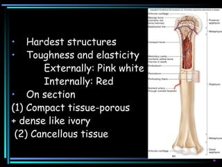
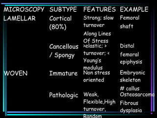
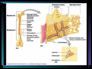
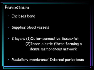
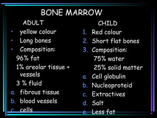
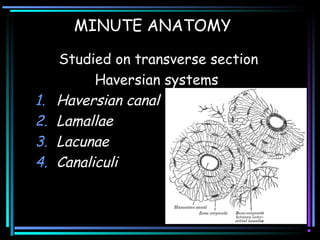
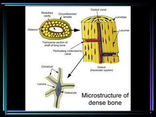
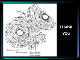
BONE ARCHITECTURE
Dnyanesh Lad
• Hardest structures
• Toughness and elasticity
Externally: Pink white
Internally: Red
• On section
(1) Compact tissue-porous
+ dense like ivory
(2) Cancellous tissue
MICROSCOPY SUBTYPE FEATURES EXAMPLE
LAMELLAR Cortical
(80%)
Strong; slow
turnover
Along Lines
Of Stress
Femoral
shaft
Cancellous
/ Spongy
>elastic; >
turnover; <
Young’s
modulus
Distal
femoral
epiphysis
WOVEN Immature Non stress
oriented
Embryonic
skeleton
# callus
Pathologic Weak,
Flexible,High
turnover,
Osteosarcoma
Fibrous
dysplasia
Periosteum
• Encloses bone
• Supplies blood vessels
• 2 layers (1)Outer-connective tissue+fat
(2)Inner-elastic fibres forming a
dense membranous network
• Medullary membrane/ Internal periosteum
Periosteum
In young
• Thick
• Vascular,
• Loosely attached
• Separated by soft
tissue with
osteoblasts
• ossification on
exterior of bone
In adults
• Thin
• Less vascular
• Closely attached
• Osteoblasts
converted to
epitheloid layer.
• Carry lymph from
bone
• RAMIFICATION OF VESSELS
• LIABILITY OF BONE
EXFOLIATION OR NECROSIS.
BONE MARROW
ADULT
• yellow colour
• Long bones
• Composition:
96% fat
1% areolar tissue +
vessels
3 % fluid
a. fibrous tissue
b. blood vessels
c. cells
CHILD
1. Red colour
2. Short flat bones
3. Composition:
75% water
25% solid matter
a. Cell globulin
b. Nucleoproteid
c. Extractives
d. Salt
e. Less fat
• TRUE “MARROW CELLS” OF KOLLIKER
red marrow
Majority of the cells
Roundish nucleated cells
smallest-erythroblasts
• GIANT CELLS-MYELOPLAQUES-
OSTEOCLASTS
more in red marrow
large
multinucleated
protoplasmic mass
‘integrin’-attach
TRAP + CA
Fn: absorbtion of bone matrix
Evacuate pits/cavities-”HOWSHIPS
LACUNAE”
VESSELS
• Compact bone- more numerous
• Cancellous bone-less numerous
larger size.
Come through compact tissue
• Largest and most numerous apertures
for veins of cancellous tissue.
MEDULLARY / NUTRIENT ARTERY
High pressure system
nutrient foramen; shaft
accompanied by 1 or 2 veins
branches in both directions to
medullary membrane
anastamoses with br. Of compact and
cancellous tissue.
• METAPHYSEAL/EPIPHYEAL VESSELS
From periarticular vascular plexus-
geniculate arteries
• PERIOSTEAL SYSTEM
Low pressure system
• Arterial flow in mature bone is centrifugal
d/t high pressure endosteal system
• In case of displaced fracture flow is
reversed-becomes centripetal.
• Arterial flow in immature bone-centripetal
since periosteum is highly vascularised.
VEINS
Emerge from 3 places
1. Accompany artery
2. At the articular extremities.
3. Arising in compact tissue
Venous flow in mature bone-centripetal
Cortical vessels-venous sinusoids-emissary
venous system.
LYMPHATICS
1. In periosteum
2. Within bone in Haversian system
NERVES
1. Freely distributed in periosteum
2. Accompany nutrient vessels to interior
3. Most numerous in –articular extremities
vertebrae
larger flat bones
MINUTE ANATOMY
Studied on transverse section
Haversian systems
1. Haversian canal
2. Lamallae
3. Lacunae
4. Canaliculi
Haversian Canal / Volkmanns Canal
• // to long axis of bone
• Branch + communicate
• Average diameter: 1/500th
of an inch
• Larger near medullary cavity
• Contains- 2 blood vessels
delicate connective tissue
nerve filaments
lymphatics
LAMELLAE
Primary / Fundamental / Circumferential
-medullary cavity of bone would be the HC
Secondary / Special
-Those laid down around actual HC
Interstitial
-between the various circular systems, filling the
irregular intervals
LACUNAE
branched bone cell / corpuscle
Processes of cell pass through canaliculi
Fn: fluids for nutrition brought in contact
with bone
CANALICULI
Do not communicate with the lacunae of
neighboring HS.
CHEMICAL STRUCTURE
• Animal Part / Organic Part: 25%
Bone Cells (4%) Matrix (20%)
• Earthy / Inorganic Part 65%
crystalline hydroxyappetite
amorphous Ca2(PO4)3
Remainder: CaCO3, CaCl2 and MgSO4
Trapped ions: citrate, fluoride, Na K Mg
OSSIFICATION
• Intramembranous
skull, maxilla, mandible, clavicle
mesenchymal tissue-scaffold for ossifn
• Intracartilaginous
• Ring of Lacroix-epiphsial ring formed. Does
not further extend to epiphysis
• Zone of Ranvier- ROL + lateral part of
physis + mesenchymal cells
Ossification Centres
• Short bones: single point; in the centre
• Long bone: central point for diaphysis (1st
)
1 or more for each extremity-epiphysis
• Union of epiphysis with shaft occurs in
reverse order to that in which ossification
began (exception : fibula)
• Regulated by direction of nutrient artery
(knee-away ; elbow-towards)
• When 1 epiphysis; medullary artery is
directed towards it.
• Eg: acromial end of clavicle
distal end of the metacarpal bone of
thumb and great toe
proximal end of other metacarpals
and metatarsals
THANK
YOU

Bone architecture

  • 1.
  • 2.
    • Hardest structures •Toughness and elasticity Externally: Pink white Internally: Red • On section (1) Compact tissue-porous + dense like ivory (2) Cancellous tissue
  • 3.
    MICROSCOPY SUBTYPE FEATURESEXAMPLE LAMELLAR Cortical (80%) Strong; slow turnover Along Lines Of Stress Femoral shaft Cancellous / Spongy >elastic; > turnover; < Young’s modulus Distal femoral epiphysis WOVEN Immature Non stress oriented Embryonic skeleton # callus Pathologic Weak, Flexible,High turnover, Osteosarcoma Fibrous dysplasia
  • 5.
    Periosteum • Encloses bone •Supplies blood vessels • 2 layers (1)Outer-connective tissue+fat (2)Inner-elastic fibres forming a dense membranous network • Medullary membrane/ Internal periosteum
  • 6.
    Periosteum In young • Thick •Vascular, • Loosely attached • Separated by soft tissue with osteoblasts • ossification on exterior of bone In adults • Thin • Less vascular • Closely attached • Osteoblasts converted to epitheloid layer. • Carry lymph from bone
  • 7.
    • RAMIFICATION OFVESSELS • LIABILITY OF BONE EXFOLIATION OR NECROSIS.
  • 8.
    BONE MARROW ADULT • yellowcolour • Long bones • Composition: 96% fat 1% areolar tissue + vessels 3 % fluid a. fibrous tissue b. blood vessels c. cells CHILD 1. Red colour 2. Short flat bones 3. Composition: 75% water 25% solid matter a. Cell globulin b. Nucleoproteid c. Extractives d. Salt e. Less fat
  • 9.
    • TRUE “MARROWCELLS” OF KOLLIKER red marrow Majority of the cells Roundish nucleated cells smallest-erythroblasts
  • 11.
    • GIANT CELLS-MYELOPLAQUES- OSTEOCLASTS morein red marrow large multinucleated protoplasmic mass ‘integrin’-attach TRAP + CA Fn: absorbtion of bone matrix Evacuate pits/cavities-”HOWSHIPS LACUNAE”
  • 12.
    VESSELS • Compact bone-more numerous • Cancellous bone-less numerous larger size. Come through compact tissue • Largest and most numerous apertures for veins of cancellous tissue.
  • 13.
    MEDULLARY / NUTRIENTARTERY High pressure system nutrient foramen; shaft accompanied by 1 or 2 veins branches in both directions to medullary membrane anastamoses with br. Of compact and cancellous tissue.
  • 14.
    • METAPHYSEAL/EPIPHYEAL VESSELS Fromperiarticular vascular plexus- geniculate arteries • PERIOSTEAL SYSTEM Low pressure system
  • 15.
    • Arterial flowin mature bone is centrifugal d/t high pressure endosteal system • In case of displaced fracture flow is reversed-becomes centripetal. • Arterial flow in immature bone-centripetal since periosteum is highly vascularised.
  • 16.
    VEINS Emerge from 3places 1. Accompany artery 2. At the articular extremities. 3. Arising in compact tissue Venous flow in mature bone-centripetal Cortical vessels-venous sinusoids-emissary venous system.
  • 17.
    LYMPHATICS 1. In periosteum 2.Within bone in Haversian system NERVES 1. Freely distributed in periosteum 2. Accompany nutrient vessels to interior 3. Most numerous in –articular extremities vertebrae larger flat bones
  • 18.
    MINUTE ANATOMY Studied ontransverse section Haversian systems 1. Haversian canal 2. Lamallae 3. Lacunae 4. Canaliculi
  • 20.
    Haversian Canal /Volkmanns Canal • // to long axis of bone • Branch + communicate • Average diameter: 1/500th of an inch • Larger near medullary cavity • Contains- 2 blood vessels delicate connective tissue nerve filaments lymphatics
  • 21.
    LAMELLAE Primary / Fundamental/ Circumferential -medullary cavity of bone would be the HC Secondary / Special -Those laid down around actual HC Interstitial -between the various circular systems, filling the irregular intervals
  • 22.
    LACUNAE branched bone cell/ corpuscle Processes of cell pass through canaliculi Fn: fluids for nutrition brought in contact with bone CANALICULI Do not communicate with the lacunae of neighboring HS.
  • 23.
    CHEMICAL STRUCTURE • AnimalPart / Organic Part: 25% Bone Cells (4%) Matrix (20%) • Earthy / Inorganic Part 65% crystalline hydroxyappetite amorphous Ca2(PO4)3 Remainder: CaCO3, CaCl2 and MgSO4 Trapped ions: citrate, fluoride, Na K Mg
  • 24.
    OSSIFICATION • Intramembranous skull, maxilla,mandible, clavicle mesenchymal tissue-scaffold for ossifn • Intracartilaginous • Ring of Lacroix-epiphsial ring formed. Does not further extend to epiphysis • Zone of Ranvier- ROL + lateral part of physis + mesenchymal cells
  • 25.
    Ossification Centres • Shortbones: single point; in the centre • Long bone: central point for diaphysis (1st ) 1 or more for each extremity-epiphysis • Union of epiphysis with shaft occurs in reverse order to that in which ossification began (exception : fibula) • Regulated by direction of nutrient artery (knee-away ; elbow-towards)
  • 26.
    • When 1epiphysis; medullary artery is directed towards it. • Eg: acromial end of clavicle distal end of the metacarpal bone of thumb and great toe proximal end of other metacarpals and metatarsals
  • 27.