Structure and function of bone powerpoint presentation .pptx
1.
STRUCTURE AND FUNCTIONOF
BONE
Presenter: Dr. Kumud Lohbariya
Moderator: Dr. Prashant Thakur
PG 1st
year Resident
NMCTH, Birgunj
2.
Introduction
• Bone isspecialised connective tissue.
• Despite its hardness and high calcium content the bone is
very much a living tissue.
• Highly vascular, with a constant turn-over of its calcium
content. It shows a characteristic pattern of growth.
• It is subjected to disease and heals after a fracture. It has
greater regenerative power than any other tissue of the body,
except blood.
• Can mould itself according to changes in stress and strain it
bears.
• Shows disuse atrophy and overuse hypertrophy.
3.
• Despite itshardness and high calcium content the bone is
very much a living tissue.
• It is highly vascular, with a constant turn-over of its calcium
content. It shows a characteristic pattern of growth.
• It is subjected to disease and heals after a fracture. It has
greater regenerative power than any other tissue of the
body, except blood.
• It can mould itself according to changes in stress and
strain it bears.
• It shows disuse atrophy and overuse hypertrophy.
4.
Cellular structure ofbone
• Composition
• Water (10%)
• Organic matrix(osteoid matrix)(25%)
Provides flexibility and resilience
-Proteins:Type I collagen,proteoglycans,
osteocalcin,osteonectin,osteopontin
-Cells:Osteoblasts,osteoclasts,osteocytes
• Inorganic elements(65%)
Provides hardness and brittleness
-Hydroxyapatite[Ca10(Po4)6(OH)2]
-Ratio of calcium to phosphorus in bone is 2:1
5.
GROSS STRUCTURE OFAN ADULT LONG BONE
• It can be cortical or cancellous/spongy/trabeculae
• Naked eye examination of the longitudinal and transverse sections of a long
bone shows the following features.
1. Shaft: composed of periosteum, cortex and medullary cavity
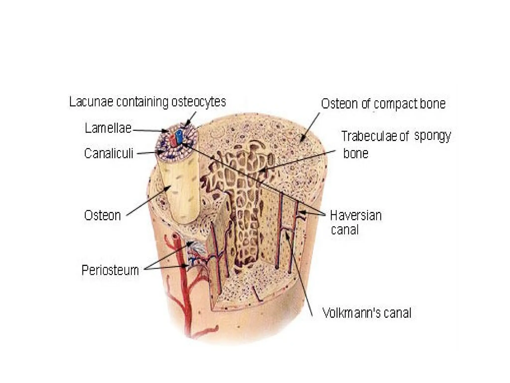
(a) Periosteum
-Thick fibrous memebrane made up of an outer fibrous layer, and an inner
cellular layer which is osteogenic in nature.
- Periosteum is united to the underlying bone by Sharpey's fibres, and the union
is particularly strong over the attachments of tendons, and ligaments.
- At articular margin,it is continuous with the capsule of the joint. The abundant
periosteal arteries nourish the outer part of the underlying cortex also.
-Periosteum has a rich nerve supply which makes it most sensitive part of the
bone.
6.
(b) Cortex ismade up of a compact bone which gives it the desired strength to
withstand all possible mechanical strains.
(c) Medullary cavity is filled with red or yellow bone marrow.
-At birth the marrow is red everywhere with widespread active haemopoiesis.
-As the age advances, the red marrow at many places atrophies and is replaced
by yellow, fatty marrow, with no power of haemopoiesis.
-Red marrow persists in the cancellous ends of long bones.
-In the sternum ribs, iliac crest, vertebrae and skull bones the red marrow is
found throughout life.
2. The two ends of a long bone are made up of cancellous bone covered with
hyaline (articular) cartilage
7.
PARTS OF AYOUNG BONE
• A typical long bone ossifies in three parts, the
two ends from secondary centres, and the
intervening shaft from a primary centre
• Before ossification is complete the following
parts of the bone can be defined.
8.
1. Epiphysis
The endsand tips of a bone which ossify from secondary centres are
called epiphyses. These are of the following types.
a) Pressure epiphysis is articular and takes part in transmission of
the weight. Examples: head of femur; lower end of radius, etc.
b) Traction epiphysis is nonarticular and does not take part in the
transmission of the weight. It always provides attachment to
one or more tendons which exert a traction on the epiphysis.
the traction epiphyses ossify later than the pressure epiphyses.
Examples: trochanters of femur and tubercles of humerus
9.
c) Atavistic epiphysisis phylogenetically an independent bone
which in man becomes fused to another bone.
Examples: coracoid process of scapula and os trigonum or
lateral tubercle of talus,
d) Aberrant epiphysis is not always present. Examples: epiphysis
at the head of the first metacarpal and at the base of other
metacarpal bones.
2. Diaphysis
• It is the elongated shaft of a long bone which ossifies from a
primary centre
10.
• 3. Metaphysis
•The epiphysial ends of a diaphysis are called metaphyses.
• Each metaphysis is the zone of active growth. Before epiphysial fusion, the
metaphysis is richly supplied with blood through end arteries forming
'hair-pin' bends.
• This is the common site of osteomyelitis in children because the bacteria
or emboli are easily trapped in the hair-pin bends, causing infarction.
• After the epiphysial fusion, vascular communications are established
between the metaphysial and epiphysial arteries.
• Now the metaphysis contains no more end-arteries and is no longer
subjected to osteomyelitis.
11.
4. Epiphysial Plateof Cartilage
• It separates epiphysis from metaphysis.
• Proliferation of cells in this cartilaginous plate is responsible
for lengthwise growth of a long bone.
• After the epiphysial fusion, the bone can no longer grow in
length.
• The growth cartilage is nourished by both the epiphysial and
metaphysial arteries.
12.
Microscopical structure ofBone
• It can be lamellar,woven,fibrous,dentine and
cement
• ARRANGEMENT OF BONY LAMELLAE
Mature bone is composed of layers and is,
therefore, known as the lamellar bone.
13.
HAVERSIAN SYSTEMS INCOMPACT BONE
• The bones are arranged in a number of cylindrical units known as the
Haversian systems or secondary osteones.
• Each system consists of a central Haversian canal, surrounded by concentric
lamellae of bony tissue.
• Between these lamellae numerous lacunae intervene, which communicate
with one another and with the central canal by numerous radiating
canaliculi.
• The central canal contains usually small vessels (artery, vein); lacunae are
filled with the osteocytes, and canaliculi contain processes of bone cells and
convey outwards the nutritive materials by diffusion from the capillaries
14.
• Each Haversiansystem is about 150 um in diameter and the central canal about
20 um in width.
• The Haversian systems run longitudinally in long bones, branch and anastomose.
• The Haversian canals communicate with medullary cavity and with the surface
of bone by numerous oblique channels, termed the Volk-mann's canals.
• The latter contain blood vessels and nerves which permeate the bones from the
periosteum or endosteum.
• The Volkmann's canals are not surrounded by concentric lamellae. About 21
million osteones are estimated to be present in adult human skeleton.
• Interstitial lamellae with their lacunae and canaliculi
• These occupy the angular intervals in between the Haversian
systems.Circumferential lamellae or primary osteones :They encircle the inner
and outer surfaces of the bone.
15.
• These lamellaeare held together by the perforating fibres
of Sharpey, which are derived fram the periosteum and run
obliquely pinning the lamellae together.
• Bony lamellae of the Haversian system :These consist of
cement substance, collagen fibres and impregnated with
bone salts.
• Each Haversian system is demarcated from the
neighbouring systems by cement line which is strongly
basophilic and is devoid of the collagen fibres.
• Successive lamella differ in orientation of collagen fibres
and in concentration of mineral salts.
16.
• Accordingly, thelamellae are of two types,
cementing and fibrillary. Cementing lamellae are rich
in minerals, and less in collagen fibres.
• Fibrillary lamellae are rich in collagen fibresand less
in minerals. Fibre arrangements in successive
lamellae vary alternately, e.g., longitudinal,
circumferential or spiral.
• With the advancement of age, longitudinal fibres
increase in number, and cirumferential fibres
decrease
18.
Volkmann’s canal
• Smallchannels in bone that connect Haversian canals to each
other and to periosteum.
• Runs parallel to long axis of bone are connected to each other
by perpendicular ducts and usually runs obtuse angles to
haversian canals.
• Allows blood vessel to enter bone from periosteum,providing
nutrition to osteons
19.
• In SpongyBone
• The bony trabeculae consist of superimposed lamellae, and usually do not
form the Haversian systems because they get nutrition from the blood
vessels of the tissues around them.
• Therefore, the trabeculae are lamellated without Haversian
system.FORMATION OF HAVERSIAN SYSTEM
• The shaft of a bone grows in width by appositional mechanism, by
depositing layers of new bone under the osteogenic layer of peri-osteum.
• Thereby circumferential lamellae are formed. After the deposition of thick
lamellae, nutrition of bone suffers and a number of longitudinal grooves
appear on the outer surface covered by the periosteum.
• Each groove contains blood vessels along the floor. The ridges of the grooves
proliferate and deposit bones till they meet and convert the grooves into
tunnels, Each tunnel is lined by osteoblasts and contains a vessel.
20.
• The continuedproliferation of the osteoblasts and
their subsequent differentiation into the osteocytes,
convert the tunnel into the Haversian systems.
• Haversian systems have limited span of life.
• Part of the systems may be dissolved and the
osteoblasts from the original canal proliferate to
form new Haversian systems.
• Interstitial lamellae are the remnants of old outer
circumferential lamellae or old Haversian systems.
21.
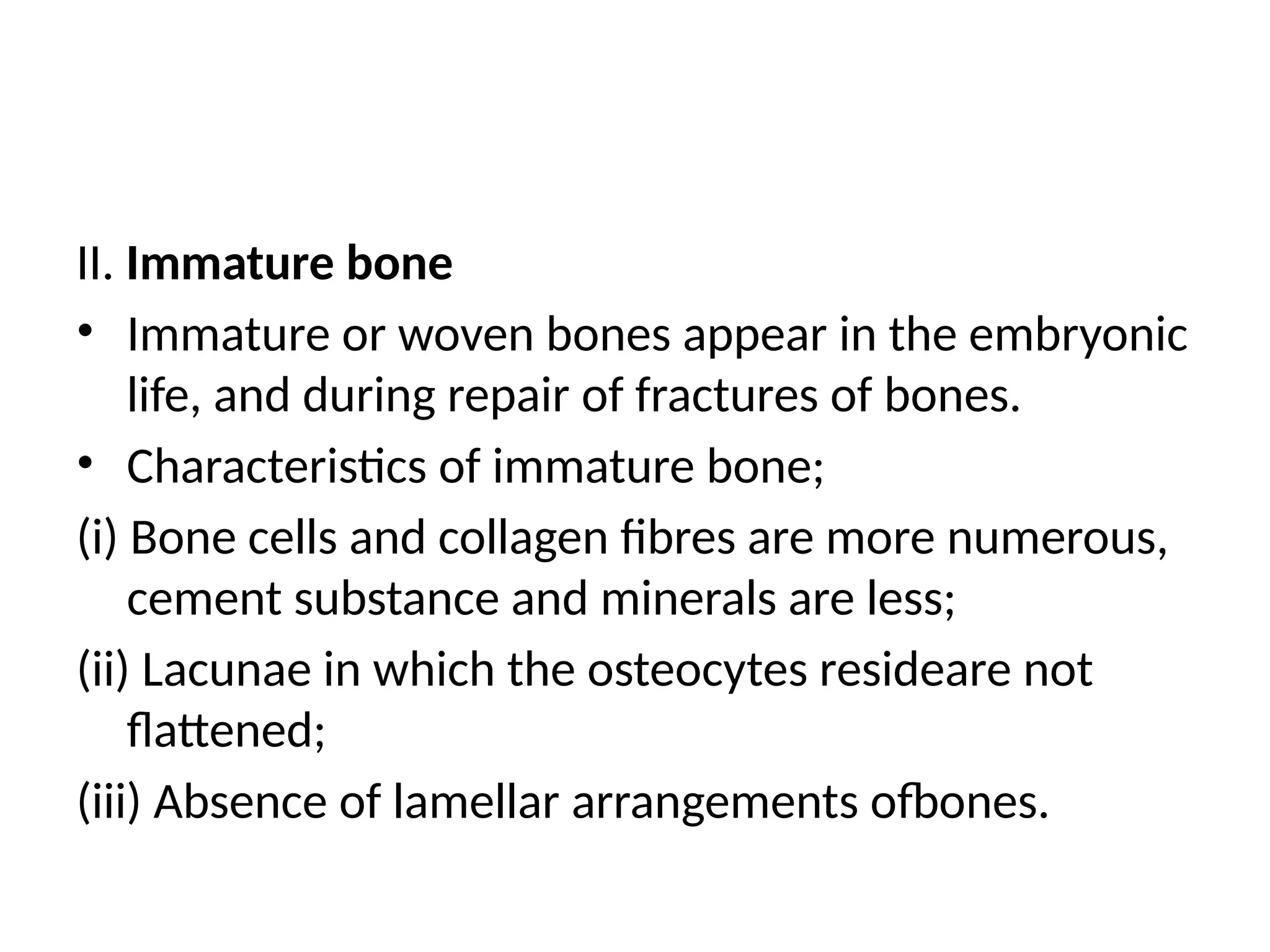
II. Immature bone
•Immature or woven bones appear in the embryonic
life, and during repair of fractures of bones.
• Characteristics of immature bone;
(i) Bone cells and collagen fibres are more numerous,
cement substance and minerals are less;
(ii) Lacunae in which the osteocytes resideare not
flattened;
(iii) Absence of lamellar arrangements ofbones.
22.
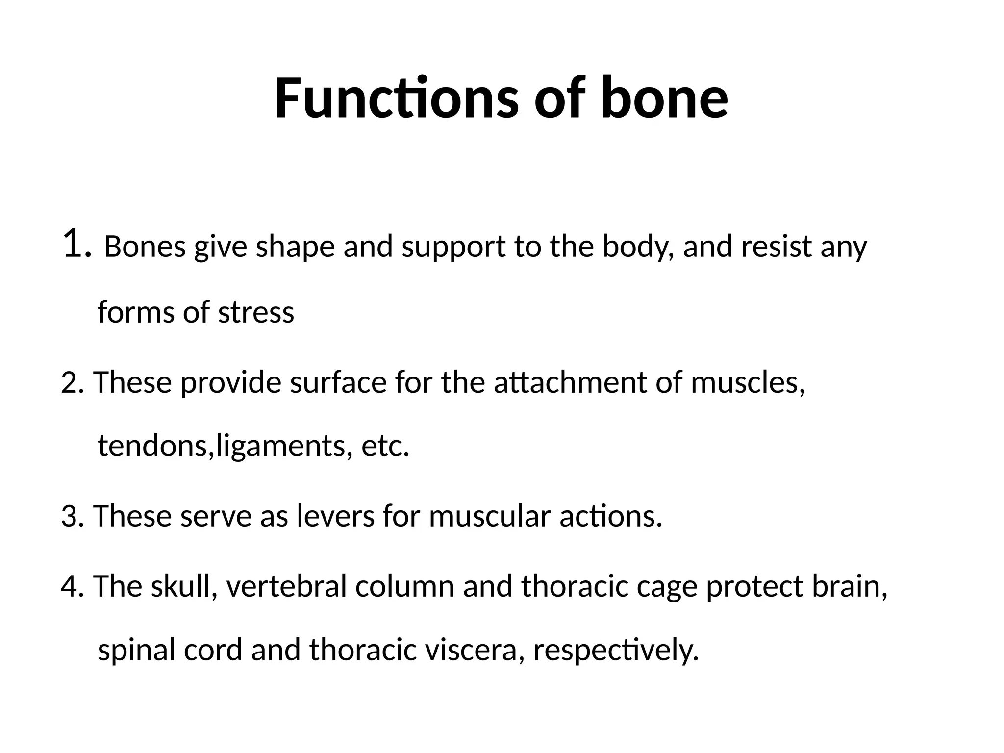
Functions of bone
1.Bones give shape and support to the body, and resist any
forms of stress
2. These provide surface for the attachment of muscles,
tendons,ligaments, etc.
3. These serve as levers for muscular actions.
4. The skull, vertebral column and thoracic cage protect brain,
spinal cord and thoracic viscera, respectively.
23.
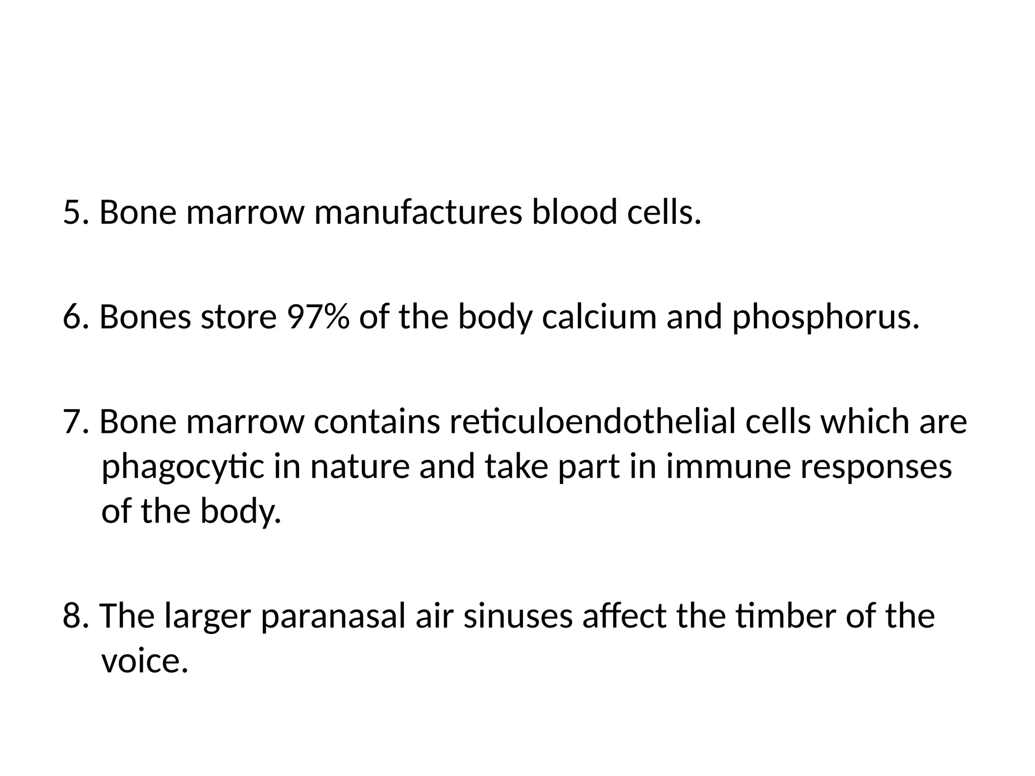
5. Bone marrowmanufactures blood cells.
6. Bones store 97% of the body calcium and phosphorus.
7. Bone marrow contains reticuloendothelial cells which are
phagocytic in nature and take part in immune responses
of the body.
8. The larger paranasal air sinuses affect the timber of the
voice.
![Cellular structure of bone
• Composition
• Water (10%)
• Organic matrix(osteoid matrix)(25%)
Provides flexibility and resilience
-Proteins:Type I collagen,proteoglycans,
osteocalcin,osteonectin,osteopontin
-Cells:Osteoblasts,osteoclasts,osteocytes
• Inorganic elements(65%)
Provides hardness and brittleness
-Hydroxyapatite[Ca10(Po4)6(OH)2]
-Ratio of calcium to phosphorus in bone is 2:1](https://image.slidesharecdn.com/structureandfunctionofbone-251111153114-060f7dc2/75/Structure-and-function-of-bone-powerpoint-presentation-pptx-4-2048.jpg)