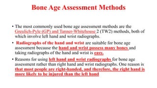
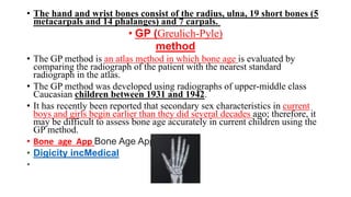
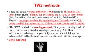
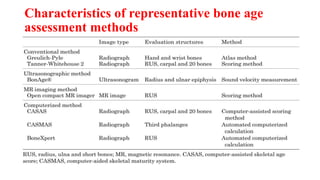
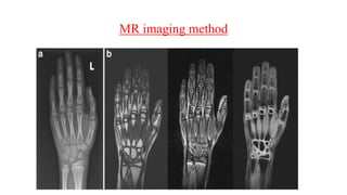
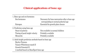
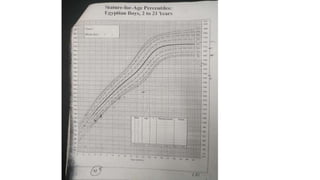
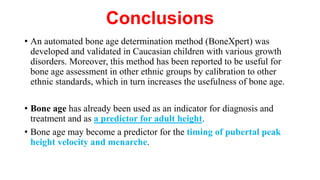
Bone age: assessment methods and
clinical applications
By
Dr . Magdy Shafik Ramadan
Senior Pediatric and Neonatology consultant
M.S, Diploma, Ph.D of Pediatrics
Introduction
• Bone age assessment is frequently performed not only in pediatric
endocrinology but also in orthodontics and pediatric orthopedics.
• Bone age is an effective indicator for diagnosing various diseases
and determining the timing of treatment.
• The aim of bone age assessment is to evaluate growth and maturity
and to diagnose and manage pediatric disorders.
• Recently, several computerized systems for bone age assessment have
been developed
What is Bone Age?
What is Bone Age?
What is Bone Age?
What is Bone Age?
• Bone age is a measure of the degree of skeletal
maturity of a child, i.e. how far the child has
advanced in its development of the skeleton.
why to determine bone age?
• 1) important in management of short stature
• bone age < chronological age = growth hormone deficiency or constititutional
delay
• bone age=chronological age = familial short stature
• bone age > chronological age = hypergonadism, cah (con. Adern. Hyper.)
• 2) in deciding when to start and stop growth hormone supplimentation.
• 3) in forensic medicine- minor cant be tried in normal courts (‫قاصر‬ ‫محاكمة‬ ‫يمكن‬ ‫ال‬
• 4) replacement therapy for hypogonadism.
• 5) gnrh for precocious puberty.(Gondortophic rleasing hormones)
• 6) prediction of final height.
In Newborns
• only indication is congenital hypothyroidis epiphysis at lower
end of femur and upper end of tibia should be present in a
normal newborn.
• Their absence indicates delayed bone age of intrapartum
cause. Mc is hypothyroidism
ossification center
• A primary ossification center is the first area of a bone to start
ossifying. It usually appears during prenatal development in the
central part of each developing bone.
• In long bones the primary centers occur in the diaphysis/shaft and in
irregular bones the primary centers occur usually in the body of the
bone.
• A secondary ossification center is the area of ossification that appears after
the primary ossification center has already appeared –
• most of which appear during the postnatal and adolescent years.
• Most bones have more than one secondary ossification center.
• In long bones, the secondary centers appear in the epiphyses.
Bone Growth and Maturation
• Bone age
• Bone age represents the degree of secondary ossification in long and short
bones, which are formed by endochondral ossification.
• A cartilaginous growth plate between the epiphysis and metaphysis is
responsible for postnatal linear bone growth .
• Chondrocytes in the growth plate not only proliferate but also differentiate.
The resting zone consists of undifferentiated chondrocyte progenitors,
which differentiate into proliferative chondrocytes and then into
hypertrophic chondrocytes.
• Finally, hypertrophic chondrocyte apoptosis occurs after blood vessels
invade into the hypertrophic zone, and osteoblasts then migrate into the
cartilage matrix to form bone.
• it has been demonstrated that estrogen is essential for growth plate
closure in both men and women.
• Delayed and accelerated bone age
• Chondrocyte proliferation and differentiation and the conversion of
chondrocytes to osteoblasts are accelerated by GH, IGF-I, thyroid
hormone, estrogen and androgen and suppressed by glucocorticoid
.Therefore, deficiencies in GH, IGF-I, thyroid hormone, estrogen and
androgen and excess glucocorticoid cause both growth impairment and
bone age delay.
• In contrast, overproduction of GH, IGF-I, thyroid hormone, estrogen
and androgen causes both overgrowth and bone age advancement. Bone
age advancement is often recognized in children with obesity.
• It has been reported that bone maturation was accelerated after puberty
onset in obese children compared with nonobese children .
• It has also been suggested that hyperestrogenemia contributes to the
advancement of bone maturation in obese boy
Bone Age Assessment Methods
• The most commonly used bone age assessment methods are the
Greulich-Pyle (GP) and Tanner-Whitehouse 2 (TW2) methods, both of
which involve left hand and wrist radiographs.
• Radiographs of the hand and wrist are suitable for bone age
assessment because the hand and wrist possess many bones and
taking radiographs of the hand and wrist is easy.
• Reasons for using left hand and wrist radiographs for bone age
assessment rather than right hand and wrist radiographs. One reason is
that most people are right-handed, and therefore, the right hand is
more likely to be injured than the left hand
• The hand and wrist bones consist of the radius, ulna, 19 short bones (5
metacarpals and 14 phalanges) and 7 carpals.
• GP (Greulich-Pyle)
method
• The GP method is an atlas method in which bone age is evaluated by
comparing the radiograph of the patient with the nearest standard
radiograph in the atlas.
• The GP method was developed using radiographs of upper-middle class
Caucasian children between 1931 and 1942.
• It has recently been reported that secondary sex characteristics in current
boys and girls begin earlier than they did several decades ago; therefore, it
may be difficult to assess bone age accurately in current children using the
GP method.
• Bone age App Bone Age App
• Digicity incMedical
•
TW2 methods
• There are actually three different TW2 methods: the radius-ulna-
short bones (RUS) method for evaluating the 13 long or short bones
(i.e., the radius, ulna and short bones of the first, third and fifth
fingers), the carpal method for evaluating the 7 carpals and the 20-
bones method for evaluating the 13 long or short bones and 7 carpals.
• The TW2 method is a scoring method. Firstly, the maturity level of
each bone is categorized into a stage (from stage A to H or I).
Afterwards, each stage is replaced by a score, and a total sore is
calculated. Finally, the total score is transformed into the bone age.
• Bone age App
Comparison between the GP and TW2
methods
• The scoring method of the TW2 method is more objective than the
atlas method, and therefore, the TW2 method is considered to have
higher reproducibility than the GP method
Other methods
• Recently, new methods for bone age assessment have been developed,
including
• ultrasonographic,
• computerized and magnetic resonance (MR) imaging method
Characteristics of representative bone age
assessment methods
Ultrasonographic method
• an arm rest between two transducers to support the subject’s hand
and wrist, and ultrasonic waves pass through the subject’s distal radius
and ulnar epiphysis. Afterwards, software is used to calculate the bone
age using an algorithm based on measurements of sound velocity and
the distance between the two transducers
Computerized method
• Several computerized systems for bone age assessment have been
reported. Some of these systems were developed based on the TW
method .One example is the computer-assisted skeletal age score
(CASAS) ,in which an image is digitized and represented by several
mathematical coefficients. These coefficients are then compared to
those generated by each stage of the TW standards, and the closest
match is determined
MR imaging method
• Terada et al. . reported the usefulness of bone age assessment using
MR images of the hand and wrist compared with radiographs. They
used an open compact MR imager with a permanent magnet that was
newly developed as a pediatric hand scanner to evaluate bone age.
Although MR imaging is a noninvasive method, one can requires a
long time (2 min and 44 sec). Therefore, this method may not be
suitable for some subjects, especially young children, due to body
movement.
MR imaging method
Clinical Applications of Bone Age
• Relationship between bone age and indicators of puberty
• Bone age at puberty onset: Flor-Cisneros et al. (41) reported that the
onset of central puberty occurred at an abnormal chronological age but
at a normal bone age in boys with congenital adrenal hyperplasia,
familial male-limited precocious puberty and idiopathic short stature.
Therefore, they suggested that the hypothalamic-pituitary-gonadal axis
maturation might be regulated by somatic growth and maturation in
the bone itself or in nonskeletal tissues that paralleled bone growth and
maturation.
• it may be difficult to predict the timing of pubertal onset in normal
children using bone age.
• Bone age at peak height velocity: Predicting the timing of peak
height velocity (PHV) is important in patients with scoliosis, kyphosis
and slipped capital femoral epiphysis for determination of the timing
of surgical treatment and in patients with growth disorders for
prediction of adult height and modification of treatment.
• Although further examinations are needed, bone age may be a
predictor for the timing of peak height velocity (PHV) .
• Bone age at menarche:
• menarche occurred after cervical vertebral maturation stage (CVMS)
III based on lateral cephalometric radiographs in orthodontic patients.
• The onset of menstruation was at a bone age of approximately 12 yr,
as evaluated by the Japanese TW2-RUS method in Japanese girls,
regardless of the chronological age at the onset of puberty.
• This bone age was equivalent to the mean chronological age (12 yr
and 4 mo) at menarche in Japanese girls.
• Bone age may be one of the indicators for predicting the timing of
menarche.
Prediction of adult height
• Prediction of adult height is very important in pediatric endocrinology.
• The commonly used methods for adult height prediction based on
bone age are the Bayley-Pinneau and TW mark II (2) methods
• new adult height prediction method based on BoneXpert bone age.
In this method, the predicted adult height (Hraw) is calculated using
height, chronological age, and bone age based on the same concept as
the Bayley-Pinneau method
• The final predicted adult height is then obtained after modifying
Hraw using the parents’ heights or the population standard height, and
the weight and body mass index (BMI) of the patient. It has been
reported that this new method performs significantly better than the
Bayley-Pinneau method in children with idiopathic short stature
• https://bonexpert.com/ahp
• The tool gives four different adult height predictions
• AHP(x-ray) is the prediction based on age, bone age and
height
• AHP (parental) is the prediction based solely on the parents’
heights. This is similar, but not identical, to the conventional
“target height” (which is mid-parental height ± 6.5 cm), but it is
more accurate, because it includes a drawing towards the
population height
• AHP(x+p) is the prediction based on age, bone age, height and
parental height
• AHP(x+m) is the prediction based on age, bone age, height
and height at menarche
Characteristics of adult height prediction methods
Clinical applications of bone age
Conclusions
• An automated bone age determination method (BoneXpert) was
developed and validated in Caucasian children with various growth
disorders. Moreover, this method has been reported to be useful for
bone age assessment in other ethnic groups by calibration to other
ethnic standards, which in turn increases the usefulness of bone age.
• Bone age has already been used as an indicator for diagnosis and
treatment and as a predictor for adult height.
• Bone age may become a predictor for the timing of pubertal peak
height velocity and menarche.
THANK YOU

Bone age assessent.pptx

  • 1.
    Bone age: assessmentmethods and clinical applications By Dr . Magdy Shafik Ramadan Senior Pediatric and Neonatology consultant M.S, Diploma, Ph.D of Pediatrics
  • 2.
    Introduction • Bone ageassessment is frequently performed not only in pediatric endocrinology but also in orthodontics and pediatric orthopedics. • Bone age is an effective indicator for diagnosing various diseases and determining the timing of treatment. • The aim of bone age assessment is to evaluate growth and maturity and to diagnose and manage pediatric disorders. • Recently, several computerized systems for bone age assessment have been developed
  • 3.
    What is BoneAge? What is Bone Age? What is Bone Age? What is Bone Age? • Bone age is a measure of the degree of skeletal maturity of a child, i.e. how far the child has advanced in its development of the skeleton.
  • 4.
    why to determinebone age? • 1) important in management of short stature • bone age < chronological age = growth hormone deficiency or constititutional delay • bone age=chronological age = familial short stature • bone age > chronological age = hypergonadism, cah (con. Adern. Hyper.) • 2) in deciding when to start and stop growth hormone supplimentation. • 3) in forensic medicine- minor cant be tried in normal courts (‫قاصر‬ ‫محاكمة‬ ‫يمكن‬ ‫ال‬ • 4) replacement therapy for hypogonadism. • 5) gnrh for precocious puberty.(Gondortophic rleasing hormones) • 6) prediction of final height.
  • 5.
    In Newborns • onlyindication is congenital hypothyroidis epiphysis at lower end of femur and upper end of tibia should be present in a normal newborn. • Their absence indicates delayed bone age of intrapartum cause. Mc is hypothyroidism
  • 7.
  • 8.
    • A primaryossification center is the first area of a bone to start ossifying. It usually appears during prenatal development in the central part of each developing bone. • In long bones the primary centers occur in the diaphysis/shaft and in irregular bones the primary centers occur usually in the body of the bone. • A secondary ossification center is the area of ossification that appears after the primary ossification center has already appeared – • most of which appear during the postnatal and adolescent years. • Most bones have more than one secondary ossification center. • In long bones, the secondary centers appear in the epiphyses.
  • 9.
    Bone Growth andMaturation • Bone age • Bone age represents the degree of secondary ossification in long and short bones, which are formed by endochondral ossification. • A cartilaginous growth plate between the epiphysis and metaphysis is responsible for postnatal linear bone growth . • Chondrocytes in the growth plate not only proliferate but also differentiate. The resting zone consists of undifferentiated chondrocyte progenitors, which differentiate into proliferative chondrocytes and then into hypertrophic chondrocytes. • Finally, hypertrophic chondrocyte apoptosis occurs after blood vessels invade into the hypertrophic zone, and osteoblasts then migrate into the cartilage matrix to form bone.
  • 10.
    • it hasbeen demonstrated that estrogen is essential for growth plate closure in both men and women.
  • 11.
    • Delayed andaccelerated bone age • Chondrocyte proliferation and differentiation and the conversion of chondrocytes to osteoblasts are accelerated by GH, IGF-I, thyroid hormone, estrogen and androgen and suppressed by glucocorticoid .Therefore, deficiencies in GH, IGF-I, thyroid hormone, estrogen and androgen and excess glucocorticoid cause both growth impairment and bone age delay. • In contrast, overproduction of GH, IGF-I, thyroid hormone, estrogen and androgen causes both overgrowth and bone age advancement. Bone age advancement is often recognized in children with obesity. • It has been reported that bone maturation was accelerated after puberty onset in obese children compared with nonobese children . • It has also been suggested that hyperestrogenemia contributes to the advancement of bone maturation in obese boy
  • 13.
    Bone Age AssessmentMethods • The most commonly used bone age assessment methods are the Greulich-Pyle (GP) and Tanner-Whitehouse 2 (TW2) methods, both of which involve left hand and wrist radiographs. • Radiographs of the hand and wrist are suitable for bone age assessment because the hand and wrist possess many bones and taking radiographs of the hand and wrist is easy. • Reasons for using left hand and wrist radiographs for bone age assessment rather than right hand and wrist radiographs. One reason is that most people are right-handed, and therefore, the right hand is more likely to be injured than the left hand
  • 14.
    • The handand wrist bones consist of the radius, ulna, 19 short bones (5 metacarpals and 14 phalanges) and 7 carpals. • GP (Greulich-Pyle) method • The GP method is an atlas method in which bone age is evaluated by comparing the radiograph of the patient with the nearest standard radiograph in the atlas. • The GP method was developed using radiographs of upper-middle class Caucasian children between 1931 and 1942. • It has recently been reported that secondary sex characteristics in current boys and girls begin earlier than they did several decades ago; therefore, it may be difficult to assess bone age accurately in current children using the GP method. • Bone age App Bone Age App • Digicity incMedical •
  • 15.
    TW2 methods • Thereare actually three different TW2 methods: the radius-ulna- short bones (RUS) method for evaluating the 13 long or short bones (i.e., the radius, ulna and short bones of the first, third and fifth fingers), the carpal method for evaluating the 7 carpals and the 20- bones method for evaluating the 13 long or short bones and 7 carpals. • The TW2 method is a scoring method. Firstly, the maturity level of each bone is categorized into a stage (from stage A to H or I). Afterwards, each stage is replaced by a score, and a total sore is calculated. Finally, the total score is transformed into the bone age. • Bone age App
  • 16.
    Comparison between theGP and TW2 methods • The scoring method of the TW2 method is more objective than the atlas method, and therefore, the TW2 method is considered to have higher reproducibility than the GP method
  • 17.
    Other methods • Recently,new methods for bone age assessment have been developed, including • ultrasonographic, • computerized and magnetic resonance (MR) imaging method
  • 18.
    Characteristics of representativebone age assessment methods
  • 19.
    Ultrasonographic method • anarm rest between two transducers to support the subject’s hand and wrist, and ultrasonic waves pass through the subject’s distal radius and ulnar epiphysis. Afterwards, software is used to calculate the bone age using an algorithm based on measurements of sound velocity and the distance between the two transducers
  • 20.
    Computerized method • Severalcomputerized systems for bone age assessment have been reported. Some of these systems were developed based on the TW method .One example is the computer-assisted skeletal age score (CASAS) ,in which an image is digitized and represented by several mathematical coefficients. These coefficients are then compared to those generated by each stage of the TW standards, and the closest match is determined
  • 21.
    MR imaging method •Terada et al. . reported the usefulness of bone age assessment using MR images of the hand and wrist compared with radiographs. They used an open compact MR imager with a permanent magnet that was newly developed as a pediatric hand scanner to evaluate bone age. Although MR imaging is a noninvasive method, one can requires a long time (2 min and 44 sec). Therefore, this method may not be suitable for some subjects, especially young children, due to body movement.
  • 22.
  • 23.
    Clinical Applications ofBone Age • Relationship between bone age and indicators of puberty • Bone age at puberty onset: Flor-Cisneros et al. (41) reported that the onset of central puberty occurred at an abnormal chronological age but at a normal bone age in boys with congenital adrenal hyperplasia, familial male-limited precocious puberty and idiopathic short stature. Therefore, they suggested that the hypothalamic-pituitary-gonadal axis maturation might be regulated by somatic growth and maturation in the bone itself or in nonskeletal tissues that paralleled bone growth and maturation. • it may be difficult to predict the timing of pubertal onset in normal children using bone age.
  • 24.
    • Bone ageat peak height velocity: Predicting the timing of peak height velocity (PHV) is important in patients with scoliosis, kyphosis and slipped capital femoral epiphysis for determination of the timing of surgical treatment and in patients with growth disorders for prediction of adult height and modification of treatment. • Although further examinations are needed, bone age may be a predictor for the timing of peak height velocity (PHV) .
  • 25.
    • Bone ageat menarche: • menarche occurred after cervical vertebral maturation stage (CVMS) III based on lateral cephalometric radiographs in orthodontic patients. • The onset of menstruation was at a bone age of approximately 12 yr, as evaluated by the Japanese TW2-RUS method in Japanese girls, regardless of the chronological age at the onset of puberty. • This bone age was equivalent to the mean chronological age (12 yr and 4 mo) at menarche in Japanese girls. • Bone age may be one of the indicators for predicting the timing of menarche.
  • 26.
    Prediction of adultheight • Prediction of adult height is very important in pediatric endocrinology. • The commonly used methods for adult height prediction based on bone age are the Bayley-Pinneau and TW mark II (2) methods • new adult height prediction method based on BoneXpert bone age. In this method, the predicted adult height (Hraw) is calculated using height, chronological age, and bone age based on the same concept as the Bayley-Pinneau method
  • 27.
    • The finalpredicted adult height is then obtained after modifying Hraw using the parents’ heights or the population standard height, and the weight and body mass index (BMI) of the patient. It has been reported that this new method performs significantly better than the Bayley-Pinneau method in children with idiopathic short stature
  • 28.
    • https://bonexpert.com/ahp • Thetool gives four different adult height predictions • AHP(x-ray) is the prediction based on age, bone age and height • AHP (parental) is the prediction based solely on the parents’ heights. This is similar, but not identical, to the conventional “target height” (which is mid-parental height ± 6.5 cm), but it is more accurate, because it includes a drawing towards the population height • AHP(x+p) is the prediction based on age, bone age, height and parental height • AHP(x+m) is the prediction based on age, bone age, height and height at menarche
  • 29.
    Characteristics of adultheight prediction methods
  • 30.
  • 32.
    Conclusions • An automatedbone age determination method (BoneXpert) was developed and validated in Caucasian children with various growth disorders. Moreover, this method has been reported to be useful for bone age assessment in other ethnic groups by calibration to other ethnic standards, which in turn increases the usefulness of bone age. • Bone age has already been used as an indicator for diagnosis and treatment and as a predictor for adult height. • Bone age may become a predictor for the timing of pubertal peak height velocity and menarche.
  • 33.