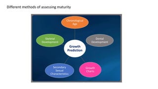
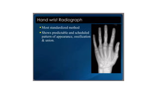
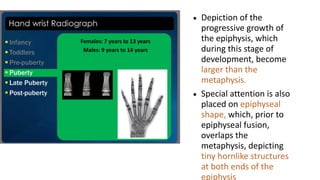
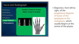
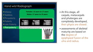
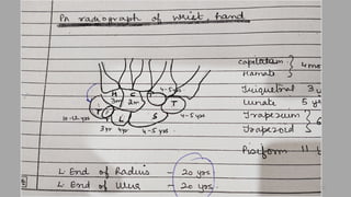
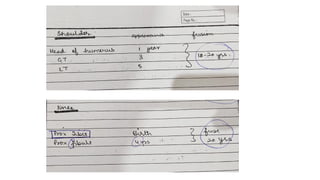
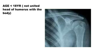
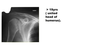
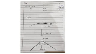
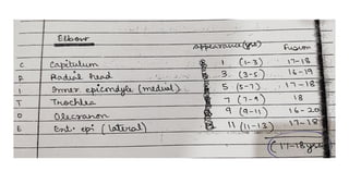
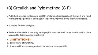
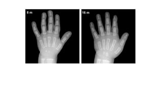
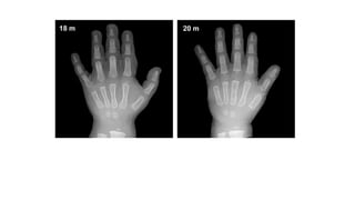
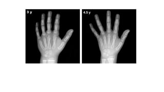
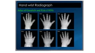
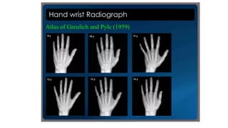
BONE AGE ESTIMATION
Need of Bone Age Estimation!!
• Medico-legal
• Short stature with Growth Delay
• Monitoring growth hormone and anabolic steroid response
• Monitoring treatment in endocrinopathy
• D/D of sexual precocity
• Prediction of Adult height
• Selection of children in sports
• Comparison of environmental, dietary factors
Different methods of assessing maturity
VARIOUS METHODS
• Hand Wrist Radiograph
• Cervical vertebrae
• Sternum
• Pelvic bones
• Frontal sinus
Methods of skeletal maturity assessment
• Appearance of fusion and ossification centres
• Gruelich Pyle method (G-P)
• Tanner and Whitehouse II
Appearance and fusion of ossification centres
• Sets of radiographs taken-
1. PA view of wrist ,Hand
2. Elbow
3. knee
4. shoulder
5. Pelvis AP for iliac crest fusion and appearance
• TO Facilitate bone age
assessment , skeletal
development is divided in
six major categories and
have been highlighted the
specific ossifications centres
that are best predictors of
skeletal maturity for each
group.
1. Infancy- Birth to 10 month(carpal bones and
radial epiphysis).
2. Toddlers-10 month to 2yr (the number of
epiphysis visible in the long bones of the
hand).
3. Pre-Puberty- 2yrs to 7yrs of age (the size of
phalangeal epiphysis).
4. Early and mid puberty- 7yrs to 13 yrs of age
(the size of phalangeal epiphysis).
5. Late Puberty- 13 to 15 yrs of age ( the degree
of epiphyseal fusion).
6. Post puberty-15 to 18yrs of age ( the degree of
epiphyseal fusion of radius and ulna).
7
The ossification centres
for capitate and hamate
become apparent at
about 3 months of age
and remain only useful
observable feature for
next six months.
• Bone age
determinations are
primarily based on the
assessment of the
number of identifiable
epiphyseal ossification
centres.
• The ossification centres
for the epiphyses of all
phalanges and
metacarpals become
recognizable during this
stage, usually in the
middle finger first, and
the fifth finger last.
• During this stage of
development, the
ossification centers for the
epiphyses increase in
width and thickness, and
eventually assume a
transverse diameter as
wide as the metaphyses.
• Depiction of the
progressive growth of the
width of the epiphyses,
which, during this stage of
development, become as
wide as the metaphysis.
• Depiction of the
progressive growth of
the epiphysis, which
during this stage of
development, become
larger than the
metaphysis.
• Special attention is also
placed on epiphyseal
shape, which, prior to
epiphyseal fusion,
overlaps the
metaphysis, depicting
tiny hornlike structures
at both ends of the
epiphysis
• Depiction, from left to
right, of the
progressive degrees
of fusion of the
epiphyses to the
metaphysis, which
usually begins at the
centre of the physis.
Assessments of
skeletal maturity in
this stage are
primarily based on
the degree of
epiphyseal fusion of
the distal phalanges.
• At this stage, all
carpals, metacarpals
and phalanges are
completely developed,
their physis are closed.
• assessments of skeletal
maturity are based on
the degree of
epiphyseal fusion of the
ulna and radius.
Age from hip bone examination
• Two pubic rami of the hip (6y)
• Suture at the acetabulum(15yr)
• Ischeal tuberosity with the ischium (21yr)
• Iliac crest with the ilium(23yr)
AGE > 4YRS ( Ossific centre at greater
trochanter ) <15yr ( triradiate suture is
opened.
22
AGE < 18YR ( not united
head of humerus with the
body)
> 19yrs
( united
head of
humerus).
(B) Greulich and Pyle method (G-P)
• Published an atlas containing a set (30) of standard radiographs of the wrist and hand
representing a particular bone age at the some 30 points along the maturity scale.
• Standard for boys and girls.
• To determine skeletal maturity, radiograph is matched with those in atlas and as close
as possible determination is selected
• LIMITATIONS-:
1. Subjectivity of matching.
2. Scale used for expressing maturity i.e as close to as possible.
(C) Tanner and Whitehouse method
• Principle-
1.Each bone develops to a regional constant functional shape.
2. A sequence of recognized state defined right from appearance to functional shape all along development journey is stated.
3. Both visual and descriptive information is used for assigning the stage.
Technique-
PA xray of Left hand and wrist taken.
Long axis of middle finger , forearm and arm in direct line.
Centering on 3rd metacarpal
20 Bones assessed and score is assigned.
( Carpals score- 7 excluding pisiform
Radius, Ulna, 1,3,5 metacarpal and phalanges of 1,3,5 metacarpals)
Once all the bones have been scored a total score is generated by adding them all and then plotted on a radiograph to
Determine how bone age relates to chronological age.
New Methods-
1. Computer assisted Skeletal age scores
2. USG based age bone scores

BONE AGE ESTIMATION

  • 1.
  • 2.
    Need of BoneAge Estimation!! • Medico-legal • Short stature with Growth Delay • Monitoring growth hormone and anabolic steroid response • Monitoring treatment in endocrinopathy • D/D of sexual precocity • Prediction of Adult height • Selection of children in sports • Comparison of environmental, dietary factors
  • 3.
    Different methods ofassessing maturity
  • 4.
    VARIOUS METHODS • HandWrist Radiograph • Cervical vertebrae • Sternum • Pelvic bones • Frontal sinus
  • 5.
    Methods of skeletalmaturity assessment • Appearance of fusion and ossification centres • Gruelich Pyle method (G-P) • Tanner and Whitehouse II
  • 6.
    Appearance and fusionof ossification centres • Sets of radiographs taken- 1. PA view of wrist ,Hand 2. Elbow 3. knee 4. shoulder 5. Pelvis AP for iliac crest fusion and appearance
  • 7.
    • TO Facilitatebone age assessment , skeletal development is divided in six major categories and have been highlighted the specific ossifications centres that are best predictors of skeletal maturity for each group. 1. Infancy- Birth to 10 month(carpal bones and radial epiphysis). 2. Toddlers-10 month to 2yr (the number of epiphysis visible in the long bones of the hand). 3. Pre-Puberty- 2yrs to 7yrs of age (the size of phalangeal epiphysis). 4. Early and mid puberty- 7yrs to 13 yrs of age (the size of phalangeal epiphysis). 5. Late Puberty- 13 to 15 yrs of age ( the degree of epiphyseal fusion). 6. Post puberty-15 to 18yrs of age ( the degree of epiphyseal fusion of radius and ulna). 7
  • 9.
    The ossification centres forcapitate and hamate become apparent at about 3 months of age and remain only useful observable feature for next six months.
  • 10.
    • Bone age determinationsare primarily based on the assessment of the number of identifiable epiphyseal ossification centres. • The ossification centres for the epiphyses of all phalanges and metacarpals become recognizable during this stage, usually in the middle finger first, and the fifth finger last.
  • 11.
    • During thisstage of development, the ossification centers for the epiphyses increase in width and thickness, and eventually assume a transverse diameter as wide as the metaphyses. • Depiction of the progressive growth of the width of the epiphyses, which, during this stage of development, become as wide as the metaphysis.
  • 12.
    • Depiction ofthe progressive growth of the epiphysis, which during this stage of development, become larger than the metaphysis. • Special attention is also placed on epiphyseal shape, which, prior to epiphyseal fusion, overlaps the metaphysis, depicting tiny hornlike structures at both ends of the epiphysis
  • 13.
    • Depiction, fromleft to right, of the progressive degrees of fusion of the epiphyses to the metaphysis, which usually begins at the centre of the physis.
  • 14.
    Assessments of skeletal maturityin this stage are primarily based on the degree of epiphyseal fusion of the distal phalanges.
  • 15.
    • At thisstage, all carpals, metacarpals and phalanges are completely developed, their physis are closed. • assessments of skeletal maturity are based on the degree of epiphyseal fusion of the ulna and radius.
  • 18.
    Age from hipbone examination • Two pubic rami of the hip (6y) • Suture at the acetabulum(15yr) • Ischeal tuberosity with the ischium (21yr) • Iliac crest with the ilium(23yr)
  • 20.
    AGE > 4YRS( Ossific centre at greater trochanter ) <15yr ( triradiate suture is opened.
  • 22.
  • 24.
    AGE < 18YR( not united head of humerus with the body)
  • 25.
  • 28.
    (B) Greulich andPyle method (G-P) • Published an atlas containing a set (30) of standard radiographs of the wrist and hand representing a particular bone age at the some 30 points along the maturity scale. • Standard for boys and girls. • To determine skeletal maturity, radiograph is matched with those in atlas and as close as possible determination is selected • LIMITATIONS-: 1. Subjectivity of matching. 2. Scale used for expressing maturity i.e as close to as possible.
  • 34.
    (C) Tanner andWhitehouse method • Principle- 1.Each bone develops to a regional constant functional shape. 2. A sequence of recognized state defined right from appearance to functional shape all along development journey is stated. 3. Both visual and descriptive information is used for assigning the stage. Technique- PA xray of Left hand and wrist taken. Long axis of middle finger , forearm and arm in direct line. Centering on 3rd metacarpal 20 Bones assessed and score is assigned. ( Carpals score- 7 excluding pisiform Radius, Ulna, 1,3,5 metacarpal and phalanges of 1,3,5 metacarpals) Once all the bones have been scored a total score is generated by adding them all and then plotted on a radiograph to Determine how bone age relates to chronological age.
  • 35.
    New Methods- 1. Computerassisted Skeletal age scores 2. USG based age bone scores