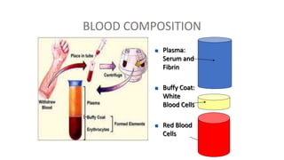
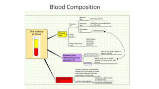
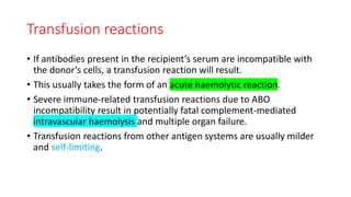
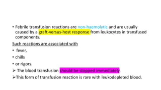
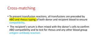
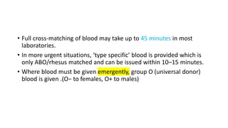
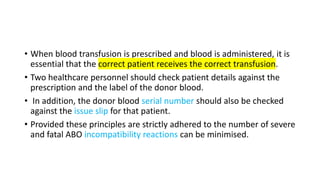
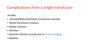
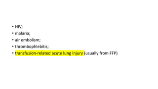
BLOOD PRODUCTS AND
TRANSFUSION
DR SS KAMBLE
TRANSFUSION
• The transfusion of blood and blood products has become
commonplace since the first successful transfusion in 1818.
• Although the incidence of severe transfusion reactions and infections
is now very low, in recent years it has become apparent that there is
an immunological price to be paid from the transfusion of
heterologous blood, leading to increased morbidity and decreased
survival in certain population groups (trauma, malignancy).
• Supplies are also limited, and therefore the use of blood and blood
products must always be judicious and justifiable for clinical need
(Table 2.5)
Blood and blood products
• Blood is collected from donors who have been previously screened
before donating, to exclude any donor whose blood may have the
potential to harm the patient, or to prevent possible harm that
donating a unit of blood may have on the donor.
• In the UK, up to 450 mL of blood is drawn, a maximum of three times
each year.
Each unit is tested for evidence of
• hepatitis B,
• hepatitis C,
• HIV-1,
• HIV-2
• and syphilis.
• Donations are leukodepleted as a precaution against variant
Creutzfeldt– Jakob disease (this may also reduce the immunogenicity
of the transfusion).
• The ABO and rhesus D blood groups are determined, as well as the
presence of irregular red cell antibodies.
• The blood is then processed into subcomponents.
BLOOD COMPOSITION
 Plasma:
Serum and
Fibrin
 Buffy Coat:
White
Blood Cells
 Red Blood
Cells
11
Whole blood
• Whole blood is now rarely available in civilian practice because it has
been seen as an inefficient use of the limited resource.
• However, whole blood transfusion has significant advantages over
packed cells as it is coagulation factor rich and, if fresh, more
metabolically active than stored blood.
Blood Composition
Packed red cells
• Packed red blood cells are spun-down and concentrated packs of red
blood cells.
• Each unit is approximately 330 mL and has a haematocrit of 50–70%.
• Packed cells are stored in a SAG-M solution (saline–adenine–glucose–
mannitol) to increase shelf life to 5 weeks at 2–6°C.
• (Older storage regimes included storage in CPD: citrate–phosphate–
dextrose solutions, which have a shelf life of 2–3 weeks.)
Fresh-frozen plasma
• Fresh-frozen plasma (FFP) is rich in coagulation factors and is
removed from fresh blood and stored at −40 to −50°C with a 2-year
shelf life.
• It is the first-line therapy in the treatment of coagulopathic
haemorrhage.
• Rhesus D-positive FFP may be given to a rhesus D-negative woman
although it is possible for seroconversion to occur with large volumes
owing to the presence of red cell fragments, and Rh-D immunisation
should be considered.
Cryoprecipitate
• Cryoprecipitate is a supernatant precipitate of FFP and is rich in factor
VIII and fibrinogen.
• It is stored at −30°C with a 2-year shelf life.
• It is given in low fibrinogen states or factor VIII deficiency.
Platelets
• Platelets are supplied as a pooled platelet concentrate and contain
about 250 × 109 /L.
• Platelets are stored on a special agitator at 20–24°C and have a shelf
life of only 5 days.
• Platelet transfusions are given to patients with thrombocytopenia or
with platelet dysfunction who are bleeding or undergoing surgery.
• Patients are increasingly presenting on antiplatelet therapy such as
aspirin or clopidogrel for reduction of cardiovascular risk.
• Aspirin therapy rarely poses a problem but control of haemorrhage
on the more potent platelet inhibitors can be extremely difficult.
• Patients on clopidogrel who are actively bleeding and undergoing
major surgery may require almost continuous infusion of platelets
during the course of the procedure.
• Arginine vasopressin or its analogues (DDAVP) have also been used in
this patient group, although with limited success.
Prothrombin complex concentrates
• Prothrombin complex concentrates (PCC) are highly purified
concentrates prepared from pooled plasma.
• They contain factors II, IX and X.
• Factor VII may be included or produced separately.
• It is indicated for the emergency reversal of anticoagulant (warfarin)
therapy in uncontrolled haemorrhage.
Autologous blood
• It is possible for patients undergoing elective surgery to predonate
their own blood up to 3 weeks before surgery for retransfusion during
the operation.
• Similarly, during surgery blood can be collected in a cell-saver which
washes and collects red blood cells which can then be returned to the
patient
Indications for blood transfusion
• Blood transfusions should be avoided if possible, and many previous
uses of blood and blood products are now no longer considered
appropriate.
The indications for blood transfusion are as follows:
● Acute blood loss, to replace circulating volume and maintain oxygen
delivery;
● Perioperative anaemia, to ensure adequate oxygen delivery during
the perioperative phase;
● Symptomatic chronic anaemia, without haemorrhage or impending
surgery.
Transfusion trigger
• Historically, patients were transfused to achieve a haemoglobin >10
g/dL.
• This has now been shown not only to be unnecessary but also to be
associated with an increased morbidity and mortality compared with
lower target values.
• A haemoglobin level of 6 g/dL is acceptable in patients who are not
actively bleeding, not about to undergo major surgery and are not
symptomatic.
There is some controversy as to the optimal haemoglobin level in some
patient groups, such as those with
• cardiovascular disease,
• sepsis
• and traumatic brain injury.
Although, conceptually, a higher haemoglobin level improves oxygen
delivery, there is little clinical evidence at this stage to support higher
levels in these groups (Table 2.6)
Blood groups and cross-matching
• Human red cells have on their cell surface many different antigens.
• Two groups of antigens are of major importance in surgical practice –
the ABO and rhesus systems.
ABO system
• These proteins are strongly antigenic and are associated with
naturally occurring antibodies in the serum.
• The system consists of three allelic genes – A, B and O – which control
synthesis of enzymes that add carbohydrate residues to cell surface
glycoproteins.
• A and B genes add specific residues while the O gene is an amorph
and does not transform the glycoprotein.
• The system allows for six possible genotypes although there are only
four phenotypes.
• Naturally occurring antibodies are found in the serum of those
lacking the corresponding antigen. (Table 2.7)
• Blood group O is the universal donor type as it contains no antigens
to provoke a reaction.
• Conversely, group AB individuals are ‘universal recipients’ and can
receive any ABO blood type because they have no circulating
antibodies.
• Rhesus system The rhesus D (Rh(D)) antigen is strongly antigenic and
is present in approximately 85% of the population in the UK.
• Antibodies to the D antigen are not naturally present in the serum of
the remaining 15% of individuals, but their formation may be
stimulated by the transfusion of Rh-positive red cells, or acquired
during delivery of a Rh(D)-positive baby.
• Acquired antibodies are capable, during pregnancy, of crossing the
placenta and, if present in a Rh(D)-negative mother, may cause severe
haemolytic anaemia and even death (hydrops fetalis) in a Rh(D)-
positive fetus in utero.
• The other minor blood group antigens may be associated with
naturally occurring antibodies, or may stimulate the formation of
antibodies on relatively rare occasions.
Transfusion reactions
• If antibodies present in the recipient’s serum are incompatible with
the donor’s cells, a transfusion reaction will result.
• This usually takes the form of an acute haemolytic reaction.
• Severe immune-related transfusion reactions due to ABO
incompatibility result in potentially fatal complement-mediated
intravascular haemolysis and multiple organ failure.
• Transfusion reactions from other antigen systems are usually milder
and self-limiting.
• Febrile transfusion reactions are non-haemolytic and are usually
caused by a graft-versus-host response from leukocytes in transfused
components.
Such reactions are associated with
• fever,
• chills
• or rigors.
 The blood transfusion should be stopped immediately.
This form of transfusion reaction is rare with leukodepleted blood.
Cross-matching
• To prevent transfusion reactions, all transfusions are preceded by
ABO and rhesus typing of both donor and recipient blood to ensure
compatibility.
• The recipient’s serum is then mixed with the donor’s cells to confirm
ABO compatibility and to test for rhesus and any other blood group
antigen–antibody reaction.
• Full cross-matching of blood may take up to 45 minutes in most
laboratories.
• In more urgent situations, ‘type specific’ blood is provided which is
only ABO/rhesus matched and can be issued within 10–15 minutes.
• Where blood must be given emergently, group O (universal donor)
blood is given .(O− to females, O+ to males)
• When blood transfusion is prescribed and blood is administered, it is
essential that the correct patient receives the correct transfusion.
• Two healthcare personnel should check patient details against the
prescription and the label of the donor blood.
• In addition, the donor blood serial number should also be checked
against the issue slip for that patient.
• Provided these principles are strictly adhered to the number of severe
and fatal ABO incompatibility reactions can be minimised.
Complications of blood transfusion
• Complications from blood transfusion can be categorised as those
arising from a single transfusion and those related to massive
transfusion.
Complications from a single transfusion
Include:
• incompatibility haemolytic transfusion reaction;
• febrile transfusion reaction;
• allergic reaction;
• infection:
• bacterial infection (usually due to faulty storage);
• hepatitis;
• HIV;
• malaria;
• air embolism;
• thrombophlebitis;
• transfusion-related acute lung injury (usually from FFP)
Complications from massive transfusion
Include:
• coagulopathy;
• hypocalcaemia;
• hyperkalaemia;
• hypokalaemia;
• hypothermia.
• In addition, patients who receive repeated transfusions over long
periods of time (e.g. patients with thalassaemia) may develop iron
overload.
• (Each transfused unit of red blood cells contains approximately 250
mg of elemental iron.)
Management of coagulopathy
• Correction of coagulopathy is not necessary if there is no active
bleeding and haemorrhage is not anticipated (not due for surgery).
• However, coagulopathy following or during massive transfusion
should be anticipated and managed aggressively.
• Prevention of dilutional coagulopathy is central to the damage control
resuscitation of patients who are actively bleeding.
• This is the prime reason for delivering balanced transfusion regimes
matching red blood cell packs with plasma and platelets.
• Based on moderate evidence, when red cells are transfused for active
haemorrhage, it is best to match each red cell unit with one unit of
FFP and one of platelets (1:1:1).
• This will reduce the incidence and severity of subsequent dilutional
coagulopathy.
• Crystalloids and colloids should be avoided for the same reason.
• The balanced transfusion approach cannot, however, correct
coagulopathy.
• Therefore, coagulation should be monitored routinely, either with
point-of-care testing (thromboelastometry) or with laboratory tests
(fibrinogen, clotting times).
• Underlying coagulopathies should be treated in addition to the
administration of 1:1:1 balanced transfusions.
• There are pharmacological adjuncts to blood component therapy.
• The antifibrinolytic tranexamic acid is the most commonly
administered.
• It is usually administered empirically to bleeding patients because
effective point-of-care tests of fibrinolysis are not yet routinely
available.
• There is little evidence to support the use of other coagulation factor
concentrates at this time.
Blood substitutes
Blood substitutes are an attractive alternative to the costly process of
• donating,
• checking,
• storing
• and administering blood,
• especially given the immunogenic and potential infectious
complications associated with transfusion.
• There are several oxygen-carrying blood substitutes under
investigation in experimental animal or early clinical trials.
• Blood substitutes are either biomimetic or abiotic.
• Biomimetic substitutes mimic the standard oxygen-carrying capacity
of the blood and are haemoglobin based.
• Abiotic substitutes are synthetic oxygen carriers and are currently
primarily perfluorocarbon based.
• Haemoglobin is seen as the obvious candidate for developing an
effective blood substitute.
• Various engineered molecules are under clinical trials, and are based
on human, bovine or recombinant technologies.
• Second-generation perfluorocarbon emulsions are also showing
potential in clinical trials.
THANK YOU

Blood transfusion

  • 1.
  • 2.
    TRANSFUSION • The transfusionof blood and blood products has become commonplace since the first successful transfusion in 1818. • Although the incidence of severe transfusion reactions and infections is now very low, in recent years it has become apparent that there is an immunological price to be paid from the transfusion of heterologous blood, leading to increased morbidity and decreased survival in certain population groups (trauma, malignancy).
  • 3.
    • Supplies arealso limited, and therefore the use of blood and blood products must always be judicious and justifiable for clinical need (Table 2.5)
  • 6.
    Blood and bloodproducts • Blood is collected from donors who have been previously screened before donating, to exclude any donor whose blood may have the potential to harm the patient, or to prevent possible harm that donating a unit of blood may have on the donor. • In the UK, up to 450 mL of blood is drawn, a maximum of three times each year.
  • 7.
    Each unit istested for evidence of • hepatitis B, • hepatitis C, • HIV-1, • HIV-2 • and syphilis.
  • 8.
    • Donations areleukodepleted as a precaution against variant Creutzfeldt– Jakob disease (this may also reduce the immunogenicity of the transfusion). • The ABO and rhesus D blood groups are determined, as well as the presence of irregular red cell antibodies. • The blood is then processed into subcomponents.
  • 10.
    BLOOD COMPOSITION  Plasma: Serumand Fibrin  Buffy Coat: White Blood Cells  Red Blood Cells
  • 11.
  • 12.
    Whole blood • Wholeblood is now rarely available in civilian practice because it has been seen as an inefficient use of the limited resource. • However, whole blood transfusion has significant advantages over packed cells as it is coagulation factor rich and, if fresh, more metabolically active than stored blood.
  • 15.
  • 16.
    Packed red cells •Packed red blood cells are spun-down and concentrated packs of red blood cells. • Each unit is approximately 330 mL and has a haematocrit of 50–70%. • Packed cells are stored in a SAG-M solution (saline–adenine–glucose– mannitol) to increase shelf life to 5 weeks at 2–6°C. • (Older storage regimes included storage in CPD: citrate–phosphate– dextrose solutions, which have a shelf life of 2–3 weeks.)
  • 17.
    Fresh-frozen plasma • Fresh-frozenplasma (FFP) is rich in coagulation factors and is removed from fresh blood and stored at −40 to −50°C with a 2-year shelf life. • It is the first-line therapy in the treatment of coagulopathic haemorrhage. • Rhesus D-positive FFP may be given to a rhesus D-negative woman although it is possible for seroconversion to occur with large volumes owing to the presence of red cell fragments, and Rh-D immunisation should be considered.
  • 19.
    Cryoprecipitate • Cryoprecipitate isa supernatant precipitate of FFP and is rich in factor VIII and fibrinogen. • It is stored at −30°C with a 2-year shelf life. • It is given in low fibrinogen states or factor VIII deficiency.
  • 21.
    Platelets • Platelets aresupplied as a pooled platelet concentrate and contain about 250 × 109 /L. • Platelets are stored on a special agitator at 20–24°C and have a shelf life of only 5 days. • Platelet transfusions are given to patients with thrombocytopenia or with platelet dysfunction who are bleeding or undergoing surgery.
  • 23.
    Platelets • Platelets aresupplied as a pooled platelet concentrate and contain about 250 × 109 /L. • Platelets are stored on a special agitator at 20–24°C and have a shelf life of only 5 days. • Platelet transfusions are given to patients with thrombocytopenia or with platelet dysfunction who are bleeding or undergoing surgery.
  • 24.
    • Patients areincreasingly presenting on antiplatelet therapy such as aspirin or clopidogrel for reduction of cardiovascular risk. • Aspirin therapy rarely poses a problem but control of haemorrhage on the more potent platelet inhibitors can be extremely difficult. • Patients on clopidogrel who are actively bleeding and undergoing major surgery may require almost continuous infusion of platelets during the course of the procedure.
  • 25.
    • Arginine vasopressinor its analogues (DDAVP) have also been used in this patient group, although with limited success.
  • 26.
    Prothrombin complex concentrates •Prothrombin complex concentrates (PCC) are highly purified concentrates prepared from pooled plasma. • They contain factors II, IX and X. • Factor VII may be included or produced separately. • It is indicated for the emergency reversal of anticoagulant (warfarin) therapy in uncontrolled haemorrhage.
  • 36.
    Autologous blood • Itis possible for patients undergoing elective surgery to predonate their own blood up to 3 weeks before surgery for retransfusion during the operation. • Similarly, during surgery blood can be collected in a cell-saver which washes and collects red blood cells which can then be returned to the patient
  • 37.
    Indications for bloodtransfusion • Blood transfusions should be avoided if possible, and many previous uses of blood and blood products are now no longer considered appropriate. The indications for blood transfusion are as follows: ● Acute blood loss, to replace circulating volume and maintain oxygen delivery;
  • 38.
    ● Perioperative anaemia,to ensure adequate oxygen delivery during the perioperative phase; ● Symptomatic chronic anaemia, without haemorrhage or impending surgery.
  • 39.
    Transfusion trigger • Historically,patients were transfused to achieve a haemoglobin >10 g/dL. • This has now been shown not only to be unnecessary but also to be associated with an increased morbidity and mortality compared with lower target values. • A haemoglobin level of 6 g/dL is acceptable in patients who are not actively bleeding, not about to undergo major surgery and are not symptomatic.
  • 40.
    There is somecontroversy as to the optimal haemoglobin level in some patient groups, such as those with • cardiovascular disease, • sepsis • and traumatic brain injury. Although, conceptually, a higher haemoglobin level improves oxygen delivery, there is little clinical evidence at this stage to support higher levels in these groups (Table 2.6)
  • 42.
    Blood groups andcross-matching • Human red cells have on their cell surface many different antigens. • Two groups of antigens are of major importance in surgical practice – the ABO and rhesus systems.
  • 43.
    ABO system • Theseproteins are strongly antigenic and are associated with naturally occurring antibodies in the serum. • The system consists of three allelic genes – A, B and O – which control synthesis of enzymes that add carbohydrate residues to cell surface glycoproteins. • A and B genes add specific residues while the O gene is an amorph and does not transform the glycoprotein.
  • 44.
    • The systemallows for six possible genotypes although there are only four phenotypes. • Naturally occurring antibodies are found in the serum of those lacking the corresponding antigen. (Table 2.7)
  • 47.
    • Blood groupO is the universal donor type as it contains no antigens to provoke a reaction. • Conversely, group AB individuals are ‘universal recipients’ and can receive any ABO blood type because they have no circulating antibodies.
  • 49.
    • Rhesus systemThe rhesus D (Rh(D)) antigen is strongly antigenic and is present in approximately 85% of the population in the UK. • Antibodies to the D antigen are not naturally present in the serum of the remaining 15% of individuals, but their formation may be stimulated by the transfusion of Rh-positive red cells, or acquired during delivery of a Rh(D)-positive baby.
  • 50.
    • Acquired antibodiesare capable, during pregnancy, of crossing the placenta and, if present in a Rh(D)-negative mother, may cause severe haemolytic anaemia and even death (hydrops fetalis) in a Rh(D)- positive fetus in utero. • The other minor blood group antigens may be associated with naturally occurring antibodies, or may stimulate the formation of antibodies on relatively rare occasions.
  • 51.
    Transfusion reactions • Ifantibodies present in the recipient’s serum are incompatible with the donor’s cells, a transfusion reaction will result. • This usually takes the form of an acute haemolytic reaction. • Severe immune-related transfusion reactions due to ABO incompatibility result in potentially fatal complement-mediated intravascular haemolysis and multiple organ failure. • Transfusion reactions from other antigen systems are usually milder and self-limiting.
  • 52.
    • Febrile transfusionreactions are non-haemolytic and are usually caused by a graft-versus-host response from leukocytes in transfused components. Such reactions are associated with • fever, • chills • or rigors.  The blood transfusion should be stopped immediately. This form of transfusion reaction is rare with leukodepleted blood.
  • 53.
    Cross-matching • To preventtransfusion reactions, all transfusions are preceded by ABO and rhesus typing of both donor and recipient blood to ensure compatibility. • The recipient’s serum is then mixed with the donor’s cells to confirm ABO compatibility and to test for rhesus and any other blood group antigen–antibody reaction.
  • 54.
    • Full cross-matchingof blood may take up to 45 minutes in most laboratories. • In more urgent situations, ‘type specific’ blood is provided which is only ABO/rhesus matched and can be issued within 10–15 minutes. • Where blood must be given emergently, group O (universal donor) blood is given .(O− to females, O+ to males)
  • 55.
    • When bloodtransfusion is prescribed and blood is administered, it is essential that the correct patient receives the correct transfusion. • Two healthcare personnel should check patient details against the prescription and the label of the donor blood. • In addition, the donor blood serial number should also be checked against the issue slip for that patient. • Provided these principles are strictly adhered to the number of severe and fatal ABO incompatibility reactions can be minimised.
  • 60.
    Complications of bloodtransfusion • Complications from blood transfusion can be categorised as those arising from a single transfusion and those related to massive transfusion.
  • 61.
    Complications from asingle transfusion Include: • incompatibility haemolytic transfusion reaction; • febrile transfusion reaction; • allergic reaction; • infection: • bacterial infection (usually due to faulty storage); • hepatitis;
  • 62.
    • HIV; • malaria; •air embolism; • thrombophlebitis; • transfusion-related acute lung injury (usually from FFP)
  • 63.
    Complications from massivetransfusion Include: • coagulopathy; • hypocalcaemia; • hyperkalaemia; • hypokalaemia; • hypothermia.
  • 64.
    • In addition,patients who receive repeated transfusions over long periods of time (e.g. patients with thalassaemia) may develop iron overload. • (Each transfused unit of red blood cells contains approximately 250 mg of elemental iron.)
  • 65.
    Management of coagulopathy •Correction of coagulopathy is not necessary if there is no active bleeding and haemorrhage is not anticipated (not due for surgery). • However, coagulopathy following or during massive transfusion should be anticipated and managed aggressively. • Prevention of dilutional coagulopathy is central to the damage control resuscitation of patients who are actively bleeding.
  • 66.
    • This isthe prime reason for delivering balanced transfusion regimes matching red blood cell packs with plasma and platelets. • Based on moderate evidence, when red cells are transfused for active haemorrhage, it is best to match each red cell unit with one unit of FFP and one of platelets (1:1:1). • This will reduce the incidence and severity of subsequent dilutional coagulopathy. • Crystalloids and colloids should be avoided for the same reason.
  • 67.
    • The balancedtransfusion approach cannot, however, correct coagulopathy. • Therefore, coagulation should be monitored routinely, either with point-of-care testing (thromboelastometry) or with laboratory tests (fibrinogen, clotting times). • Underlying coagulopathies should be treated in addition to the administration of 1:1:1 balanced transfusions.
  • 68.
    • There arepharmacological adjuncts to blood component therapy. • The antifibrinolytic tranexamic acid is the most commonly administered. • It is usually administered empirically to bleeding patients because effective point-of-care tests of fibrinolysis are not yet routinely available. • There is little evidence to support the use of other coagulation factor concentrates at this time.
  • 69.
    Blood substitutes Blood substitutesare an attractive alternative to the costly process of • donating, • checking, • storing • and administering blood, • especially given the immunogenic and potential infectious complications associated with transfusion.
  • 70.
    • There areseveral oxygen-carrying blood substitutes under investigation in experimental animal or early clinical trials. • Blood substitutes are either biomimetic or abiotic. • Biomimetic substitutes mimic the standard oxygen-carrying capacity of the blood and are haemoglobin based. • Abiotic substitutes are synthetic oxygen carriers and are currently primarily perfluorocarbon based.
  • 72.
    • Haemoglobin isseen as the obvious candidate for developing an effective blood substitute. • Various engineered molecules are under clinical trials, and are based on human, bovine or recombinant technologies. • Second-generation perfluorocarbon emulsions are also showing potential in clinical trials.
  • 74.