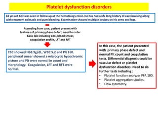
2. Bleeding :
• spontaneous, excessive, or delayed in onset following tissue injury
results from a localized pathologic process or a disorder of the
hemostatic process.
Pathophysiology of hemostasis
6. Clinical manifestation
Disorders of Plt or blood vessels (primary
phase) :
• Cutaneous bleeding characterize by :
- petechiae.
- Ecchymoses
• mucosal bleeding including epistaxis and/or
gingival bleeding.
• Menorrhagia (menstrual flow that does not
taper after more than three days) or
metrorrhagia (bleeding in between periods).
Coagulation disorders (secondary
phase):
• large palpable ecchymoses and
large, spreading, deep soft tissue
hematomas.
• Hemorrhage into synovial joints
(hemarthrosis)
Petechiae ecchymoses Gingival
bleeding
8. History of bleeding
Personal history Family history :
• Ask about any
blood or bleeding
disorders in
family.Bleeding episodes:
• Onset.
• Sites of bleeding (as
cutaneous or mucosal
and deep)
• Amount of bleeding
• Duration of bleeding
• Frequency and
severity of each one.
• Medication
Hemostasis challenging
as any bleeding during :
• Immunization
• Tooth extraction
• Circumcision
• Trauma
• Surgery
• Menstrual history
9. Diagnostic approach
Laboratory test including :
• CBC for Plt count.
• Peripheral blood smear to confirm Plt count and look for structural
abnormalities as size.
• Coagulation profile :
- PT for extrinsic pathway.
- aPTT for intrinsic pathway.
- Thrombin time (TT) for common pathway
- Fibrinogen
• RFT and LFT
Primary hemostasis disorders :
• Abnormal Plt count.
• Abnormal structural Plt on
blood film.
• Could be normal results in case
of functional Plt disorders or
vascular disorders.
Secondary hemostasis
disorders:
• Abnormal coagulation
profile.
• Normal CBC and blood
smear result.
The result will differentiate
between
10. Then other test depend on results of
previous tests
1- primary hemostasis disorders
others depend on differential diagnosis for that patient (see page 4)
11. 2- secondary hemostasis disorders
Abnormal result in coagulation profile
Do mixing study to differentiate between factors deficiency or inhibitors by adding
normal plasma into patient plasma at temperature of 37C and repeat coagulation profile
If coagulation profile results
have been corrected indicate
factors deficiency
If coagulation profile results not corrected
indicate factors inhibitors caused by :
- Acquired coagulation factor inhibitor
- Antiphospholipid antibodies
- Heparin
Then, do coagulation factor assays to
identify which factors have been defect.
Need be specific which factors you want
to test which depend on :
• If PT only prolong, test for VII.
• If aPTT only prolong, test for VIII, IX, XI,
XII.
• If both PT and aPTT prolong test for all
factors including common pathway.
Other test like von willebrand profile
including vWF antigen level and
ristocetin activity
15. Von willebrand disease
22 yrs old lady was seen for evaluation of menorrhagia. She reported that her menstrual periods had
always been heavy. She also gave history of intermittent epistaxis and gum bleeding. She had a
prolonged bleed following tooth extraction in the past. Examination was unremarkable except for
the pallor.
According from case, patient present with
primary phase defect (muco-cutaneous bleeding
with menorrhagia)
Need to do basic laboratory tests including CBC,
blood smear, coagulation profile, LFT and RFT
CBC showed Hb 9.6 g/dL, WBC of 6.8 and Plt 360.
blood smear was normal. PT was 11s, APTT was 42s,
thrombin time was 15s and fibrinogen was 3.2 g/L.
liver function test and creatinine were within normal
limits.
DD
1- Plt dysfunction.
2- vWD
3- secondary hemostasis
disorders.
4- vascular disorders
Need further test including
mixing study with coagulation
factors assay, platelet function
test and vW profile.
16. vWD :
• caused by mutations that lead to an impairment in the synthesis or function of
von Willebrand factor (VWF) present with :
- Usually mucocutaneous
- Heavy menstrual bleeding
- mimics the findings seen in classical hemophilia, including soft tissue, joint, and
urinary bleeding, and bleeding after invasive procedures as associated with low
factor VIII especially in type 2N VWD.
classification of VWD :
17.
18. Diagnostic approach :
To confirm the diagnosis of VWD :
• Plasma VWF antigen.
• Plasma VWF activity (ristocetin cofactor activity, VWF:RCo and VWF
collagen binding, VWF:CB).
• Factor VIII activity (FVIII).
Then other test to
determine the type of VWD
• VWF multimer distribution using gel electrophoresis.
• Ristocetin-induced platelet aggregation (RIPA)
21. Hemophilia
6 yrs old boy presented with the emergency room with painful swollen right knee. He had prolonged
bleeding after circumcision previously. He also had multiple bruises following minor injuries. He was
admitted with a swollen right knee a few times in the past. Examination showed warm, tender,
swollen right knee with restriction of movement. The rest of his examination was unremarkable.
According from case, patient present with
secondary phase defect (deep tissue bleeding ).
Need to do basic laboratory tests including CBC,
blood smear, coagulation profile, LFT and RFT
CBC and blood smear were normal. Coagulation
showed PT 11s and APTT of 68s. LFT AND RFT were
normal.
So mixing study was done which showed full
correction of APTT after mixing with normal donor
plasma (indicate factors deficiency).
Then factors assay for intrinsic was done which
showed :
- Factor VIII < 1%
- Factor IX 90%
- Factor XI 102%
- Factor XII 98%
DD
Hemophilia type A
Confirmed by genetic molecular
test
22. Hemophilia
• an inherited bleeding disorder caused by deficiency of coagulation factors including :
- Hemophilia A (commonly) – Inherited deficiency of factor VIII an X-linked recessive disorder.
- Hemophilia B – Inherited deficiency of factor IX an X-linked recessive disorder.
- Hemophilia C – Inherited deficiency of factor XI an autosomal recessive disorder.
Clinical presentation
• Onset and clinical manifestation Depend on
severity.
• Sites of bleeding :
- Joints and muscle (most common)
- GI bleeding
- Genitourinary tract.
- Intracranial bleeding (rare).
• Late complication :
- Hemophilic arthropathy.
- Infection from plasma derived products.
- Development of factors inhibitors.
- Cardiovascular disease.
24. Management
Routine care :
• Newborn care for those with high risk from known
female carrier as :
- multidisciplinary planning is required to address maternal
and fetal bleeding risks, method of delivery, and avoidance
of interventions such as fetal scalp electrodes, fetal venous
sampling, and operative vaginal delivery (eg, use of
forceps, vacuum extraction).
• Immunization for hepatitis.
• Dental care to prevent gum and tooth disease, which
increase the risk of bleeding.
• Exercise and athletic participation as avoid high impact
activities.
• Avoid Medicines that increase the risk of bleeding,
including anticoagulants, aspirin, and other nonsteroidal
anti-inflammatory drugs (NSAIDs).
• Planning for invasive procedures.
• Desmopressin might help in mild cases as promotes
release of factor VIII and its carrier protein von
Willebrand factor (VWF) from storage pools in platelet
granules and endothelial cells.
• Obstetric care and preconception counseling.
Prophylaxis by factor
replacement therapy :
• Give as second line if the
bleeding not control.
25.
26. Immune thrombocytopenia (ITP)
24 yrs old lady went to her doctor having noticed bruising on her arms and legs for 4 days. The
bruises were not painful but were increasing in number every day. Examination was unremarkable
apart from the bruises.
DD Low Plt count for further
evaluation (DD in page 4)
According from case, patient present with
primary phase defect (muco-cutaneous bleeding
with menorrhagia)
Need to do basic laboratory tests including CBC,
blood smear, coagulation profile, LFT and RFT
CBC showed Hb 11 g/dL, WBC of 5 and Plt 4. blood
smear showed low Plt count. PT and APTT were
normal. liver function test and creatinine were within
normal limits.
27. ITP :
• an acquired thrombocytopenia caused by autoantibodies against platelet
antigens which enhance the destruction of Plt by spleen. Present with :
- Could be asymptomatic.
- Mucocutaneous bleeding including petechiae, purpura and epistaxis.
- In severe cases (rare) present with severe hemorrhage as intracranial hemorrhage
(ICH), overt gastrointestinal bleeding, and hematuria.
classification of ITP :
Newly diagnosed :
• Up to three months
since diagnosis.
Chronic :
• More than 12 months
since diagnosis.
Persistent :
• 3 to 12 months since
diagnosis.
28. Diagnostic approach :
ITP is essentially a diagnosis of exclusion. But there are many DD which require a lot of
workup.
Clinical diagnosis by history and examination.
Laboratory tests to rule in/out other causes:
• CBC and blood smear with low Plt.
• Coagulation profile with normal result.
• LFT and RFT.
• HIV and HCV testing.
• H.pylori testing.
• Thyroid function test
• Immunologic studies for ANA and RF.
• Vitamin B12 and folate levels.
• Bone marrow examination in case of :
- If associated with neutropenia
- Or more than two cell line affected.
- If > 60 yrs of age.
- If not respond to ITP therapy.
31. Management
Emergency treatment if associated with
active bleeding in CNS, GI or GU :
• Stop drugs reducing Plt function, control BP
and minimize trauma.
• Start with steroid as prednisone (1mg/Kg) or
methylprednisolone or dexamethasone.
• Antifibrinolytic as tranexamic acid.
• IVIG 2g/Kg over 5days.
• Plt transfusion
• Emergency splenectomy.
Non-urgent treatment :
1st line :
• Steroid by dexamethasone 40mg/d for 4
weeks or prednisone 1mg/Kg/d.
• IVIG at 1g/Kg for one or two days.
• Anti-Rh(D) with 50 to 75 mcg/Kg IV for
those who have Rh+ blood type.
2nd line :
• Immunosuppressant (azathioprine,
cyclophosphamide).
• Retuximab.
• Thrombopoietin receptor agonist
(romiplostim).
• Splenectomy in case :
- Adult with more than 6 months persistent.
- In pediatric for selective cases if long
duration and symptomatic.
- If not response for previous treatment.
33. Thrombotic thrombocytopenia purpura (TTP)
40 year old female, presented with a history of fever, pain in the abdomen, vomiting and loose motions since 8
days. The fever was intermittent, high grade and the loose motions were watery, large in quantity and occurred 5 to
6 times daily. This was followed by reddish, small, patchy, skin lesions which started three days before admission.
Two days before admission, the patient developed diminishing urine output and deteriorating consciousness.
On admission, the pulse was 100/minute and regular; the respiratory rate was 28/minute and B.P. was 130/80 mm
Hg in the right upper limb in supine position. There was a petechial and ecchymotic rash all over the body but more
prominent on the extremities. She also had puffiness of the face and oedema of the feet. There was no icterus.
Examination of the respiratory system revealed crepitations in the right infra-mammary region. Central nervous
system examination showed that the patient was unconscious. She responded to painful stimulus with purposeful
movements of all four limbs. Planters were bilaterally extensor. The rest of the examination was normal and
revealed no focal deficit.
DD
Low Plt count for further
evaluation (DD in page 4)
According from case, patient present with primary
phase defect (muco-cutaneous bleeding)
associated with renal and neurological complaints
Need to do basic laboratory tests including CBC,
blood smear, coagulation profile, LFT and RFT
CBC showed Hb 8 g/dL with retics 3%, WBC of 6 and Plt 56. blood smear
showed low Plt count and schistocytes . PT and APTT were normal. liver
function test was normal and creatinine was 160.
34. Thrombotic microangiopathy syndromes (TMA):
• specific pathologic lesion in which abnormalities in the vessel wall of arterioles
and capillaries lead to microvascular thrombosis. TMA syndromes include :
1- thrombotic thrombocytopenia purpura (TTP).
2- Shiga toxin-mediated HUS (hemolytic-uremic syndrome [ST-HUS]).
3- drug-induced TMA (DITMA) syndromes.
4- complement-mediated TMA.
• TTP associated with Deficiency of
ADAMTS13 which can be hereditary or
acquired. This enzyme break down
ultra large von willrbrand factor
multimers (ULVWF) which become
inactive. Once reduce ADAMTS13 will
lead to increase ULVWF causing more
Plt aggregation and form large
occlusive platelet thrombi which are
capable of embolizing to microvessels
contributing to organ ischemia.
• HUS associated with direct affect of
toxin (Shiga toxin producing E. Coli )
into Plt and RBC.
Once
this
occur,
will
cause
Microangiopathic hemolytic
anemia (MAHA) :
• intravascular red blood cell
fragmentation that produces
schistocytes on the peripheral
blood smear including small
arterioles and capillaries.
thrombocytopenia
35.
36.
37. Disseminated intravascular coagulation (DIC)
A 46-year old Caucasian female with a history of intravenous drug abuse, hypertension and traumatic splenectomy
(after a motor vehicle accident 12 years prior), presented to our institution with altered mental status for the past
one day. She was also a chronic smoker and had a history of noncompliance to vaccinations. On arrival to the
emergency department, she had multiple episodes of coffee ground emesis and tarry colored stools.
On examination, she was febrile up to a temperature 101.7 F and persistently hypotensive with blood pressure (BP)
74/22, and heart rate 70 beats per min (bpm). A petechial rash was noted initially on her face, but then rapidly
became generalized spreading to her arms, legs and trunk. She also had multiple track marks on both upper
extremities.
DD
both primary and
secondary phases are
affected with feature of
hemodynamically
unstable. So, could be
DIC
According from case, patient present with features
of septic shock associated with primary phase
defect (muco-cutaneous bleeding)
Need to do basic laboratory tests including CBC,
blood smear, coagulation profile, LFT and RFT
CBC showed Hb 7 g/dL. WBC of 15 and Plt 35. blood smear showed low
Plt count and schistocytes . PT and APTT were prolonged with low
fibrinogen. liver function test was normal and creatinine were 159
38. DIC :
• Widespread inappropriate intravascular deposition of fibrin with consumption
of coagulation factors and platelets occurs as consequences of many disorders.
Causes of DIC :
40. Laboratory finding :
• Low Hb and Plt.
• Low fibrinogen concentration.
• Prolong PT and APTT
• High level of fibrin degradation products in serum and urine
as D-dimer.
• Blood smear show schistocytes as MAHA.
41. Management
• Stabilize the patient and treat the underlying cause.
• Prevent/treat the bleeding :
- Fresh frozen plasma and Plt transfusion in patient with extensive
bleeding.
- Cryoprecipitate and red cell transfusion.
• Prevent/ treat the thrombosis :
- Use of heparin or anti-platelet drugs to inhibit the coagulation
process if no active bleeding.
- Anti-thrombin concentrate or recombinant activated human
protein C might use to inhibit DIC.
42. Heparin induce thrombocytopenia (HIT)
58 yrs old lady with history of HTN and DM II, was admitted with a hip fracture that she sustained following a fall at
home. Plain Xray showed a sub-trochanteric right hip fracture. After stabilization , she underwent open reduction
and internal fixation. 12hrs following surgery, she was started on LWMH enoxaparin 40mg subcutaneously daily. On
the 6th day post-op, CBC showed Hb 9, WBC8, Plt 98. further investigation didn’t reveal any other abnormality. Her
Plt count improved over the subsequent few days.
From the case, Plt drop after administration of
LWMH with no significant symptoms. Possible
causes
HIT :
• is a life-threatening complication of
exposure to heparin (ie, unfractionated
heparin, low molecular weight [LMW]
heparin) regardless of the dose,
schedule, or route of administration.
• More occur in exposure of
unfractionated heparin.
44. Need to rule
out type 2
immediately
as more
severe form
compared
with type 1
Mechanism
45. Clinical manifestation
Thrombocytopenia :
• A platelet count drop of >50 percent of baseline
is typical; the mean nadir is
approximately 60,000/microL; and
counts <20,000/microare or bleeding are rare.
• The typical onset of thrombocytopenia occurs 5
to 10 days after the initiation of heparin
therapy. If occur within first 24 hrs of exposure,
seen in patient who has been exposed to
heparin previous 1-3 months.
• The resolution of thrombocytopenia following
withdrawal of heparin typically occurs within
seven days.
Thrombosis :
• thrombosis occurs in up to 50 percent
of individuals with HIT, with venous
more common than arterial thrombi.
• Common sites of venous thrombosis
include leg veins, cardiac vessels, and
skin.
When one of these
symptoms occur
after administration
of heparin, do 4T
score
46. If intermediate or
high probability,
confirm the
diagnosis by
• Positive ELISA with an optical density (OD)
>2.00 or
• Positive functional assay for HIT antibodies by :
- Serotonin release assay.
- Heparin induced Plt aggregation (HIPA).
47. Other differential diagnosis with bleeding during administration of
heparin, consider heparin overdose :
• Patient will has high APTT and TT.
• Mixing study will not correcting as anti-coagulation act as inhibitors.
• TT normalize once you given anti-dote (protamine).
48. Vit K deficiency
a 45-year-old man known case of chron’s disease, presented to the Emergency Room (ER) with sudden onset of
localized ecchymosis upper and lower limbs and widespread hematomas. No history of spontaneous or induced
bleeding episodes, antibiotic use or non-steroidal anti-inflammatory drugs use was reported. The patient presented
with hypertension, which was treated with ACE inhibitors.
According from case, patient present with
features of secondary phase defect, need to
order basic lab including CBC, blood smear,
coagulation profile, LFT and RFT
CBC and blood smear were normal. Coagulation
showed prolonged PT and APTT. LFT and RFT were
normal.
So mixing study was done which showed full
correction of PT and APTT after mixing with normal
donor plasma (indicate factors deficiency).
Then factors assay for intrinsic was done which
showed decreased level of FII, FVII, FIX and FX.
Based on clinical presentation,
the background of the patient
(chron’s disease) and finding of
laboratory tests which going
into diagnosis of Vit K
deficiency.
49. Vit K deficiency :
• Fat soluble vit K is obtained from green vegetables and bacterial synthesis in the gut.
Deficiency may present in newborn or later life caused by inadequate diet, malabsorption
or inhibition of vit K by drugs as warfarin and chronic use of antibiotic.
Mechanism
Diagnosis :
• easy bruisability, mucosal bleeding, splinter hemorrhages, melena,
hematuria, or any other manifestations of impaired coagulation.
• Prolonged PT and APTT with low plasma level of factor II, VII. IX, X.
50. Other differential diagnosis with bleeding in patient taking
warfarin is warfarin overdose :
• Patient will has high PT, APTT and INR.
• Management depend on clinical presentation and INR :
- INR > 4.5 without active bleeding, stopped warfarin for 1 or 2days and
adjust the dose.
- INR > 8 without bleeding or without bleeding, give oral dose of 0.5 to 2.5
mg Vit K and stopped warfarin for 1 or 2day and adjust the dose.
- Serious bleeding, stopped warfarin and give IV 2.5mg vit K or infusion of
prothrombin complex concentrate or fresh frozen plasma.
Management :
• In case of active bleeding, give Vit K 10 mg slowly IV. Some correction of PT is usual
within 6 hrs. the dose should be repeated on the next 2 days after optimal correction
occur.
• Rapid correction may be achieved by infusion of prothrombin complex concentrate.
• Prophylaxis with 5 mg/day orally of Vit K.
51. Platelet dysfunction disorders
18 yrs old boy was seen in follow up at the hematology clinic. He has had a life long history of easy bruising along
with recurrent epistaxis and gum bleeding. Examination showed multiple bruises on his arms and legs.
According from case, patient present with
features of primary phase defect, need to order
basic lab including CBC, blood smear,
coagulation profile, LFT and RFT
CBC showed Hb8.9g/dL, WBC 5.2 and Plt 160.
peripheral smear showed a microcytic hypochromic
picture and Plt were normal in count and
morphology. Coagulation, LFT and RFT were
normal.
In this case, the patient presented
with primary phase defect and
normal Plt count and coagulation
tests. Differential diagnosis could be
vascular defect or platelet
dysfunction disorders. Need to do
further tests including :
• Platelet function analyzer PFA 100.
• Platelet aggregation studies.
• Flow cytometry.
52. Platelet function
If these steps are
affected
Platelet dysfunction disorders
Inherited disorders :
• Giant platelet diroders
• Wiskott-aldrich syndrome.
• Glanzmann disease
• Other as storage pool disorders, glycoprotein VI
defect.
Acquired disorders :
• Anti-platelet medication as aspirin and
clopidgrel.
• Liver disease, uremia,
myeloproflerative disorders, diabetes
mellitus and other.
53. Examples of inherited Platelet dysfunction disorders
Giant platelet disorders :
• These include platelet glycoprotein abnormalities
(eg, Bernard-Soulier syndrome ), deficiency of
platelet alpha granules (eg, gray platelet
syndrome) and others.
• These syndromes characterize by large size of
platelet than normal.
• Usually associated with defective adherence.
• Treat with Plt transfusion and non-peptide
thrombopoietin receptor agonist (eltrombopag)
Glanzmann’s disease :
• autosomal recessive bleeding disorder
characterized by a defect in the
platelet integrin αIIbβ3 leading to
failure of primary platelet aggregation.
• It usually presents in the neonatal
period.
54. Approach to patient with platelet dysfunction disorders
• Need to assess the patient clinically to exclude possible
acquired causes especially in uremia or drug induce.
• Order basic lab including CBC, blood smear, coagulation
profile, LFT and RFT as to rule out other differential diagnosis.
• In peripheral blood smear, look for any giant size of platelet.
Do platelet function analyzer (PFA 100) :
• This test will tell you the abnormalities of platelet function but
will not give you specific diagnosis.
• Be careful, negative result may not rule out the plt dysfunction
disorders.
• the PFA 100 records the closure time which a duration to form
a hemostatic plug when the platelet are exposed within
citrated whole blood to high shear (5000 to 6000/second).
Other test to specify
the diagnosis
55. Platelet aggregation assays :
• This test help to measures platelet activation and aggregation
in vitro by mixing platelet with Common agonists used in
these assays include ADP, arachidonic acid, collagen,
epinephrine, thrombin, and ristocetin.
• According to result (see the table) will give some differential
diagnosis.
56. Flow cytometric analysis of platelets and activation markers :
• can be used to measure activated platelets, platelet hypo- or
hyper-reactivity, platelet-leukocyte aggregates, platelet
microparticles, and platelet turnover.
• Example :
- In Bernard-Soulier syndrome, the test could detect the
abnormalities in the platelet membrane glycoprotein Iib/IIIa
complex.
Management :
• Treat the underlying causes of acquired disorders.
• Desmopressin
• Platelet transfusions are particularly indicated in cases of severe, uncontrolled
bleeding, when prior treatments (eg, DDAVP)
• antifibrinolytic agents (eg, tranexamic acid, epsilon aminocaproic acid) may be
helpful in reducing bleeding.
• Recombinant factor VIIa (rFVIIa) has had some success for the treatment of bleeding
in patients with congenital platelet disorders.
• Bone marrow transplantation.
57. Other differential diagnosis
Disorders of Contact factors :
• Patient present with secondary phase defect.
• PT and APTT will be affected according which type of contact factors and mixing
study required to differentiate between inhibitors or deficiency.
Factor VIII inhibitors (acquired hemophilia) :
• Patient present with same features of inherited hemophilia but with different
mechanism.
• APTT will be prolonged and mixing study will show correction after 2 hrs in
temperature of 37C (compared with inherited hemophilia which show correction
immediate ).
• Treat with steroid and immunosuppression as cyclophosphamide.
Factor XIII deficiency :
• Patient present with secondary phase defect, recurrent miscarriage.
• PT, APTT and TT will be normal. To diagnosis, assess the clot solubility by mixing in
5M urea or 1% monochloroacetic acid and see the time of lysis. There are other tests
for qualitative and quantitative testing.
• Treat by XIII replacement therapy.