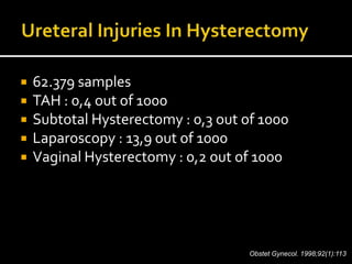
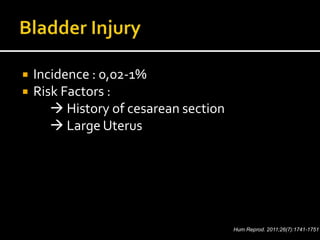
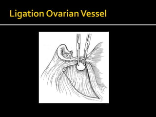
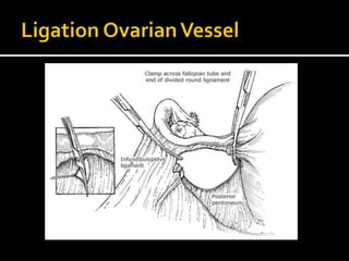
BrahmanaAskandarTjokroprawiro
 The first abdominal hysterectomy was
performed by Charles Clay in Manchester,
England in 1843
 Ellis Burnham from Lowell, Massachusetts
achieved the first successful abdominal
hysterectomy
 Women should be counseled before surgery
about the planned type of abdominal incision
 Vaginal examnination may help determine
the types of incision
 There are no proven medical or surgical benefits of
performing subtotal hysterectomy if the cervix can be easily
removed with the corpus
 Retaining the cervix commits the patient to continued
cervical cancer screening
 The only absolute contraindication to subtotal hysterectomy
is the presence of a malignant or premalignant condition of
the uterine corpus or cervix.
European Journal of Obstetrics & Gynecology and Reproductive Biology 193 (2015) 40–45
European Journal of Obstetrics & Gynecology and Reproductive Biology 193 (2015) 40–
 Although supracervical (subtotal) hysterectomy preserves
the cervix, upper vagina, and pelvic attachments, it does not
prevent subsequent prolapse.
 Randomized trials comparing total abdominal versus
supracervical hysterectomy have reported no difference in
vaginal support, regardless of cervical preservation or
removal
Obstet Gynecol. 2003;102(3):453.
N Engl J Med. 2002;347(17):1318.
 Position in the dorsal supine or lithotomy position (preferred
by some surgeons so that a second assistant can stand
between the patient's legs)
 Perform an examination under anesthesia (helps to confirm
pelvic findings and guide the final choice of incision)
 Insert Foley bladder catheter
 Perform sterile preparation of the abdomen and vagina
 Place surgical draping.
 The skin incision may be transverse or midline vertical and is
determined by a variety of factors, such as presence of prior
surgical scar, need for exploration of the upper abdomen,
size and mobility of the uterus, and desired cosmetic results.
 If a prior incision exists, most surgeons prefer to use this
incision.
 If the prior scar is cosmetically unacceptable, it may be
excised at the beginning or end of the procedure
 Most surgeons prefer to use a self-retaining retractor for an
abdominal hysterectomy
 The type of self-retaining retractor used depends on surgeon
preference.
 When positioning retractors, it is important to avoid placing
the lateral blades over a femoral nerve as it emerges lateral
to the psoas muscle, since this can lead to a peripheral
neuropathy
 The key of successful surgery
 Communication with anesthesiologist
 Use retractor may be helpfull
 Traditionally, a large Kelly clamp is placed across each
uterine cornu  cut  suture
 Electrocauter can also be used
 A common error is to divide the round ligament too close to
the uterus
 The round ligament is best divided at its mid portion, or
more laterally, and then the ligament can be easily lifted to
facilitate peritoneal dissection and division.
 The incision in the round ligament is then
carried inferiorly through the peritoneum of
the broad ligament to the level of the uterine
artery, and then medially along the
vesicouterine fold, separating the bladder
peritoneum from the lower uterine segment
 Open the retroperitoneum and visualize the ureter on the
posterior leaf of the broad ligament peritoneum to prevent
ureteral injury
 The visualization of ureteral peristalsis confirms its identity
 Elevating the infundibulopelvic ligaments prior to division
creates a space between the ureter and ovarian vessels and
ensures that the ureter is not included in the clamp
 62.379 samples
 TAH : 0,4 out of 1000
 Subtotal Hysterectomy : 0,3 out of 1000
 Laparoscopy : 13,9 out of 1000
 Vaginal Hysterectomy : 0,2 out of 1000
Obstet Gynecol. 1998;92(1):113.
 Incidence : 0,02-1%
 Risk Factors :
 History of cesarean section
 Large Uterus
Hum Reprod. 2011;26(7):1741-1751
 Be carefull if there is history of cesarean section
 Sharp dissection is recommended as the use of a
blunt dissection with a sponge stick may lead to a
cystostomy
 Incision into the bladder caused by sharp dissection
is more easily repaired than a tear from blunt
dissection
 The bladder must be reflected inferiorly with sharp
dissection prior to dividing the uterine arteries.
 A curved clamp is placed perpendicular to the
uterine artery at the junction of the cervix and lower
uterine segment
 Single / double clamps can be used
Extrafascial technique :
 The cervicovaginal junction at the level of the
external cervical os is palpated, and an
incision is made, entering the vaginal apex
 A circumferential vaginal incision is made
with scissors, amputating the cervix and
uterus
Intrafascial technique :
 Transverse incisions are made on the anterior and posterior
surfaces of the cervix, below the level of the uterine
vasculature
 The pubovesicocervical fascia is then dissected off the lower
uterine segment and cervix with the handle of the scalpel or
with gauze-covered index finger
 The vagina is incised and the cervix and uterus are then
resected using heavy curved scissors
 Numerous techniques have been described
for management of the vaginal cuff closure
 Randomized trials have found no difference
in postoperative infectious morbidity with an
open or closed cuff technique
AmJ Obstet Gynecol. 1995;173(6):1807.
Int J Gynaecol Obstet. 1998;63(1):29
 An alternative approach minimizes blood loss and avoids
spillage of vaginal content into the peritoneal cavity
 Curved Heaney clamps are placed from lateral to medial at
the level of the external cervical os
 The cervix is amputated with a scalpel or scissors
 Using a size 0 absorbable suture, a running stitch is placed
from medial to lateral on each side, oversewing the clamp
 The clamps are then removed and the sutures pulled tight.
 Leaving the cuff open to heal secondarily
 A running suture is used for hemostasis along the
cuff edge and the peritoneal defect superior to the
cuff is sutured closed.
 There appears to be no difference in postoperative
febrile morbidity whether the vaginal cuff is closed
or remains open
 The association between hysterectomy and subsequent
pelvic organ prolapse is controversial
 Experts agree that the vaginal apex should be suspended at
the time of hysterectomy to minimize subsequent apical
support loss
 Common techniques for vaginal apex suspension include:
intrafascial hysterectomy (to preserve the uterosacral-
cardinal ligament complex) and incorporating the
uterosacral ligaments into the vaginal cuff angle at the time
of closure Obstet Gynecol. 1982;59(4):435
J Am CollSurg. 1994;178(5):507
Best Pract Res Clin Obstet Gynaecol. 2005;19(3):403.
Courtesy of Thomas Lyons, MD.
The lateral vaginal cuff is attached to the uterosacral ligament and tied
into place to support the vaginal cuff
 The pelvis is thoroughly irrigated with warm
saline or Ringer's lactate solution.
 Meticulous hemostasis at all pedicles is
confirmed
 The bladder and ureters are inspected
 It is not necessary or desirable to
reapproximate the visceral or parietal
peritoneum
 The fascia and skin are reapproximated in
standard fashion
Uptodate 2015
PERITONEAL
CLOSURE
“The incidence of adhesion : Peritoneal closure (22,2%) vs No peritoneal closure (15,8%),
stastistically not significant”
No Difference in :
• Incisional hernia
• Intestinal obstruction
• Reoperation rate
• Length of hospital stay
FASCIAL CLOSURE
 Fascial closure should reapproximate the
wound edges without undue tension or tissue
ischemia
 Interrupted  tissue ischemia due to an
uneven distribution of tension
 Continuous closure distributes tension evenly
along the entire length of the incision, allows
better tissue perfusion, and saves time.
SUBCUTANEOUS
CLOSURE
 A systematic review identified eight trials
evaluating subcutaneous closure for non-
cesarean delivery, concluding that the low-
quality evidence available was insufficient to
support or refute subcutaneous closure
1. Patient positioning, examination under anesthesia,
and sterile preparation
2. Incision, exploration, and adhesiolysis
3. Round ligament ligation
4. Broad ligament dissection
5. Adnexal removal (if indicated or elected by patient)
6. Perivesical and perirectal dissection
7. Cervical amputation or removal (subtotal versus total
AH)
8. Treatment of the vaginal cuff
9. Final examination and closure
www.uptodate.com
 Surgical planning for (abdominal hysterectomy) AH includes
patient and surgeon decision-making regarding choice of
incision, salpingo-oophorectomy, and subtotal versus total
hysterectomy.
 In women undergoing AH, we recommend antibiotics for
surgical site infection prevention rather than no antibiotics
(Grade 1A).
 In women planning AH who have bacterial vaginosis, we
recommend treatment for eight days, starting four days
preoperatively with metronidazole rather than no treatment
(Grade 1A)
 To prevent ureteral injury, open the retroperitoneum and
visualize the ureter
 Dissecting the perivesical and perirectal spaces helps to
avoid injury of ureter and bowel
 Numerous techniques have been described for management
of the vaginal cuff closure. High quality studies have found
no difference in postoperative infectious morbidity with an
open or closed cuff technique.
 In patients undergoing laparotomy who have
a 2 cm or greater subcutaneous fat layer, we
recommend closure of the subcutaneous
layer (Grade 1A).
 Careful inspection of all pedicles before
abdominal closure is the best method to
prevent intraoperative and postoperative
hemorrhage
evidence base steps hysterectomy

evidence base steps hysterectomy

  • 1.
  • 2.
     The firstabdominal hysterectomy was performed by Charles Clay in Manchester, England in 1843  Ellis Burnham from Lowell, Massachusetts achieved the first successful abdominal hysterectomy
  • 3.
     Women shouldbe counseled before surgery about the planned type of abdominal incision  Vaginal examnination may help determine the types of incision
  • 4.
     There areno proven medical or surgical benefits of performing subtotal hysterectomy if the cervix can be easily removed with the corpus  Retaining the cervix commits the patient to continued cervical cancer screening  The only absolute contraindication to subtotal hysterectomy is the presence of a malignant or premalignant condition of the uterine corpus or cervix.
  • 6.
    European Journal ofObstetrics & Gynecology and Reproductive Biology 193 (2015) 40–45
  • 7.
    European Journal ofObstetrics & Gynecology and Reproductive Biology 193 (2015) 40–
  • 9.
     Although supracervical(subtotal) hysterectomy preserves the cervix, upper vagina, and pelvic attachments, it does not prevent subsequent prolapse.  Randomized trials comparing total abdominal versus supracervical hysterectomy have reported no difference in vaginal support, regardless of cervical preservation or removal Obstet Gynecol. 2003;102(3):453. N Engl J Med. 2002;347(17):1318.
  • 10.
     Position inthe dorsal supine or lithotomy position (preferred by some surgeons so that a second assistant can stand between the patient's legs)  Perform an examination under anesthesia (helps to confirm pelvic findings and guide the final choice of incision)  Insert Foley bladder catheter  Perform sterile preparation of the abdomen and vagina  Place surgical draping.
  • 11.
     The skinincision may be transverse or midline vertical and is determined by a variety of factors, such as presence of prior surgical scar, need for exploration of the upper abdomen, size and mobility of the uterus, and desired cosmetic results.  If a prior incision exists, most surgeons prefer to use this incision.  If the prior scar is cosmetically unacceptable, it may be excised at the beginning or end of the procedure
  • 12.
     Most surgeonsprefer to use a self-retaining retractor for an abdominal hysterectomy  The type of self-retaining retractor used depends on surgeon preference.  When positioning retractors, it is important to avoid placing the lateral blades over a femoral nerve as it emerges lateral to the psoas muscle, since this can lead to a peripheral neuropathy
  • 13.
     The keyof successful surgery  Communication with anesthesiologist  Use retractor may be helpfull
  • 15.
     Traditionally, alarge Kelly clamp is placed across each uterine cornu  cut  suture  Electrocauter can also be used  A common error is to divide the round ligament too close to the uterus  The round ligament is best divided at its mid portion, or more laterally, and then the ligament can be easily lifted to facilitate peritoneal dissection and division.
  • 17.
     The incisionin the round ligament is then carried inferiorly through the peritoneum of the broad ligament to the level of the uterine artery, and then medially along the vesicouterine fold, separating the bladder peritoneum from the lower uterine segment
  • 20.
     Open theretroperitoneum and visualize the ureter on the posterior leaf of the broad ligament peritoneum to prevent ureteral injury  The visualization of ureteral peristalsis confirms its identity  Elevating the infundibulopelvic ligaments prior to division creates a space between the ureter and ovarian vessels and ensures that the ureter is not included in the clamp
  • 23.
     62.379 samples TAH : 0,4 out of 1000  Subtotal Hysterectomy : 0,3 out of 1000  Laparoscopy : 13,9 out of 1000  Vaginal Hysterectomy : 0,2 out of 1000 Obstet Gynecol. 1998;92(1):113.
  • 24.
     Incidence :0,02-1%  Risk Factors :  History of cesarean section  Large Uterus Hum Reprod. 2011;26(7):1741-1751
  • 30.
     Be carefullif there is history of cesarean section  Sharp dissection is recommended as the use of a blunt dissection with a sponge stick may lead to a cystostomy  Incision into the bladder caused by sharp dissection is more easily repaired than a tear from blunt dissection
  • 32.
     The bladdermust be reflected inferiorly with sharp dissection prior to dividing the uterine arteries.  A curved clamp is placed perpendicular to the uterine artery at the junction of the cervix and lower uterine segment  Single / double clamps can be used
  • 34.
    Extrafascial technique : The cervicovaginal junction at the level of the external cervical os is palpated, and an incision is made, entering the vaginal apex  A circumferential vaginal incision is made with scissors, amputating the cervix and uterus
  • 35.
    Intrafascial technique : Transverse incisions are made on the anterior and posterior surfaces of the cervix, below the level of the uterine vasculature  The pubovesicocervical fascia is then dissected off the lower uterine segment and cervix with the handle of the scalpel or with gauze-covered index finger  The vagina is incised and the cervix and uterus are then resected using heavy curved scissors
  • 38.
     Numerous techniqueshave been described for management of the vaginal cuff closure  Randomized trials have found no difference in postoperative infectious morbidity with an open or closed cuff technique AmJ Obstet Gynecol. 1995;173(6):1807. Int J Gynaecol Obstet. 1998;63(1):29
  • 40.
     An alternativeapproach minimizes blood loss and avoids spillage of vaginal content into the peritoneal cavity  Curved Heaney clamps are placed from lateral to medial at the level of the external cervical os  The cervix is amputated with a scalpel or scissors  Using a size 0 absorbable suture, a running stitch is placed from medial to lateral on each side, oversewing the clamp  The clamps are then removed and the sutures pulled tight.
  • 41.
     Leaving thecuff open to heal secondarily  A running suture is used for hemostasis along the cuff edge and the peritoneal defect superior to the cuff is sutured closed.  There appears to be no difference in postoperative febrile morbidity whether the vaginal cuff is closed or remains open
  • 45.
     The associationbetween hysterectomy and subsequent pelvic organ prolapse is controversial  Experts agree that the vaginal apex should be suspended at the time of hysterectomy to minimize subsequent apical support loss  Common techniques for vaginal apex suspension include: intrafascial hysterectomy (to preserve the uterosacral- cardinal ligament complex) and incorporating the uterosacral ligaments into the vaginal cuff angle at the time of closure Obstet Gynecol. 1982;59(4):435 J Am CollSurg. 1994;178(5):507 Best Pract Res Clin Obstet Gynaecol. 2005;19(3):403.
  • 46.
    Courtesy of ThomasLyons, MD. The lateral vaginal cuff is attached to the uterosacral ligament and tied into place to support the vaginal cuff
  • 48.
     The pelvisis thoroughly irrigated with warm saline or Ringer's lactate solution.  Meticulous hemostasis at all pedicles is confirmed  The bladder and ureters are inspected
  • 50.
     It isnot necessary or desirable to reapproximate the visceral or parietal peritoneum  The fascia and skin are reapproximated in standard fashion Uptodate 2015
  • 51.
  • 52.
    “The incidence ofadhesion : Peritoneal closure (22,2%) vs No peritoneal closure (15,8%), stastistically not significant”
  • 54.
    No Difference in: • Incisional hernia • Intestinal obstruction • Reoperation rate • Length of hospital stay
  • 55.
  • 56.
     Fascial closureshould reapproximate the wound edges without undue tension or tissue ischemia  Interrupted  tissue ischemia due to an uneven distribution of tension  Continuous closure distributes tension evenly along the entire length of the incision, allows better tissue perfusion, and saves time.
  • 62.
  • 64.
     A systematicreview identified eight trials evaluating subcutaneous closure for non- cesarean delivery, concluding that the low- quality evidence available was insufficient to support or refute subcutaneous closure
  • 66.
    1. Patient positioning,examination under anesthesia, and sterile preparation 2. Incision, exploration, and adhesiolysis 3. Round ligament ligation 4. Broad ligament dissection 5. Adnexal removal (if indicated or elected by patient) 6. Perivesical and perirectal dissection 7. Cervical amputation or removal (subtotal versus total AH) 8. Treatment of the vaginal cuff 9. Final examination and closure www.uptodate.com
  • 67.
     Surgical planningfor (abdominal hysterectomy) AH includes patient and surgeon decision-making regarding choice of incision, salpingo-oophorectomy, and subtotal versus total hysterectomy.  In women undergoing AH, we recommend antibiotics for surgical site infection prevention rather than no antibiotics (Grade 1A).  In women planning AH who have bacterial vaginosis, we recommend treatment for eight days, starting four days preoperatively with metronidazole rather than no treatment (Grade 1A)
  • 68.
     To preventureteral injury, open the retroperitoneum and visualize the ureter  Dissecting the perivesical and perirectal spaces helps to avoid injury of ureter and bowel  Numerous techniques have been described for management of the vaginal cuff closure. High quality studies have found no difference in postoperative infectious morbidity with an open or closed cuff technique.
  • 69.
     In patientsundergoing laparotomy who have a 2 cm or greater subcutaneous fat layer, we recommend closure of the subcutaneous layer (Grade 1A).  Careful inspection of all pedicles before abdominal closure is the best method to prevent intraoperative and postoperative hemorrhage