The document discusses various topics related to protein metabolism, including:
- Protein digestion and absorption in the stomach and small intestine.
- The amino acid pool and its sources from dietary protein, protein turnover, and biosynthesis.
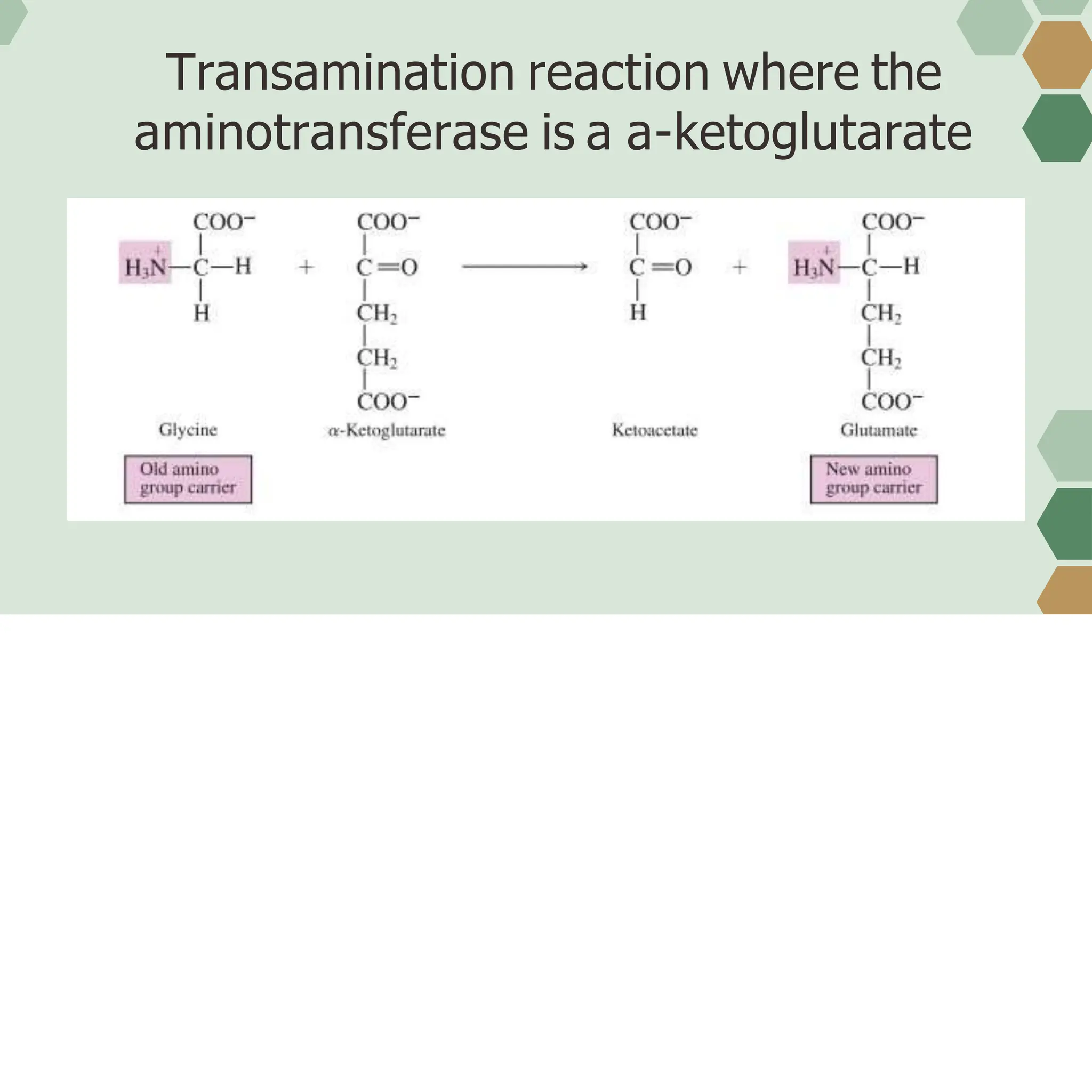
- Transamination and oxidative deamination, which are the first steps in amino acid degradation and involve transferring amino groups to alpha-ketoglutarate to form glutamate.
- The urea cycle, a cyclic pathway in the liver that involves several reactions to convert ammonia and carbon dioxide into less toxic urea for excretion.
- The fates of amino acid carbon skeletons after removal of the amino group, which can become precursors for glucose, enter the TCA cycle,


![REFERENCES:
MEDSimplified. (2020, March 10). UREA CYCLE MADE EASY 2020 - METABOLISMS MADE
SIMPLE [Video]. YouTube. https://www.youtube.com/watch?v=vhCF-dN6WYQ
General, Organic, and Biological Chemistry by Stephen H. Stoker](https://image.slidesharecdn.com/group-3-protein-metabolism1-231209163759-f819d59f/75/Biochemistry-protein-metabolism-1-pptx-118-2048.jpg)


