Downloaded 131 times
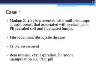
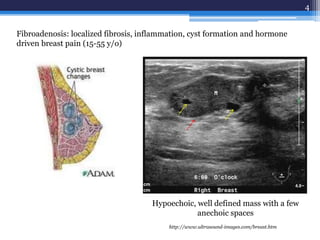
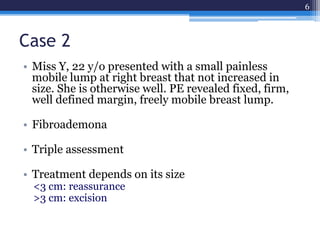
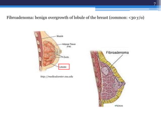
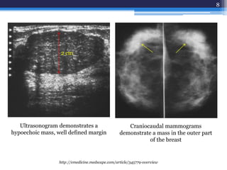
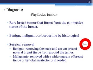
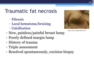
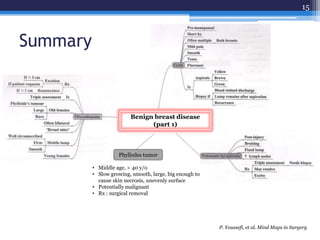


This document discusses several types of benign breast diseases including fibroadenosis/fibrocystic disease, fibroadenoma, giant fibroadenoma, phyllodes tumor, and traumatic fat necrosis. It presents three case studies: a 40-year-old woman with cyclical breast pain and lumps diagnosed with fibroadenosis; a 22-year-old with a painless mobile lump diagnosed with fibroadenoma; and a 48-year-old with an enlarging lump over many years diagnosed with a phyllodes tumor, a rare breast tumor that can be benign, malignant, or borderline.


![Hypothalamus short ppt by Dr. Neha [PT].pptx](https://cdn.slidesharecdn.com/ss_thumbnails/hypothalamusbydr-260124145759-b9f94a93-thumbnail.jpg?width=640&height=640&fit=bounds)


