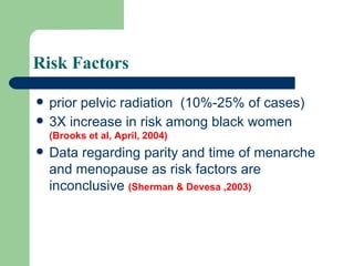
Uterine Sarcoma
Challenging Rare  ->  Limited data Rapidly growing (doubling time is 4 weeks) tend to be increasing  (Nordal & Thoresen 1997)
Epidemiology Rare  2% to 5% of all uterine malignancies 17 per million women annually [Platz, & Benda, 1995]  Between 1989-1999, 2677 women were diagnosed with uterine sarcoma  (Brooks et al, April, 2004)
Risk Factors prior pelvic radiation  (10%-25% of cases) 3X increase in risk among black women  (Brooks et al, April, 2004) Data regarding parity and time of menarche and menopause as risk factors are inconclusive  (Sherman & Devesa ,2003)
long-term adjuvant tamoxifen An increase in the risk of uterine sarcomas appears to accompany the use of long-term adjuvant tamoxifen in women with breast cancer  [Wickerham et al, 2002, Wysowski et al, 2002].  Since 1978, when tamoxifen was first marketed in the United States, 159 cases of uterine sarcoma worldwide have been reported
Histologic Classification Mixed mesodermal sarcoma Carcinosarcoma Mixed Liposarcoma (ii) Endometrial stromal sarcoma Osteosarcoma (i) endolymphatic stromal sarcoma Chondrosarcoma Stromal sarcoma Rhabdomyosarcoma Leimyosarcoma Pure Heterologous Homologous Type
GOG , 1993 Mixed mullerian sarcomas - 50%  homologous heterologous Leiomyosarcoma (30%).  Endometrial stromal sarcoma (15%) Others (Adenosarcoma 5%)
Leiomyosarcoma Arise from smooth muscles of the uterus usually de novo  appear grossly as a large (>10 cm) yellow or tan solitary mass with soft, fleshy cut surfaces exhibiting hemorrhage and necrosis  [Viereck et al, 2002].
Leiomyosarcoma
Leiomyosarcoma: Low or high grade Frequent mitotic figures significant nuclear atypia, presence of coagulative necrosis of tumor cells. [ Bell et al, 1994  ]
Endometrial stromal Sarcoma :  A pure homologous neoplasm  Subtypes: low and high grade  Low grade : slow growing tumors with infrequent metastasis or recurrence after therapy.  [Oliva, et al, 2000].  high grade : enlarge and metastasize quickly and are often fatal.
Mixed mullerian sarcomas  Both carcinomatous and sarcomatous elements must be present in this type of sarcoma.  metastasize early in the course of the disease via hematogenous and lymphatic pathways  grows as a polypoidal mass with a broad base
Subdivided homologous ( carcinosarcoma ) contain only tissue elements that are native to the uterus.  heterologous ( mixed mesodermal sarcoma ) contain tissue element that is foreign to the uterus.
MMT
Adenosarcoma  both malignant stromal and benign epithelial components  a significantly increased occurrence of this tumor  (Seidman et al, 1999) present as polypoid masses
Clinical Diagnosis Vaginal bleeding is the most common presenting symptom of a uterine sarcoma.  On pelvic examination, the uterus is enlarged and, in some patients, part of the tumor may protrude from the uterine cavity through the cervical os.
Rapidly growing!! Among 341 women with a rapidly growing uterus by clinical or ultrasound examination, only one (0.27 percent) had a uterine sarcoma . [Parker et al, 1994].
Should be considered in postmenopausal women with a pelvic mass, abnormal bleeding, and pelvic pain, where the incidence of sarcoma is 1 to 2 percent  [Leibsohn   et al, 1990]
Evaluation Ultrasound examination, MRI, or CT scan  cannot reliably distinguish between a sarcoma, leiomyoma or endometrial cancer  [Rha et al, 2003].   The diagnosis of uterine sarcomas is made from histologic examination of the entire uterus
Staging: surgical Sarcoma has spread outside the true pelvis IV Sarcoma has spread outside the uterus but is confined to the true pelvis III Sarcoma is confined to the corpus and cervix II Sarcoma is confined to the corpus I Description Stage based on FIGO staging for endometrial cancer
Lymph node Biopsy patients with uterine sarcoma grossly confined to the uterus/cervix showed lymph node metastases in 5 of 101 patients should be reserved for women with clinically suspicious nodes  [Leitao et al, 2003 ]
Further support In one series of 208 women with uterine leiomyosarcoma, only four of 36 who underwent lymph node sampling had positive nodes  [Giuntoli et al, 2003].
Treatment because of their rarity, uterine sarcomas are not suitable for screening.  (Levenback, 1996)
Surgery is the only curative therapy for uterine sarcomas  [Morice et al, 2003]
Modalities Surgery (total abdominal hysterectomy, bilateral salpingo-oophorectomy).  Surgery plus adjuvant chemotherapy.  Surgery plus adjuvant irradiation
Is it beneficial !! Interpretation of the possible benefit of different modalities is hampered by the difficulty in comparing outcomes from series in which patients of varying stages and histologies were reported
The five year survivals Surgery alone (46 %) Surgery and radiotherapy (62 %) Surgery and chemotherapy  (43 %) Radiation alone (8 %) Weitmann et al, 2001
three-year local recurrence rates     No adjuvant treatment  62 %   Whole pelvis external beam radiation therapy  31 % Chemotherapy alone  71 percent [Livi et al, 2003]
Massachusetts General Hospital  1990-1999 Adjuvant therapy after optimal cytoreduction does not decrease the rate of recurrence  [Dinh et al, 2004]
   Adjuvant radiation therapy  Some studies of postoperative radiation suggest a survival benefit  [Moskovic et al, 1993  Knocke et al, 1998, Weitmann et al, 2001].   Other studies showed cure rate was similar for those treated with surgery alone or followed by radiation, regardless of the stage of disease  [Giuntoli et al, 2003]
Complications of Radiation Tx: Acute: Perforation Fever Diarrhea Bladder spasm Chronic: Proctitis Cystitis (a/w UTI) Fistula Enteritis
Adjuvant chemotherapy   Current studies consist primarily of Phase II chemotherapy trials for advanced disease  The role of chemotherapy in the treatment of uterine sarcomas has been limited
This is because Adjuvant chemotherapy following complete resection (stage I and II) has not been established to be effective in RCT  But some nonrandomized trials have reported improved survival following adjuvant chemotherapy with or without radiation therapy  Piver  et al, 1988 ,van Nagell , et al, 1986, Peters et al, 1989
   Leiomyosarcoma  doxorubicin is an effective drug for advanced leiomyosarcoma  combinations with doxorubicin increase the objective response rate but add substantial toxicity  A very recent small trial showed promising results with gemcitabine plus docetaxel  [Hensley et al, 2002].
   Carcinosarcoma  benefit from cisplatin-based chemotherapy particularly combinations of cisplatin with doxorubicin and ifosfamide, or single agent paclitaxel [ Gallup et al, 2003 , van Rijswijk  et al, 2003, Harris et al, 2003]
Mixed mesodermal tumors  Cisplatin and ifosfamide appear to have greater activity than does doxorubicin alone  [Ramondetta et al, 2003].   In a very small uncontrolled trial : cisplatin, doxorubicin, and dacarbazine give three year survivals of 51 %  [Baker et al, 1991].
Hormone therapy  Estrogen and/or progesterone receptors are present in leiomyosarcomas and endometrial stromal sarcomas but do not predict hormone responsiveness.  In fact, only one of 28 patients with residual or recurrent disease following surgery had an objective response to hormone therapy  (Wade 1994)
Recurrent Disease Most relapses occur in the pelvis, followed by lung and abdomen  currently no standard therapy for patients with recurrent disease  Doxorubcin in leiomyosarcoma ? Cisplatin in carcinosarcoma ?
In a recent RCT 2000 ifosfamide with or without cisplatin for recurrent  sarcoma demonstrated a higher response rate on the combination arm  However,use of the combination was not justified because of increased toxic effects  [Sutton et al, 2000]
Prognosis poor prognosis  the 5-year survival : stage I  less than 50%  remaining stages : 0% to 20%. strongest predictor of survival was menopausal status at time of diagnosis [Major et al, 1993]
leiomyosarcoma age over 50 years was a poor prognostic factor, as was size greater than 5 cm  [Giuntoli et al, 2003].
Conclusion  Aggressive surgical cytoreduction at the time of initial diagnosis offers the best survival  More RCTs are needed to determine the value and regime of adjuvant therapy
Thank You

Uterine sarcoma

  • 1.
  • 2.
    Challenging Rare -> Limited data Rapidly growing (doubling time is 4 weeks) tend to be increasing (Nordal & Thoresen 1997)
  • 3.
    Epidemiology Rare 2% to 5% of all uterine malignancies 17 per million women annually [Platz, & Benda, 1995] Between 1989-1999, 2677 women were diagnosed with uterine sarcoma (Brooks et al, April, 2004)
  • 4.
    Risk Factors priorpelvic radiation (10%-25% of cases) 3X increase in risk among black women (Brooks et al, April, 2004) Data regarding parity and time of menarche and menopause as risk factors are inconclusive (Sherman & Devesa ,2003)
  • 5.
    long-term adjuvant tamoxifenAn increase in the risk of uterine sarcomas appears to accompany the use of long-term adjuvant tamoxifen in women with breast cancer [Wickerham et al, 2002, Wysowski et al, 2002]. Since 1978, when tamoxifen was first marketed in the United States, 159 cases of uterine sarcoma worldwide have been reported
  • 6.
    Histologic Classification Mixedmesodermal sarcoma Carcinosarcoma Mixed Liposarcoma (ii) Endometrial stromal sarcoma Osteosarcoma (i) endolymphatic stromal sarcoma Chondrosarcoma Stromal sarcoma Rhabdomyosarcoma Leimyosarcoma Pure Heterologous Homologous Type
  • 7.
    GOG , 1993Mixed mullerian sarcomas - 50% homologous heterologous Leiomyosarcoma (30%). Endometrial stromal sarcoma (15%) Others (Adenosarcoma 5%)
  • 8.
    Leiomyosarcoma Arise fromsmooth muscles of the uterus usually de novo appear grossly as a large (>10 cm) yellow or tan solitary mass with soft, fleshy cut surfaces exhibiting hemorrhage and necrosis [Viereck et al, 2002].
  • 9.
  • 10.
    Leiomyosarcoma: Low orhigh grade Frequent mitotic figures significant nuclear atypia, presence of coagulative necrosis of tumor cells. [ Bell et al, 1994 ]
  • 11.
    Endometrial stromal Sarcoma: A pure homologous neoplasm Subtypes: low and high grade Low grade : slow growing tumors with infrequent metastasis or recurrence after therapy. [Oliva, et al, 2000]. high grade : enlarge and metastasize quickly and are often fatal.
  • 12.
    Mixed mullerian sarcomas Both carcinomatous and sarcomatous elements must be present in this type of sarcoma. metastasize early in the course of the disease via hematogenous and lymphatic pathways grows as a polypoidal mass with a broad base
  • 13.
    Subdivided homologous (carcinosarcoma ) contain only tissue elements that are native to the uterus. heterologous ( mixed mesodermal sarcoma ) contain tissue element that is foreign to the uterus.
  • 14.
  • 15.
    Adenosarcoma bothmalignant stromal and benign epithelial components a significantly increased occurrence of this tumor (Seidman et al, 1999) present as polypoid masses
  • 16.
    Clinical Diagnosis Vaginalbleeding is the most common presenting symptom of a uterine sarcoma. On pelvic examination, the uterus is enlarged and, in some patients, part of the tumor may protrude from the uterine cavity through the cervical os.
  • 17.
    Rapidly growing!! Among341 women with a rapidly growing uterus by clinical or ultrasound examination, only one (0.27 percent) had a uterine sarcoma . [Parker et al, 1994].
  • 18.
    Should be consideredin postmenopausal women with a pelvic mass, abnormal bleeding, and pelvic pain, where the incidence of sarcoma is 1 to 2 percent [Leibsohn et al, 1990]
  • 19.
    Evaluation Ultrasound examination,MRI, or CT scan cannot reliably distinguish between a sarcoma, leiomyoma or endometrial cancer [Rha et al, 2003]. The diagnosis of uterine sarcomas is made from histologic examination of the entire uterus
  • 20.
    Staging: surgical Sarcomahas spread outside the true pelvis IV Sarcoma has spread outside the uterus but is confined to the true pelvis III Sarcoma is confined to the corpus and cervix II Sarcoma is confined to the corpus I Description Stage based on FIGO staging for endometrial cancer
  • 21.
    Lymph node Biopsypatients with uterine sarcoma grossly confined to the uterus/cervix showed lymph node metastases in 5 of 101 patients should be reserved for women with clinically suspicious nodes [Leitao et al, 2003 ]
  • 22.
    Further support Inone series of 208 women with uterine leiomyosarcoma, only four of 36 who underwent lymph node sampling had positive nodes [Giuntoli et al, 2003].
  • 23.
    Treatment because oftheir rarity, uterine sarcomas are not suitable for screening. (Levenback, 1996)
  • 24.
    Surgery is theonly curative therapy for uterine sarcomas [Morice et al, 2003]
  • 25.
    Modalities Surgery (totalabdominal hysterectomy, bilateral salpingo-oophorectomy). Surgery plus adjuvant chemotherapy. Surgery plus adjuvant irradiation
  • 26.
    Is it beneficial!! Interpretation of the possible benefit of different modalities is hampered by the difficulty in comparing outcomes from series in which patients of varying stages and histologies were reported
  • 27.
    The five yearsurvivals Surgery alone (46 %) Surgery and radiotherapy (62 %) Surgery and chemotherapy (43 %) Radiation alone (8 %) Weitmann et al, 2001
  • 28.
    three-year local recurrencerates   No adjuvant treatment 62 %   Whole pelvis external beam radiation therapy 31 % Chemotherapy alone 71 percent [Livi et al, 2003]
  • 29.
    Massachusetts General Hospital 1990-1999 Adjuvant therapy after optimal cytoreduction does not decrease the rate of recurrence [Dinh et al, 2004]
  • 30.
       Adjuvant radiationtherapy Some studies of postoperative radiation suggest a survival benefit [Moskovic et al, 1993 Knocke et al, 1998, Weitmann et al, 2001]. Other studies showed cure rate was similar for those treated with surgery alone or followed by radiation, regardless of the stage of disease [Giuntoli et al, 2003]
  • 31.
    Complications of RadiationTx: Acute: Perforation Fever Diarrhea Bladder spasm Chronic: Proctitis Cystitis (a/w UTI) Fistula Enteritis
  • 32.
    Adjuvant chemotherapy Current studies consist primarily of Phase II chemotherapy trials for advanced disease The role of chemotherapy in the treatment of uterine sarcomas has been limited
  • 33.
    This is becauseAdjuvant chemotherapy following complete resection (stage I and II) has not been established to be effective in RCT But some nonrandomized trials have reported improved survival following adjuvant chemotherapy with or without radiation therapy Piver et al, 1988 ,van Nagell , et al, 1986, Peters et al, 1989
  • 34.
       Leiomyosarcoma doxorubicin is an effective drug for advanced leiomyosarcoma combinations with doxorubicin increase the objective response rate but add substantial toxicity A very recent small trial showed promising results with gemcitabine plus docetaxel [Hensley et al, 2002].
  • 35.
       Carcinosarcoma benefit from cisplatin-based chemotherapy particularly combinations of cisplatin with doxorubicin and ifosfamide, or single agent paclitaxel [ Gallup et al, 2003 , van Rijswijk et al, 2003, Harris et al, 2003]
  • 36.
    Mixed mesodermal tumors Cisplatin and ifosfamide appear to have greater activity than does doxorubicin alone [Ramondetta et al, 2003]. In a very small uncontrolled trial : cisplatin, doxorubicin, and dacarbazine give three year survivals of 51 % [Baker et al, 1991].
  • 37.
    Hormone therapy Estrogen and/or progesterone receptors are present in leiomyosarcomas and endometrial stromal sarcomas but do not predict hormone responsiveness. In fact, only one of 28 patients with residual or recurrent disease following surgery had an objective response to hormone therapy (Wade 1994)
  • 38.
    Recurrent Disease Mostrelapses occur in the pelvis, followed by lung and abdomen currently no standard therapy for patients with recurrent disease Doxorubcin in leiomyosarcoma ? Cisplatin in carcinosarcoma ?
  • 39.
    In a recentRCT 2000 ifosfamide with or without cisplatin for recurrent sarcoma demonstrated a higher response rate on the combination arm However,use of the combination was not justified because of increased toxic effects [Sutton et al, 2000]
  • 40.
    Prognosis poor prognosis the 5-year survival : stage I less than 50% remaining stages : 0% to 20%. strongest predictor of survival was menopausal status at time of diagnosis [Major et al, 1993]
  • 41.
    leiomyosarcoma age over50 years was a poor prognostic factor, as was size greater than 5 cm [Giuntoli et al, 2003].
  • 42.
    Conclusion Aggressivesurgical cytoreduction at the time of initial diagnosis offers the best survival More RCTs are needed to determine the value and regime of adjuvant therapy
  • 43.