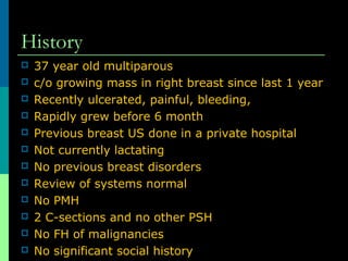
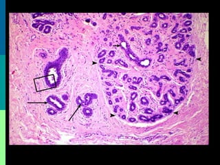
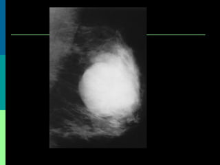
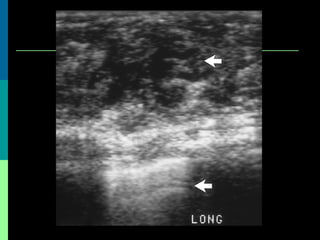
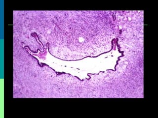
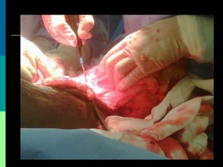
8. History
37 year old multiparous
c/o growing mass in right breast since last 1 year
Recently ulcerated, painful, bleeding,
Rapidly grew before 6 month
Previous breast US done in a private hospital
Not currently lactating
No previous breast disorders
Review of systems normal
No PMH
2 C-sections and no other PSH
No FH of malignancies
No significant social history
9.
10. Examination
30 X 20 cm
OQ right breast
Overlaying skin
Shinny, dilated veins, stretched skin
Shallow 5 X 4 cm ulcer upper outer
quadrant
Redness, swollen, pus in the ulcer base
No nipple discharges
11. Palpation
Non-tender, firm, well demarcated,
Irregular, lobulated borders, freely mobile,
smooth surface
Area around the ulcer warm
Not fluctuant, not compressible,
Normal contralateral breast
No LN were palpable
16. Benign Vs. Malignant
Benign
Tendency to grow aggressively
Recurs locally
Malignant
Recurrent malignant tumors seem to be more aggressive
Metastasis to lungs, skeleton, heart, and liver
hemtogenously
Metastasis to axillary lymph nodes occurred in only 2%
Symptoms from metastasis seen as early as a few
months to as late as 12 years after the initial therapy
30% of patients with malignant phyllodes tumors die from
the disease
Mostly within 3 years of the initial treatment
No cures for systemic metastases exist
40. Radiotherapy & Chemotherapy
Role of radiotherapy remains unclear
May be considered for high-risk phyllodes
tumors, including those >5cm, with
stromal overgrowth, with >10
mitoses/high-power field, or with
infiltrating margins
Doxorubicin and ifosfamide-based
chemotherapies
41. Outcomes
Locar recurrences of approximately 26%
Stromal overgrowth, larger tumor size,
and involved margin were all significantly
correlated with LR.
5-year survival rate malignant phyllodes
tumour ranges from 54% to 82%
10-year ranges from 23% to 42%
History
Patients typically present with a firm, mobile, well-circumscribed, nontender breast mass.
A small mass may rapidly increase in size in the few weeks before the patient seeks medical attention.
Tumors rarely involve the nipple-areola complex or ulcerate to the skin.
Patients with metastases may present with such symptoms as dyspnea, fatigue, and bone pain.
A 62-year-old female noticed a tumor in her right breast in January, 1995. She had no abnormality in the family history. She had a past history of diabetes mellitus. The tumor grew rapidly and she visited our out-patient clinic in February 1995. On physical examination, a 10 × 8 cm, well defmed and movable mass with a smooth surface was palpated in the upper outer quadrant of the right breast. Slight dimple was not detected. Mammography showed a large tumor shadow in the upper outer quadrant of the right breast without any microcalcification, Ultrasonography revealed a large cystic shadow with a low echoic lesion and solid component with heterogeneous intemal echo in the cyst (Fig. 1). Old bloody fluid was aspirated by fine needle aspiration cytology, resulting in no malignant cells. CEA, TPA and CA 15-3 in the cystic fluid were 25.3 ng/ml, 251 344 U/ml and 59 U/ml, respectively. c-erbB-2 overexpression was not detected (using DNA extracted from the aspiration fluid). Serum CEA, TPA and CA 15-3 were 1.0 ng/ml, 55.5 V/ml and 11 U/ml, respectively. Under general anesthesia, the tumor was widely excised with about a 2 em margin. The resected specimen was 11.5 × 11 × 11 cm in size and the tumor was not invasive to surrounding tissues. Old bloody fluid was contained within the cyst. The gross appearance showed papillary process protrusions into a central cystic cavity (Fig. 2). Microscopically leaf-like papillary protrusions of stromal connective tissues lined with the epithelium extended into the cystic cavity (Fig. 3). The tumor margin was well demarcated. Slight stromal cellularity and stromal cell atypia were observed (Fig. 4). Mitoses were occasionally approved. The histological diagnosis was a borderline phyllodes tumor. Immunohistochemically, estrogen receptor (ER), c-erbB-2 and p53 protein were negative, and progesterone receptor (PgR) was positive in the epithelial cells but negative in the stromal cells. The proliferating cell nuclear antigen (PCNA) labeling index was 72% in the stromal cells and 4% in the epithelial cells (Table 1, Figs 5 and 6). Two years after the operation, she is doing well without any recurrence.
A 35-year-old multiparous woman presented with massive enlargement of both breasts. She reported that the growth had been slow over many years. The examination revealed multiple firm, hard lumps filling all quadrants of both breasts, with stretched skin, a bosselated surface, dilated veins, and restricted mobility over the chest wall probably due to the size of the tumor (Figure 1). There was a 3.0 x 4.0 cm firm, nontender, and mobile axillary lymph node on the left side. No lymphadenopathy were detected elsewhere in the body. A clinical diagnosis of bilateral phyllodes tumour was made.
Background
Cystosarcoma phyllodes is a rare, predominantly benign tumor that occurs almost exclusively in the female breast.1 Its name is derived from the Greek words sarcoma, meaning fleshy tumor, and phyllo, meaning leaf. Grossly, the tumor displays characteristics of a large, malignant sarcoma, takes on a leaflike appearance when sectioned, and displays epithelial, cystlike spaces when viewed histologically (hence the name). Because most tumors are benign, the name may be misleading. Thus, the favored terminology is now phyllodes tumor.
Pathophysiology
Phyllodes tumor is the most commonly occurring nonepithelial neoplasm of the breast, although it represents only about 1% of tumors in the breast.2 It has a smooth, sharply demarcated texture and typically is freely movable. It is a relatively large tumor, with an average size of 5 cm. However, lesions of more than 30 cm have been reported.
Frequency
United States
No difference in phyllodes tumor frequency appears to exist between patients from the United States and those from other countries. Phyllodes tumors account for approximately 1% of all breast neoplasms.2
Mortality/Morbidity
Because of limited data, the percentage of benign vs malignant phyllodes tumors is not well defined. Reports suggest, however, that about 85-90% of phyllodes tumors are benign and that approximately 10-15% are malignant.3 Although the benign tumors do not metastasize, they have a tendency to grow aggressively and can recur locally.2 Similar to other sarcomas, the malignant tumors metastasize hematogenously. Unfortunately, the pathologic appearance of a phyllodes tumor does not always predict the neoplasm's clinical behavior; in some cases, therefore, there is a degree of uncertainty about the lesion's classification. The characteristics of a malignant phyllodes tumor include the following:
Recurrent malignant tumors seem to be more aggressive than the original tumor.
The lungs are the most common metastatic site, followed by the skeleton, heart, and liver.
Symptoms from metastatic involvement can arise from as early as a few months to as late as 12 years after the initial therapy.
Most patients with metastases die within 3 years of the initial treatment.4
No cures for systemic metastases exist.
Roughly 30% of patients with malignant phyllodes tumors die from the disease.
Race
A racial predilection does not appear to exist for phyllodes tumors.
Sex
Phyllodes tumors occur almost exclusively in females. Rare case reports have been described in males.
Age
Phyllodes tumors can occur in people of any age; however, the median age is the fifth decade of life.Some juvenile fibroadenomas in teenagers can look histologically like phyllodes tumors; however, they behave in a benign fashion similar to that of other fibroadenomas.
Although the benign tumors do not metastasize, they have a tendency to grow aggressively and can recur locally.2 Similar to other sarcomas, the malignant tumors metastasize hematogenously. Unfortunately, the pathologic appearance of a phyllodes tumor does not always predict the neoplasm's clinical behavior; in some cases, therefore, there is a degree of uncertainty about the lesion's classification. The characteristics of a malignant phyllodes tumor include the following:
Recurrent malignant tumors seem to be more aggressive than the original tumor.
The lungs are the most common metastatic site, followed by the skeleton, heart, and liver.
Symptoms from metastatic involvement can arise from as early as a few months to as late as 12 years after the initial therapy.
Most patients with metastases die within 3 years of the initial treatment.4
No cures for systemic metastases exist.
Roughly 30% of patients with malignant phyllodes tumors die from the disease.
Workup
Laboratory Studies
No specific hematologic tumor markers or other blood tests can be used to diagnose cystosarcoma phyllodes.
Imaging Studies
Although mammography and ultrasonography generally are important in the diagnosis of breast lesions, they are notoriously unreliable in differentiating benign cystosarcoma phyllodes (CSP) from the malignant form of the condition or from fibroadenomas. Thus, findings on imaging studies are not definitively diagnostic of CSP.6
Procedures
Fine-needle aspiration for cytologic examination usually is inadequate for the diagnosis of phyllodes tumors. Core biopsy is more reliable, but there still can be sampling errors and difficulty in distinguishing the lesion from a fibroadenoma.
Open excisional breast biopsy for smaller lesions or incisional biopsy for large lesions is the definitive method for diagnosing phyllodes tumors.
Histologic Findings
All phyllodes tumors contain a stromal component that can vary significantly in histologic appearance from one lesion to another. In general, benign phyllodes tumors demonstrate a markedly increased number of regular fusiform fibroblasts in the stroma. Occasionally, highly anaplastic cells with myxoid changes are observed. A high degree of cellular atypia, with increased stromal cellularity and an increased mitotic count, is almost always observed in the malignant form of cystosarcoma phyllodes. Ultrastructurally, in the benign and malignant forms of phyllodes tumors, nucleoli may reveal a coarsely meshed nucleolonema and abundant cisternae in the endoplasmic reticulum.
Imaging Studies
Although mammography and ultrasonography generally are important in the diagnosis of breast lesions, they are notoriously unreliable in differentiating benign cystosarcoma phyllodes (CSP) from the malignant form of the condition or from fibroadenomas. Thus, findings on imaging studies are not definitively diagnostic of CSP.6
Most frequent anomaly is regular oval margines
On ultrasound the tumours were lobulated in most of the cases. Heterogeneous internal echoes and ntramural cysts are also said to be suggestive of phyllodes tumours5. Phyllodes tumour on mammography is described as a sharply defined round or oval mass with lobulation5
reported mammo-graphic and sonographic findings between benign and malignant phyllodes tumors. Cystic areas at sonography were more common in malignant than benign tumors. These data suggested that a malignant phyllodes tumor was likely to undergo cystic formation. There is possible malignancy in phyllodes tumors showing intracystic growth.
Role of Mri not fully elucidated
Effort to find corelation eith malignancy
Signal intensity higher than normal breast tissue in T1
Cystic changes with irregular wall
Signal intensity loweer than normal breast tissue in T2
Malignant phyllodes tumor of right breast in 17-year-old girl. (a) Coronal nonenhanced T1-weighted MR image (6.4/2.5) shows low-signal-intensity circumscribed mass (arrow).
Transverse short inversion time inversion-recovery T2-weighted image (5970/103/150) shows circumscribed mass of heterogeneous signal intensity. Note the isointense area (arrow) in the lateral aspect of the tumor relative to the surrounding breast tissue (*).
Sagittal multiplanar reconstruction image constructed from contrast-enhanced subtraction image shows heterogeneously enhancing mass. Note the cystic change with an irregular wall (arrow). An ROI (circle) was placed in superior part of the tumor.
Procedures
Fine-needle aspiration for cytologic examination usually is inadequate for the diagnosis of phyllodes tumors. Core biopsy is more reliable, but there still can be sampling errors and difficulty in distinguishing the lesion from a fibroadenoma.
Open excisional breast biopsy for smaller lesions or incisional biopsy for large lesions is the definitive method for diagnosing phyllodes tumors.
Difficulties with diagnosis of phyllodes tumour by FNAC have been reported. The cytologist reported phyllodes tumour in only 23% of cases where FNAC was done at the time of diagnosis. In cases where core biopsy was done at the visit where the diagnosis was made, the core biopsy correctly diagnosed 65% of phyllodes tumours5
The diagnostic findings on needle biopsy consist of abundant stromal cells, which appear as bare bipolar nuclei, throughout the aspirate; sheets of fairly uniform sized epithelial cells which are typically arranged in either an antler-like pattern, or a honeycomb pattern. These epithelial sheets tend to show typical metachromatic blue staining on DiffQuick staining. Foam cells and apocrine cells may also be seen, although these are less diagnostic features
Fibroadenoam
Phyllodes
65% diagnoses phylloides
Histologically, a phyllodes tumor is composed of epithelial elements and a connective tissue stroma. The characteristics of the stroma alone determine whether a phyllodes tumor should be classified as benign or malignant. In general, stroma from a malignant phyllodes tumor contains cellular atypia, mitotic activity and tumor margin (1,3). Malignant areas are often focal and are overlooked if multiple samples are not observed. In this case, cell atypia was locally identified, thus indicating the diagnosis of a borderline phyllodes tumor.
Histologic Findings
All phyllodes tumors contain a stromal component that can vary significantly in histologic appearance from one lesion to another. In general, benign phyllodes tumors demonstrate a markedly increased number of regular fusiform fibroblasts in the stroma. Occasionally, highly anaplastic cells with myxoid changes are observed. A high degree of cellular atypia, with increased stromal cellularity and an increased mitotic count, is almost always observed in the malignant form of cystosarcoma phyllodes. Ultrastructurally, in the benign and malignant forms of phyllodes tumors, nucleoli may reveal a coarsely meshed nucleolonema and abundant cisternae in the endoplasmic reticulum.
Histo phylloides
Histologically, the phyllodes tumour consists of epithelial cells and connective tissue with more stromal proliferation than that of fibroadenoma, often accompanied by cellular atypia. For malignant phyllodes tumour, they are further devided into borderline, low-grade, and high-grade on the basis of the following histological criteria: tumor borders, mitotic activity, stromal atypia, and stromal overgrowth. Only the stromal cells have the potential to metastasise. The malignant character of the phyllodes tumour is therefore confirmed by the microscopic appearance of the stroma
The cut surface usually appears homogenous and firm, and is grey-white or tan in colour fibroadenoma
The preferred initial therapy for a phyllodes tumor is a wide local excision with adequate margin of normal breast tissues irrespective of histological features (9,10). Low axillary dissection is advisable if node involvement is suspected. The present tumor was widely excised with about a 2 em free margin. Axillary lymph node dissection was not added because there were no enlarged lymph nodes. As the histopathological findings revealed no malignancy, additional resection was not performed, Phyllodes tumors, even if they are not malignant, often recur in case of inadequate resection of the tumor. Hence careful follow-up of patients is recommended.
Wide local excision with adequate margin of normal breast tissues regardless of histological type
In most cases of cystosarcoma phyllodes, perform wide local excision, with a rim of normal tissue.7,8,9 No absolute rules on margin size exist. However, a 2 cm margin for small (<5 cm) tumors and a 5 cm margin for large (>5 cm) tumors have been advocated.
If the tumor to breast ratio is sufficiently high to preclude a satisfactory cosmetic result by segmental excision, total mastectomy, with or without reconstruction, is an alternative.
More radical procedures are not generally warranted.8
Perform axillary lymph node dissection only for clinically suspicious nodes. However, virtually all of these nodes are reactive and do not contain malignant cells.10
The role of radiotherapy remains unclear from published reports because of small patient numbers and a lack of controlled data. Pandey et al. suggested that adjuvant radiotherapy also improved the disease-free survival. August and Kearney recommended that adjuvant radiotherapy be considered for high-risk phyllodes tumors, including those >5cm, with stromal overgrowth, with >10 mitoses/high-power field, or with infiltrating margins3.
Different chemotherapy regimens have been applied in malignant phyllodes tumours. Doxorubicin and ifosfamide-based chemotherapies have shown some efficacy in women with metastatic spread of phyllodes tumours. In one study of 101 patients, 4 patients were treated with chemotherapy and a role for adjuvant chemotherapy in patients with stromal overgrowth was considered. This recommendation has so far not been confirmed in literature. Altogether, there is no clear indication for adjuvant chemotherapy for patients with phyllodes tumours2.
Locar recurrences (LR) are a common complication of high-grade lesions with a reported frequency of approximately 26% (12-65%)1. Stromal overgrowth, larger tumor size, and involved margin were all significantly correlated with LR. Stromal overgrowth increased the probability of LR 7-fold, whereas if the margin was <1cm, the risk of LR increased 5-fold, and if tumor size was >10 cm, then the prevalence of LR was four times greater than for smaller tumors3.
The 5- and 10-year survival rates for malignant phyllodes tumour range from 54% to 82% and from 23% to 42%, respectively2,3.