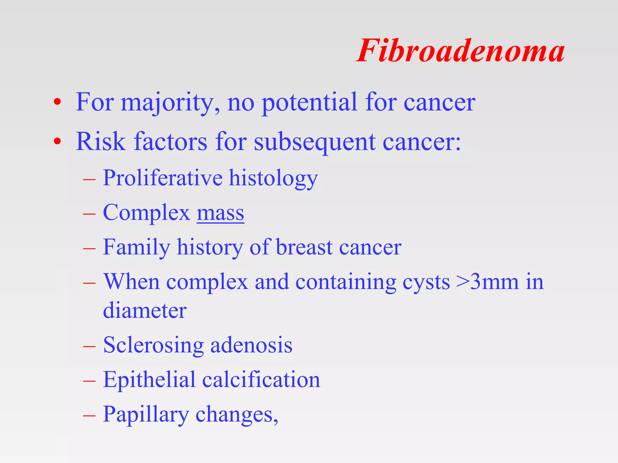
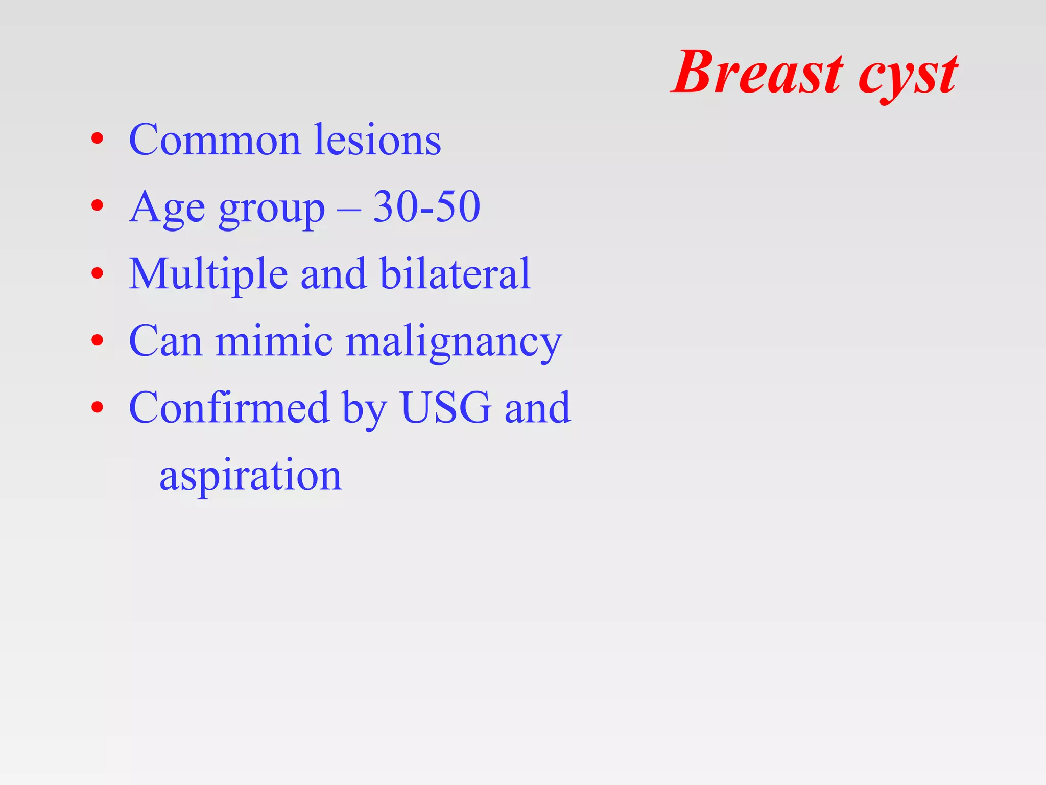
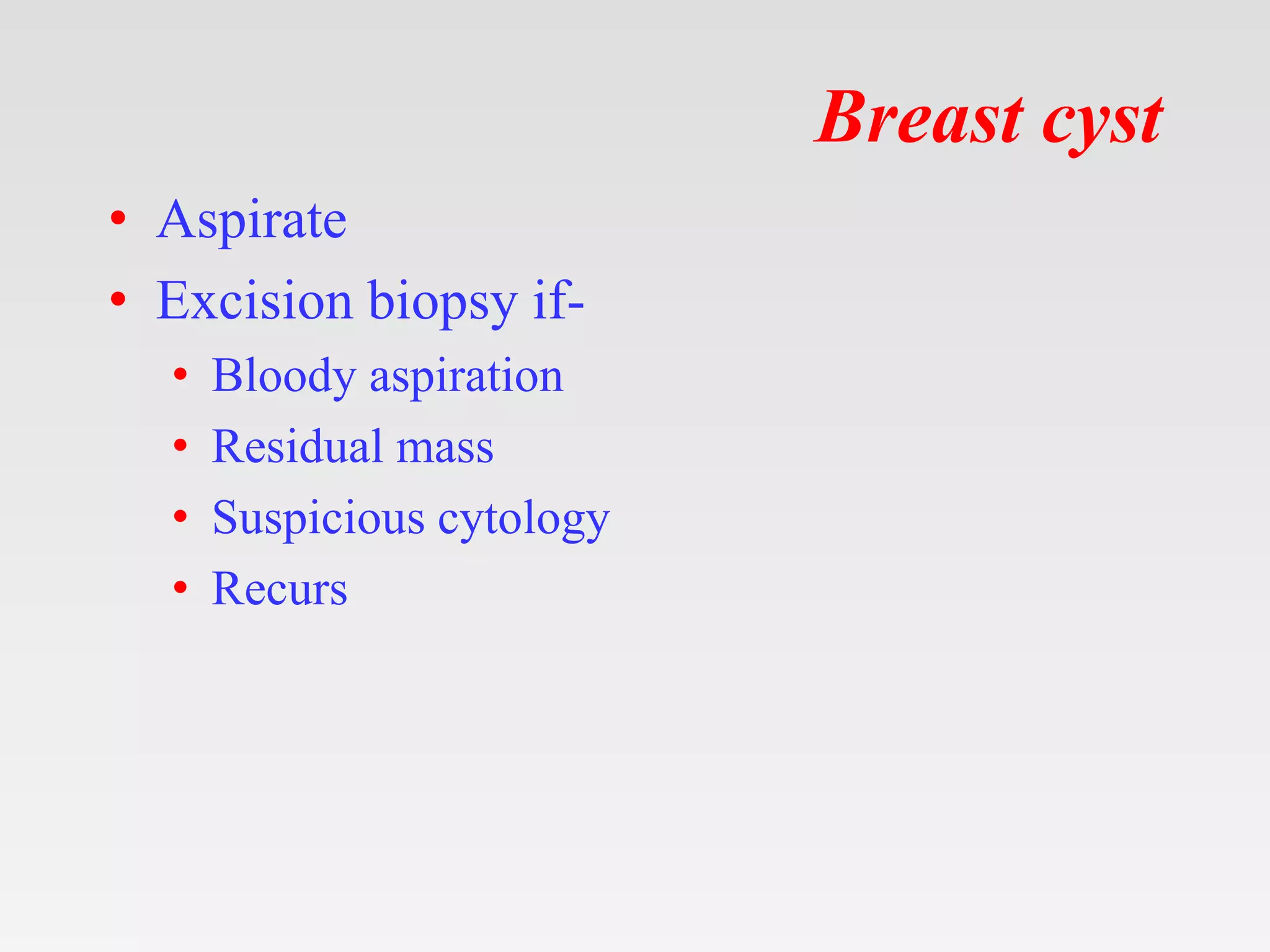
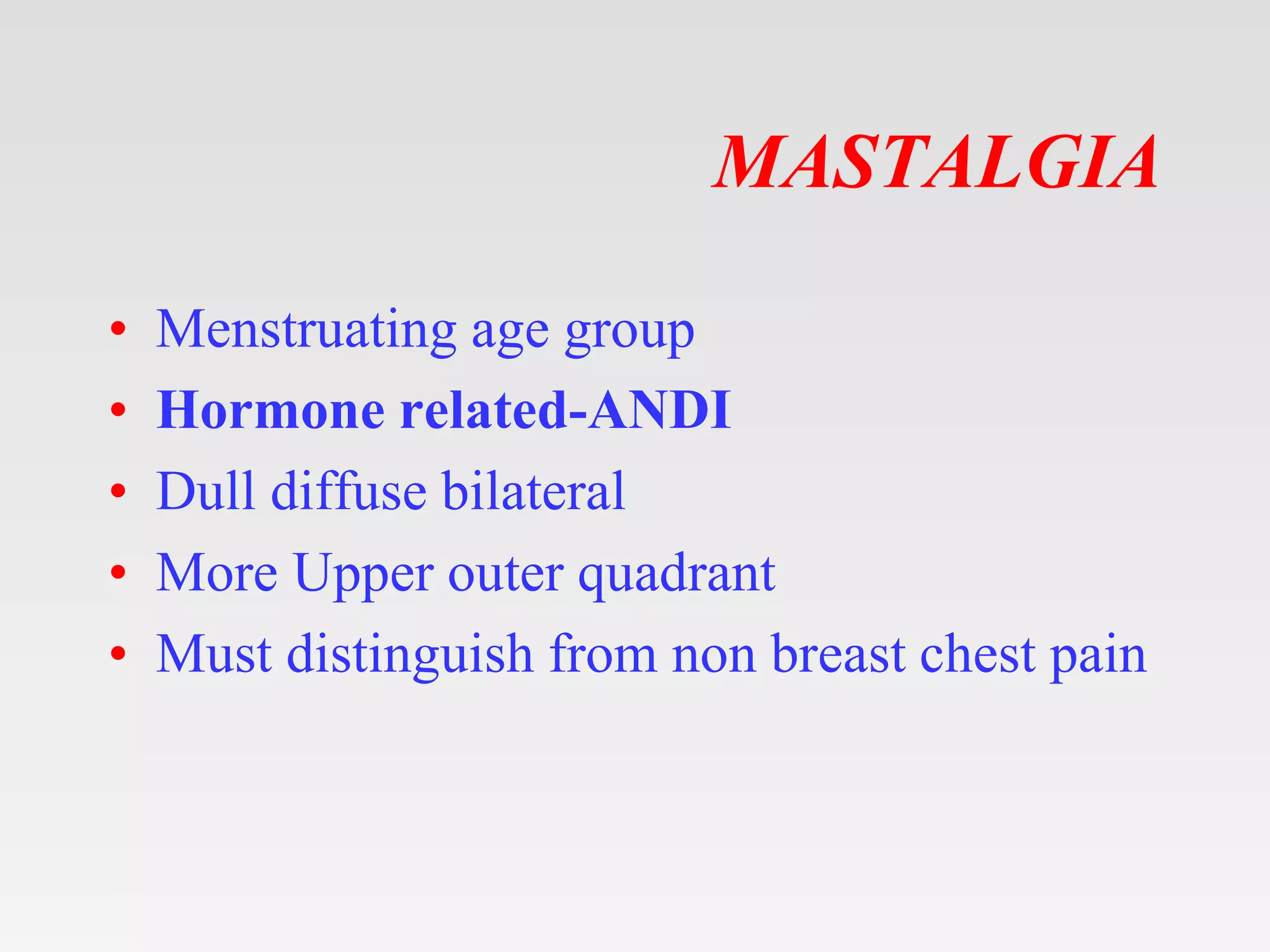
This document provides tips and instructions for using a PowerPoint presentation on benign breast conditions. It recommends asking students questions about blank slides to encourage active learning. Students should be able to describe the demography, clinical features, investigations, and management of benign breast diseases after this session. The rest of the document covers the physiology of the breast and various benign breast conditions like fibroadenoma, phyllodes tumor, cysts, and mastalgia in detail.
Seminar presentation by student under supervision of endocrinology specialist from HRPZ. References as mentioned in the slides. Mostly from Malaysia CPG.
Disclaimer: A lot from this slides were taken also from https://www.slideshare.net/babysurgeon/scrotal-swellings-1 (Dr Selvaraj Balasubramani)
This covers only :
ANATOMY
CAUSES
TORSION OF TESTIS
EPIDIDYMO-ORCHITIS
HYDROCELE
EPIDIDYMAL CYST
VARICOCELE
Disclaimer: A lot from this slides were taken also from https://www.slideshare.net/babysurgeon/scrotal-swellings-1 (Dr Selvaraj Balasubramani)
This covers only :
ANATOMY
CAUSES
TORSION OF TESTIS
EPIDIDYMO-ORCHITIS
HYDROCELE
EPIDIDYMAL CYST
VARICOCELE
Lecture class on pathology of breast for 3rd & 4th year MBBS students based on "Robbins & Cotran: Pathologic Basis of Disease'. Images are collected from internet.
Anti ulcer drugs and their Advance pharmacology ||
Anti-ulcer drugs are medications used to prevent and treat ulcers in the stomach and upper part of the small intestine (duodenal ulcers). These ulcers are often caused by an imbalance between stomach acid and the mucosal lining, which protects the stomach lining.
||Scope: Overview of various classes of anti-ulcer drugs, their mechanisms of action, indications, side effects, and clinical considerations.
These simplified slides by Dr. Sidra Arshad present an overview of the non-respiratory functions of the respiratory tract.
Learning objectives:
1. Enlist the non-respiratory functions of the respiratory tract
2. Briefly explain how these functions are carried out
3. Discuss the significance of dead space
4. Differentiate between minute ventilation and alveolar ventilation
5. Describe the cough and sneeze reflexes
Study Resources:
1. Chapter 39, Guyton and Hall Textbook of Medical Physiology, 14th edition
2. Chapter 34, Ganong’s Review of Medical Physiology, 26th edition
3. Chapter 17, Human Physiology by Lauralee Sherwood, 9th edition
4. Non-respiratory functions of the lungs https://academic.oup.com/bjaed/article/13/3/98/278874
The prostate is an exocrine gland of the male mammalian reproductive system
It is a walnut-sized gland that forms part of the male reproductive system and is located in front of the rectum and just below the urinary bladder
Function is to store and secrete a clear, slightly alkaline fluid that constitutes 10-30% of the volume of the seminal fluid that along with the spermatozoa, constitutes semen
A healthy human prostate measures (4cm-vertical, by 3cm-horizontal, 2cm ant-post ).
It surrounds the urethra just below the urinary bladder. It has anterior, median, posterior and two lateral lobes
It’s work is regulated by androgens which are responsible for male sex characteristics
Generalised disease of the prostate due to hormonal derangement which leads to non malignant enlargement of the gland (increase in the number of epithelial cells and stromal tissue)to cause compression of the urethra leading to symptoms (LUTS
New Directions in Targeted Therapeutic Approaches for Older Adults With Mantl...i3 Health
i3 Health is pleased to make the speaker slides from this activity available for use as a non-accredited self-study or teaching resource.
This slide deck presented by Dr. Kami Maddocks, Professor-Clinical in the Division of Hematology and
Associate Division Director for Ambulatory Operations
The Ohio State University Comprehensive Cancer Center, will provide insight into new directions in targeted therapeutic approaches for older adults with mantle cell lymphoma.
STATEMENT OF NEED
Mantle cell lymphoma (MCL) is a rare, aggressive B-cell non-Hodgkin lymphoma (NHL) accounting for 5% to 7% of all lymphomas. Its prognosis ranges from indolent disease that does not require treatment for years to very aggressive disease, which is associated with poor survival (Silkenstedt et al, 2021). Typically, MCL is diagnosed at advanced stage and in older patients who cannot tolerate intensive therapy (NCCN, 2022). Although recent advances have slightly increased remission rates, recurrence and relapse remain very common, leading to a median overall survival between 3 and 6 years (LLS, 2021). Though there are several effective options, progress is still needed towards establishing an accepted frontline approach for MCL (Castellino et al, 2022). Treatment selection and management of MCL are complicated by the heterogeneity of prognosis, advanced age and comorbidities of patients, and lack of an established standard approach for treatment, making it vital that clinicians be familiar with the latest research and advances in this area. In this activity chaired by Michael Wang, MD, Professor in the Department of Lymphoma & Myeloma at MD Anderson Cancer Center, expert faculty will discuss prognostic factors informing treatment, the promising results of recent trials in new therapeutic approaches, and the implications of treatment resistance in therapeutic selection for MCL.
Target Audience
Hematology/oncology fellows, attending faculty, and other health care professionals involved in the treatment of patients with mantle cell lymphoma (MCL).
Learning Objectives
1.) Identify clinical and biological prognostic factors that can guide treatment decision making for older adults with MCL
2.) Evaluate emerging data on targeted therapeutic approaches for treatment-naive and relapsed/refractory MCL and their applicability to older adults
3.) Assess mechanisms of resistance to targeted therapies for MCL and their implications for treatment selection
Prix Galien International 2024 Forum ProgramLevi Shapiro
June 20, 2024, Prix Galien International and Jerusalem Ethics Forum in ROME. Detailed agenda including panels:
- ADVANCES IN CARDIOLOGY: A NEW PARADIGM IS COMING
- WOMEN’S HEALTH: FERTILITY PRESERVATION
- WHAT’S NEW IN THE TREATMENT OF INFECTIOUS,
ONCOLOGICAL AND INFLAMMATORY SKIN DISEASES?
- ARTIFICIAL INTELLIGENCE AND ETHICS
- GENE THERAPY
- BEYOND BORDERS: GLOBAL INITIATIVES FOR DEMOCRATIZING LIFE SCIENCE TECHNOLOGIES AND PROMOTING ACCESS TO HEALTHCARE
- ETHICAL CHALLENGES IN LIFE SCIENCES
- Prix Galien International Awards Ceremony
Couples presenting to the infertility clinic- Do they really have infertility...Sujoy Dasgupta
Dr Sujoy Dasgupta presented the study on "Couples presenting to the infertility clinic- Do they really have infertility? – The unexplored stories of non-consummation" in the 13th Congress of the Asia Pacific Initiative on Reproduction (ASPIRE 2024) at Manila on 24 May, 2024.
HOT NEW PRODUCT! BIG SALES FAST SHIPPING NOW FROM CHINA!! EU KU DB BK substit...GL Anaacs
Contact us if you are interested:
Email / Skype : kefaya1771@gmail.com
Threema: PXHY5PDH
New BATCH Ku !!! MUCH IN DEMAND FAST SALE EVERY BATCH HAPPY GOOD EFFECT BIG BATCH !
Contact me on Threema or skype to start big business!!
Hot-sale products:
NEW HOT EUTYLONE WHITE CRYSTAL!!
5cl-adba precursor (semi finished )
5cl-adba raw materials
ADBB precursor (semi finished )
ADBB raw materials
APVP powder
5fadb/4f-adb
Jwh018 / Jwh210
Eutylone crystal
Protonitazene (hydrochloride) CAS: 119276-01-6
Flubrotizolam CAS: 57801-95-3
Metonitazene CAS: 14680-51-4
Payment terms: Western Union,MoneyGram,Bitcoin or USDT.
Deliver Time: Usually 7-15days
Shipping method: FedEx, TNT, DHL,UPS etc.Our deliveries are 100% safe, fast, reliable and discreet.
Samples will be sent for your evaluation!If you are interested in, please contact me, let's talk details.
We specializes in exporting high quality Research chemical, medical intermediate, Pharmaceutical chemicals and so on. Products are exported to USA, Canada, France, Korea, Japan,Russia, Southeast Asia and other countries.
Flu Vaccine Alert in Bangalore Karnatakaaddon Scans
As flu season approaches, health officials in Bangalore, Karnataka, are urging residents to get their flu vaccinations. The seasonal flu, while common, can lead to severe health complications, particularly for vulnerable populations such as young children, the elderly, and those with underlying health conditions.
Dr. Vidisha Kumari, a leading epidemiologist in Bangalore, emphasizes the importance of getting vaccinated. "The flu vaccine is our best defense against the influenza virus. It not only protects individuals but also helps prevent the spread of the virus in our communities," he says.
This year, the flu season is expected to coincide with a potential increase in other respiratory illnesses. The Karnataka Health Department has launched an awareness campaign highlighting the significance of flu vaccinations. They have set up multiple vaccination centers across Bangalore, making it convenient for residents to receive their shots.
To encourage widespread vaccination, the government is also collaborating with local schools, workplaces, and community centers to facilitate vaccination drives. Special attention is being given to ensuring that the vaccine is accessible to all, including marginalized communities who may have limited access to healthcare.
Residents are reminded that the flu vaccine is safe and effective. Common side effects are mild and may include soreness at the injection site, mild fever, or muscle aches. These side effects are generally short-lived and far less severe than the flu itself.
Healthcare providers are also stressing the importance of continuing COVID-19 precautions. Wearing masks, practicing good hand hygiene, and maintaining social distancing are still crucial, especially in crowded places.
Protect yourself and your loved ones by getting vaccinated. Together, we can help keep Bangalore healthy and safe this flu season. For more information on vaccination centers and schedules, residents can visit the Karnataka Health Department’s official website or follow their social media pages.
Stay informed, stay safe, and get your flu shot today!
The hemodynamic and autonomic determinants of elevated blood pressure in obes...
Benign Breast Diseases.pptx
1. Tips on using my ppt.
1. You can freely download, edit, modify and put your
name etc.
2. Don’t be concerned about number of slides. Half the
slides are blanks except for the title.
3. First show the blank slides (eg. Aetiology ) > Ask
students what they already know about ethology of
today's topic. > Then show next slide which enumerates
aetiologies.
4. At the end rerun the show – show blank> ask questions >
show next slide.
5. This will be an ACTIVE LEARNING SESSION x
three revisions.
6. Good for self study also.
7. See notes for bibliography.
2. Learning Objectives
• At the end of this session you shall be able
to describe the demography, clinical
features investigations and management of
Benign Breast Conditions/ Benign Breast
Diseases.
3. ANDI
Aberrations of Normal Development
and Involution of the breast
• AKA fibrocystic disease, fibrocystic changes,
fibroadenosis, chronic mastitis and mastopathy
4. Physiology
The female breast passes through 5phases
during lifetime
1. Prepubertal
2. Post pubertal
3. Pregnancy
4. Lactational
5. Menopausal
5. Physiology
• The resting (non-lactating) breast, consists mostly
of fibrous & fatty tissue
• During phases of the menstrual cycle the breast
epithelium and lobular stroma undergo cyclic
stimulation.
• Estrogen mediates development of ductal tissue;
progesterone facilitates ductal branching and
lobulo-alveolar development; and prolactin
regulates milk protein production.
• Dominant process is hypertrophy and alteration of
morphology rather than hyperplasia.
6. Physiology
• With pregnancy, there is diminution of the
fibrous stroma to accommodate the
hyperplasia of the lobular units.
• Growth is influenced by high circulating
levels of estrogen, progesterone and
prolactin .
7. Physiology
• After childbirth, there is a sudden loss of the
placental hormones.
• A continued high level of prolactin is the
principal trigger for lactation.
• The actual expulsion of milk is under
hormonal control and is caused by the
contraction of the myoepithelial cells by
hormone Oxytocin.
• Stimulation of the nipple is the physiologic
signal for both the continued pituitary
secretion of prolactin and for the acute
release of oxytocin.
8. Physiology
• When breast-feeding ceases, there is a fall
in prolactin and no stimulus for release of
oxytocin. The breast then returns to a
resting state and to the cyclic changes
induced when menstruation begins again.
9. Physiology
• After menopause progressive atrophy of
lobes & ducts takes place – Involution.
• These changes include increased fat
deposition, diminished connective tissue,
and the disappearance of lobular units.
10. ANDI
Age group :30-50 years
Aberration in normal cyclical hormonal
effects
Cyclcial mastalgia with nodularity
11. ANDI Classification of Benign Breast
Disorders
Normal Disorder Disease
Early reproductive
years (age 15–25)
Lobular development Fibroadenoma Giant fibroadenoma
Stromal development Adolescent hypertrophy Phhyllodes tumor
Nipple eversion Nipple inversion Subareolar abscess
Mammary duct fistula
Later reproductive
years (age 25–40)
Cyclical changes of
menstruation
Cyclical mastalgia Incapacitating mastalgia
Nodularity
Epithelial hyperplasia of
pregnancy
Bloody nipple discharge
Involution (age 35–55) Lobular involution Macrocysts
Sclerosing lesions
Duct involution
–Dilatation Duct ectasia Periductal mastitis
–Sclerosis Nipple retraction
Epithelial turnover Epithelial hyperplasia Epithelial hyperplasia with atypia
13. Idiopathic
• Hormonal
– receiving estrogens ± progestins increased
incidence.
– receiving tamoxifan an antioestogen reduced
incidence.
• Genetic- field effect” and more recently, a
“mutator phenotype” - predisposition to
mutations in some patients is the cause of
multiple breast lesions.
– Loss of heterozygosity (LOH), a finding caused
by deletions of small segments of DNA.
15. Congenital
Nipple
• Nipple retraction
• Cracked nipple
• Papilloma of the nipple
• Retention cyst of a gland of Montgomery
• Eczema
• Discharges from the nipple
20. ANDI Pathology
• Four features that may vary in extent and degree
in any one breast-
1. Cyst formation.
2. Fibrosis. Fat and elastic tissues disappear and are
replaced with dense white fibrous trabeculae. The
interstitial tissue is infiltrated with chronic
inflammatory cells.
3. Hyperplasia of epithelium in the lining of the
ducts and acini with or without atypia.
4. Papillomatosis. The epithelial hyperplasia may
be so extensive that it results in papillomatous
overgrowth within the ducts.
21. ANDI Pathology
• Termed fibrocystic changes.
• 50 to 60 percent of normal women may have this
pattern histologically
• Lumpy breasts or non-discrete nodules do not
have breast disease.
• Fibrocystic changes detected clinically incur no
increased risk of breast cancer.
24. Imaging Studies
• Screening vs. Diagnostic Mammography-
1. Breast density
2. Masses
3. Calcification
4. Archetectural distortion
25. Imaging Studies
•
Paramet
er
Benign Suspicious
Malignant
Density Low High
Mass Round or oval well
defined
Irregular shape with
spiculated margins
Calcifica
tions
Diffusely scattered
dystrophic
calcifications large
rod-like, popcorn,
coarse, vascular, and
milk of calcium.
clustered, linear or
variously shaped
amorphous, fine
pleomorphic, and
fine-linear branching
26. Imaging Studies BI-RADS
• Breast Imaging-Reporting and Data System
• The BI-RADS lexicon (Terminology) is a
dictionary of descriptive terms used to
describe a mammographic, ultrasound, or
MRI findings
•
28. Imaging Studies
• Sonomammography- Solid or Cystic
– For gross cysts (i.e. >4 cm) aspiration with
repeat imaging within six months.
– Suspicion of Ca.-
• If the fluid contains blood
• cyst is complex – solid component.incompletely
aspiratble.
• refilling of the same cyst after aspiration,
29. Fibroadenoma
• Represent a hyperplastic or proliferative process in a
single lobule
• Etiology is unknown, thought to be due to hormonal
influence
• Between the ages of 15-25 years & size of 2-3cm
• Painless lump- capsulated,smooth, firm, well
defined, nontender, BREAST MOUSE
• Microscopy-
intracanalicular pericanalicular
30. Fibroadenoma
• For majority, no potential for cancer
• Risk factors for subsequent cancer:
– Proliferative histology
– Complex mass
– Family history of breast cancer
– When complex and containing cysts >3mm in
diameter
– Sclerosing adenosis
– Epithelial calcification
– Papillary changes,
32. Phyllodes Tumor (Cystosarcoma
Phyllodes)
• Sarkoma -fleshy tumor
• Phyllon -leaf”
• A rare, predominantly benign tumor that
occurs almost exclusively in the
female breast.
• characterized by rapid growth often gains
huge size.
• Leaflike appearance when sectioned.
33. Pathophysiology
• Develops from connective tissue of breast.
• 85-90% of phyllodes tumors are benign and that
approximately 10-15% are malignant.
• Benign phyllodes tumors do not metastasize, grow
aggressively and recur locally.
• Like other sarcomas, malignant phyllodes tumors
metastasize hematogenously.
• Difficult to distinguish fibroadenomas, benign
phyllodes tumors, and malignant phyllodes tumors
36. Traumatic Fat Necrosis
• Clinical features - Pain & lump in the breast
• Lump is hard - extensive fibrosis caused by
tissue reaction
• D.D : Carcinoma breast
• Mammography findings - density lesion;
can have calcifications; may mimic
carcinoma breast
• Treatment - excision of the lump
37. Breast cyst
• Common lesions
• Age group – 30-50
• Multiple and bilateral
• Can mimic malignancy
• Confirmed by USG and
aspiration
38. Breast cyst
• Aspirate
• Excision biopsy if-
• Bloody aspiration
• Residual mass
• Suspicious cytology
• Recurs
39. Sclerosing adenosis
• Enlarged and distorted lobules with stromal
fibrosis and interspersed glandular cells
• Presents as palpable lump
• Diagnosis:
– Mammogram: calcifications, well-
circumscribed to spiculated mass
– True-cut biopsy
• Management:
– Small risk of subsequent malignancy
– Observation with annual breast exam and
mammogram
40. MASTALGIA
• Menstruating age group
• Hormone related-ANDI
• Dull diffuse bilateral
• More Upper outer quadrant
• Must distinguish from non breast chest pain
41. Mastalgia
• Breast pain is common and a symptom that
brings a woman to her physician. Usually it
is of functional origin and uncommonly is it
a symptom of breast cancer.
• Most patients with pain do not have breast
cancer.
43. Cyclical mastalgia
• Normal ovarian hormonal influences on breast
glandular elements frequently produce cyclical
mastalgia.
• It is most common in women in their mid-30s
• Pain is dull, diffuse
• Bilaterally symmetrical in the upper outer
quadrants.
• It is predominantly experienced in the luteal phase
of the menstrual cycle and abates
with menstruation.
46. MASTALGIA:MANAGEMENT
• Pain diary
• Reassurance
• Exclude caffeine
• Low fat diet
• Stop ocps/HRT
• Stop smoking
• Precise fitting of a bra
• Drugs
47. MASTALGIA:Drugs
1. Definitely effective-
1. Danazol, bromocriptine, and tamoxifen
2. Possibly effective-
1. Linoleic acid in the form of evening primrose
oil.
2. Iodine and vaginal progesterone
3. Definitely ineffective- vitamin E
4. Insufficiently studied-
medroxyprogesterone acetate, caffeine
avoidance, and progesterone
5. For refreactory Gnrh agonist analogues.
48. Duct Papilloma
• Proliferative breast disease without atypia
• Polyps of epithelium lined duct
• Bloody discharge
• Microdochectomy
49. Duct ectasia
• Characterized by distention of subareolar ducts
• Presence of yellowish-orange material within
these ducts.
• Penetration of the duct wall by this material may
produce acute inflammatory changes in the
surrounding tissues.
• Periductal fibrosis and nodule formation
• Stromal hyperplasia can result in nipple retraction
or in palpable lesions requiring biopsy to
distinguish from breast carcinoma.
•
50. Duct ectasia
• Histologically, crystalline oval and round
structures thought to be lipid in origin are present
in the lumen.
• Histologically, the surrounding tissue may contain
fibroblasts nearly exclusively or predominantly
fibroblasts with admixture of glandular
epithelium.
• Clear, cloudy, blue, green or black nipple
discharge
• Duct excision
51. Breast Abscess /Mastitis
• Flucloxacillin or co-amoxiclav
• Support of the breast,local heat,&
analgesics
• Incision & drainage
• Now recommended is repeated aspiration
under antibiotics
• continue breast feeding
52. MONDOR’S DISEASE
• Thromboplebitis of superficial veins of the breast
& chest wall
• Aetiology not known
• C/F – thrombosed subcutaneous cord
• DD – breast cancer
• Treatment – anti-inflammatory medication
warm compresses & support
restriction of movement
symptoms persist - excision
54. Diabetic Mastopathy
• Aka. Lymphocytic mastopathy or
lymphocytic mastitis
• Localized or diffuse areas of fibrosis
occurring in patients with diabetes
• May be due to secondary autoimmune
reaction from effects of hyperglycemia on
connective tissue
• Seen in up to 13% of patients with diabetes
mellitus type 1
•
55. Diabetic Mastopathy
• Painless mass, seen in long-standing
diabetes mellitus type 1
– Mammogram: solid mass with asymmetric
density
– Ultrasound: irregular hypoechoic mass
– CNB
• Management:
– Excision not needed as there is no increased
risk for breast cancer
– Known to recur after surgical removal
– Annual mammogram
56. Galactocele
• In lactating.
• A retention cyst containing milk.
• An obstruction of a lactiferous duct →
accumulation of epithelial cells and milk →
distention of the duct → cyst formation
• Palpable, firm mass in the subareolar
region
• no fever or pain
• needle aspiration reveals milky contents
57. Galactocele
– Mammogram:
• Complex cystic masses
• With fat/fluid levels (from the layering of portions
of retained milk)
– Ultrasound:
• Typically, a homogeneous hypoechoic lesion with
acoustic attenuation, well-defined margins, and thin
walls
– Most cases resolve spontaneously.
– Increased breastfeeding, warm compresses, and
massage
– Repeated needle aspiration or surgical excision:
for symptomatic cysts
58. Risk of Malignancy
• Benign breast epithelial lesions are grouped
histologically as -
1. Nonproliferative- associated with no
increased risk of breast cancer.
2. Proliferative without atypia increase of
1.5-2%
3. Atypical hyperplasia >2% increase .
59. Risk of Malignancy
1. No increased risk of breast cancer.
1. Fibrocystic changes
2. Periductal fibrosis
3. Hamartomas
4. Lipomas
5. Phylloides tumors
6. Neurofibromas
7. Duct ectasia
8. Hematomas and fat necrosis
9. Granulomas and mastitis
60. Risk of Malignancy
2. 1.5-2% increased risk of breast cancer.
1. Fibroadenomas
2. Hyperplasia without atypia
3. Papillomas, papillomatosis
4. Radial scar
5. Blunt duct adenosis
6. Sclerosing adenosis
61. Risk of Malignancy
3. >2% increased increased risk of breast
cancer.
1. Atypical hyperplasia
2. Lobular Carcinoma in situ:
62. Increased Risk of Malignancy
• Non-proliferative disease
• Proliferative disease without atypia
• Benign breast disease nor otherwise
specified
• Atypical hyperplasia not otherwise
specified
• Adenosis
• Atypical deutal hyperplasia
• Atypical lobular typerplasia
• Cysts not ohterwise specified
• Fibroadenoma
• Papilloma
1.17
1.76
2.07
3.93
2.00
3.28
3.92
1.55
1.41
2.06
63. Take home messages
• Benign breast diseases are common but
present diversely
• Lump, pain and nipple discharge are
common findings.
• Triple assessment
• It is important to distinguish between them
to determine the likelihood of cancer and
the best course of treatment.
• Management ranges from frequent
monitoring to surgical excision.
64. MCQ
• Discrete breast lump with tenderness over
entire breast in 18 year old female is most
likely -
• a) Fibroadenosis
• b) Fibroadenoma
• c) Ca breast
• d) Mastalgia
65. MCQ
• Discrete breast lump with tenderness over
entire breast in 18 year old female is most
likely -
• a) Fibroadenosis
• b) Fibroadenoma
• c) Ca breast
• d) Mastalgia
66. MCQ
• A 45 year old woman presents with a hard
and mobile lump in the breast. Next
investigation is :--
• A. FNAC
• B. USG
• C. Mammography
• D. Excision biopsy
67. MCQ
• A 45 year old woman presents with a hard
and mobile lump in the breast. Next
investigation is :--
• A. FNAC
• B. USG
• C. Mammography
• D. Excision biopsy
68. MCQ
• Best diagnostic method for breast lump is -
• a) USG
• b) Mammogram
• c) Biopsy
• d) FNAC
69. MCQ
• Best diagnostic method for breast lump is -
• a) USG
• b) Mammogram
• c) Biopsy
• d) FNAC
70. MCQ
• 44 years female. A mass in her right breast while taking a
shower a month ago, and it has now grown to double the
size.
• Multinodular, firm 5 cm x 5 cm mass mobile and painless.
The skin over the mass appears to be stretched and shiny
• Ultrasound well-circumscribed hypoechoic mass with
some cystic components
• What diagnosis is likely in core needle biopsy ?
A. Fibroadenoma
B. Breast abscess
C. Phyllodes tumor
D. Duct ectasia
E. Fat necrosis
71. MCQ
• 44 years female. A mass in her right breast while taking a
shower a month ago, and it has now grown to double the
size.
• Multinodular, firm 5 cm x 5 cm mass mobile and painless.
The skin over the mass appears to be stretched and shiny
• Ultrasound well-circumscribed hypoechoic mass with
some cystic components
• What diagnosis is likely in core needle biopsy ?
A. Fibroadenoma
B. Breast abscess
C. Phyllodes tumor
D. Duct ectasia
E. Fat necrosis
72. MCQ
• A 24-year-old woman, mass in 3-cm mass in the left
upper quadrant. The mass is firm, mobile, and has well-
defined margins. There are no skin or nipple changes
noted. She reports occasional tenderness and denies nipple
discharge. There is no lymphatic involvement.
Mammography shows a dense lesion. What is the most
likely cause of the patient's presentation?
• A. Ductal carcinoma in situ (DCIS)
• B. Fibroadenoma
• C. Phyllodes tumor
• D. Inflammatory carcinoma
• E. Invasive ductal carcinoma
73. MCQ
• A 24-year-old woman, mass in 3-cm mass in the left
upper quadrant. The mass is firm, mobile, and has well-
defined margins. There are no skin or nipple changes
noted. She reports occasional tenderness and denies nipple
discharge. There is no lymphatic involvement.
Mammography shows a dense lesion. What is the most
likely cause of the patient's presentation?
• A. Ductal carcinoma in situ (DCIS)
• B. Fibroadenoma
• C. Phyllodes tumor
• D. Inflammatory carcinoma
• E. Invasive ductal carcinoma
74. Get this ppt in mobile
1. Download Microsoft
PowerPoint from play
store.
2. Open Google assistant
3. Open Google lens.
4. Scan qr code from
next slide.