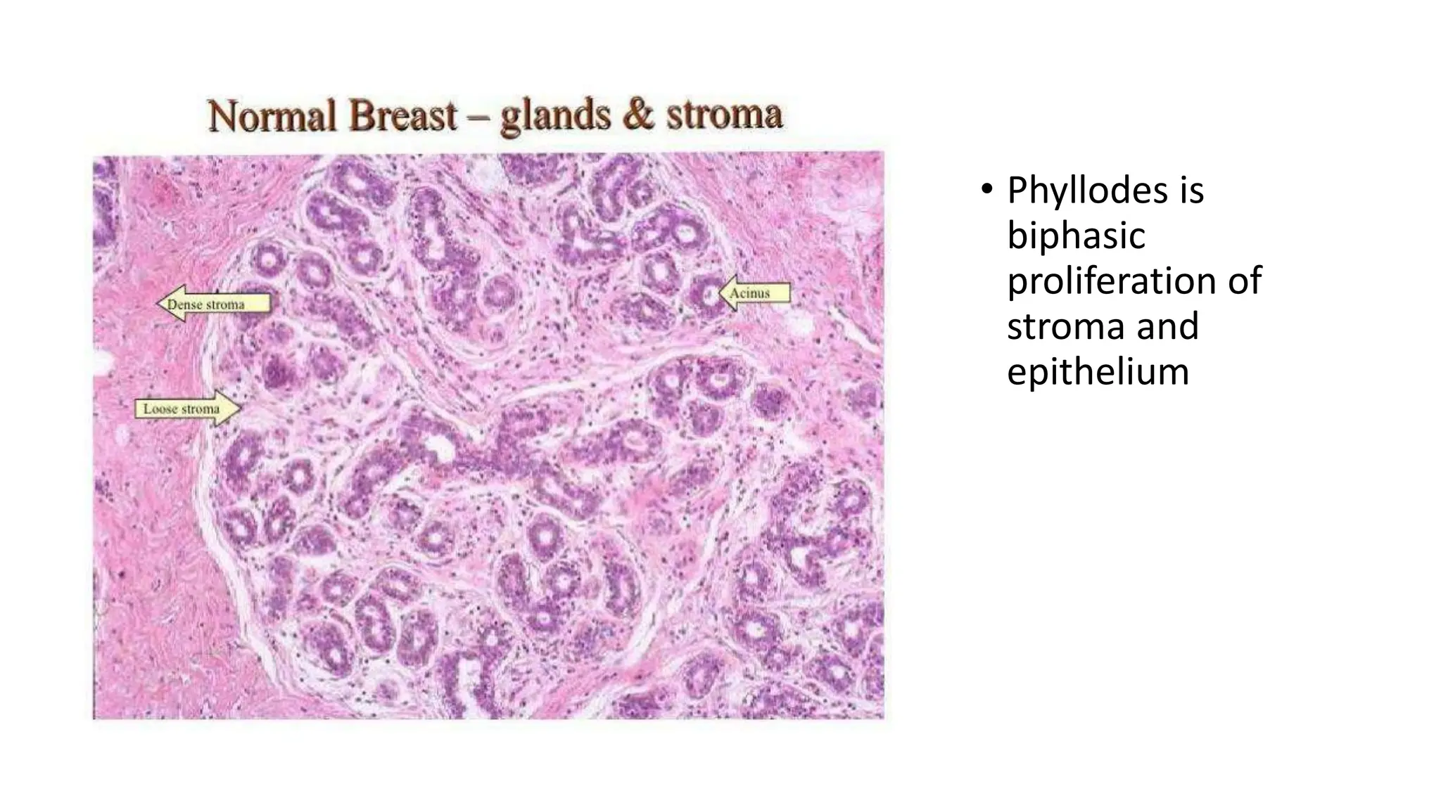
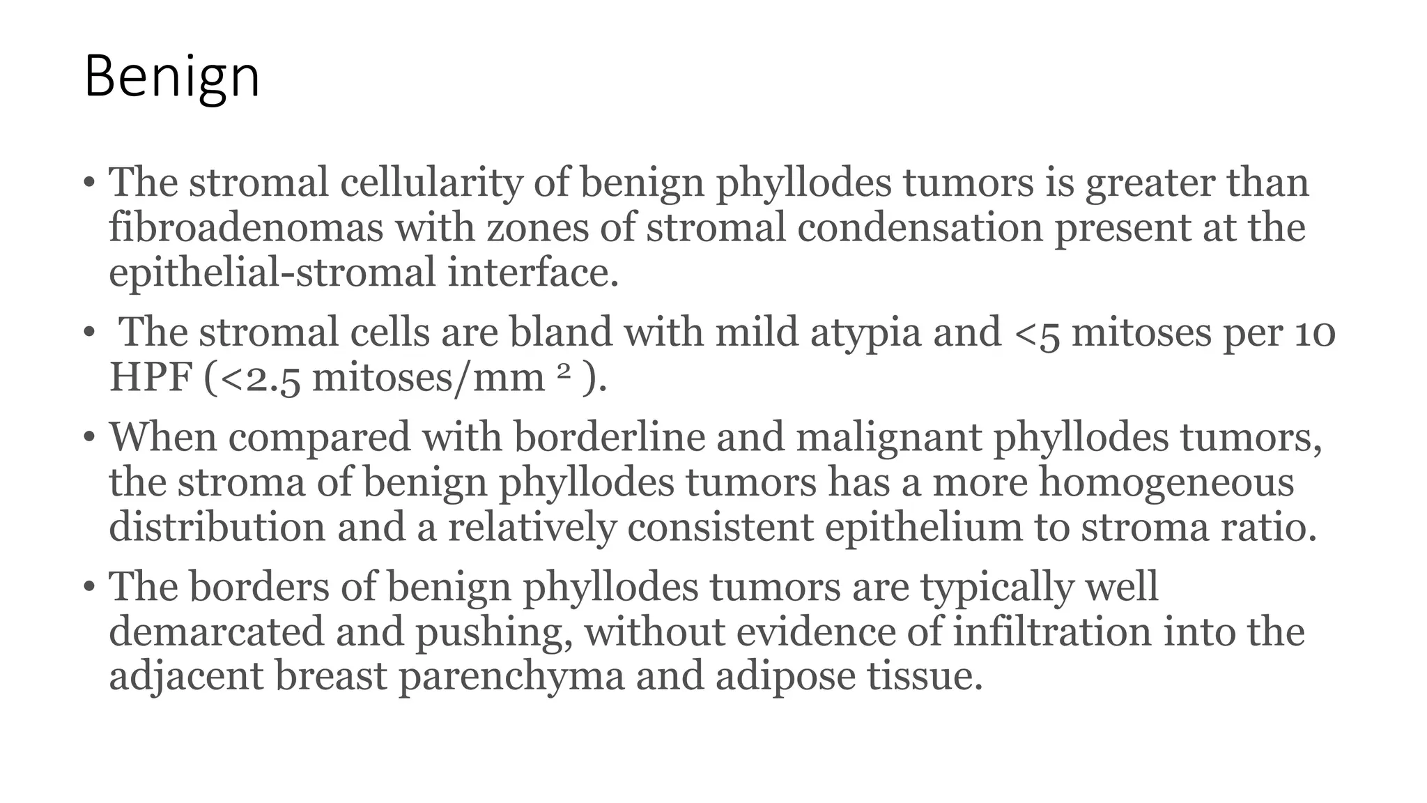
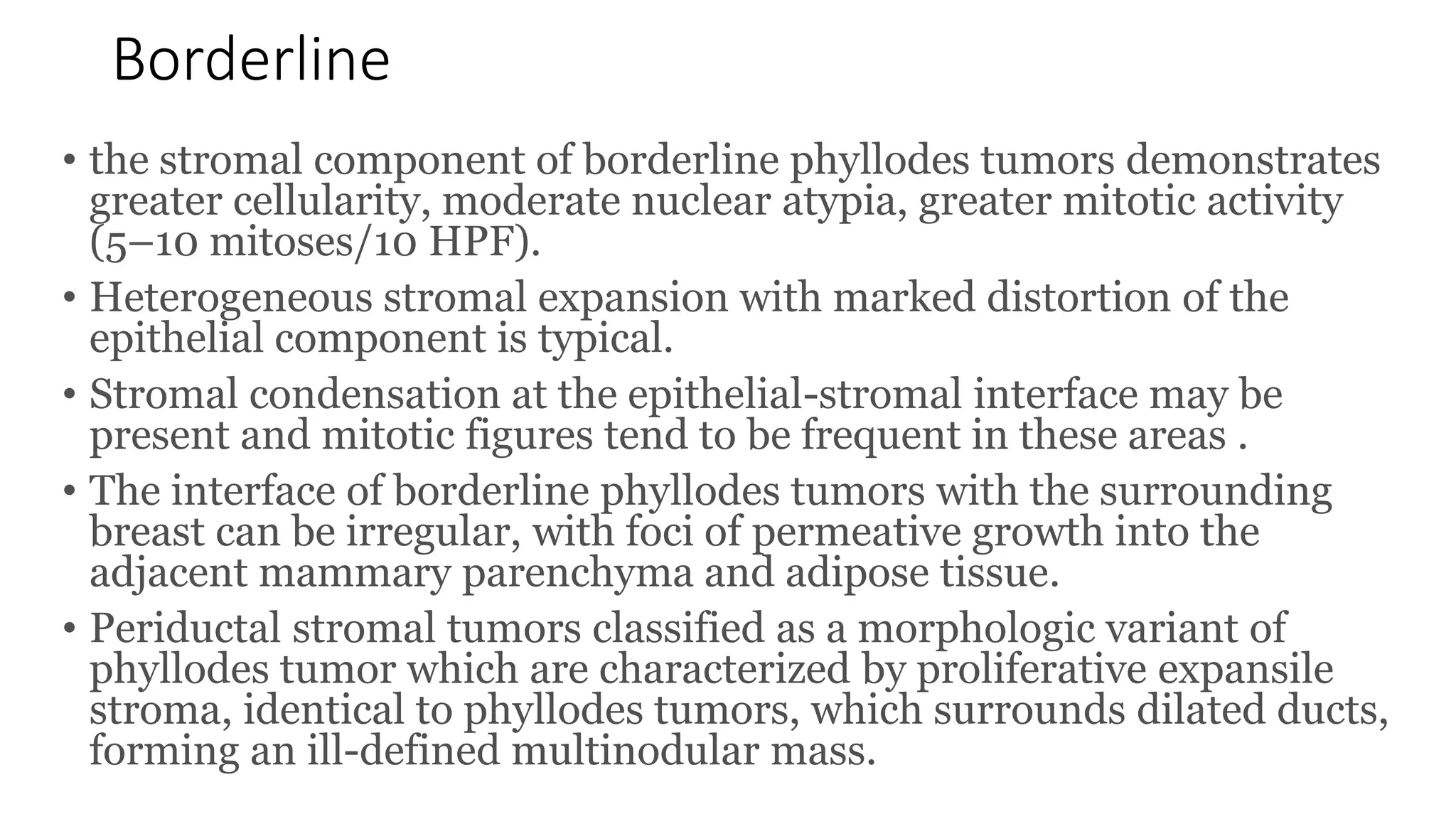
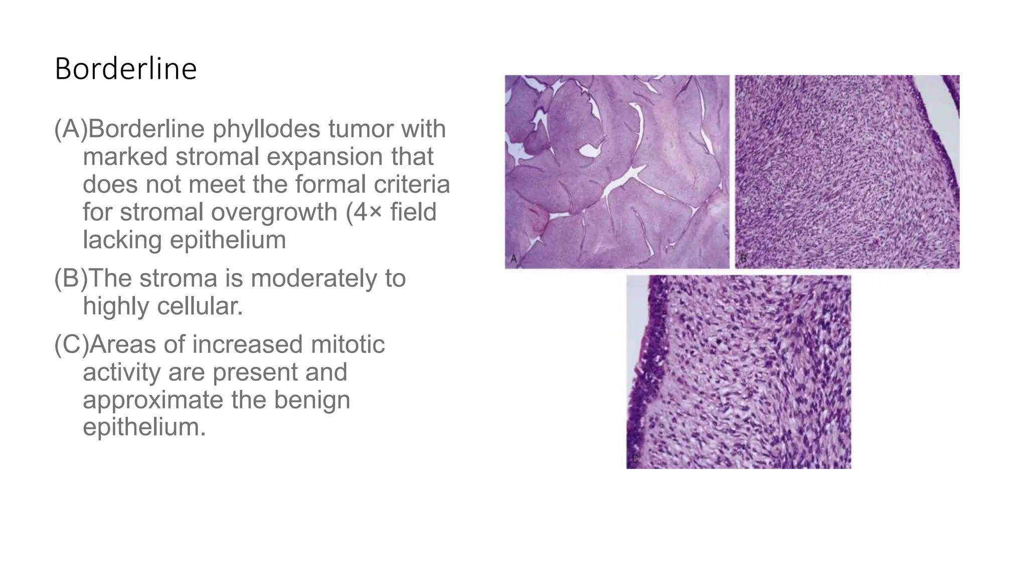
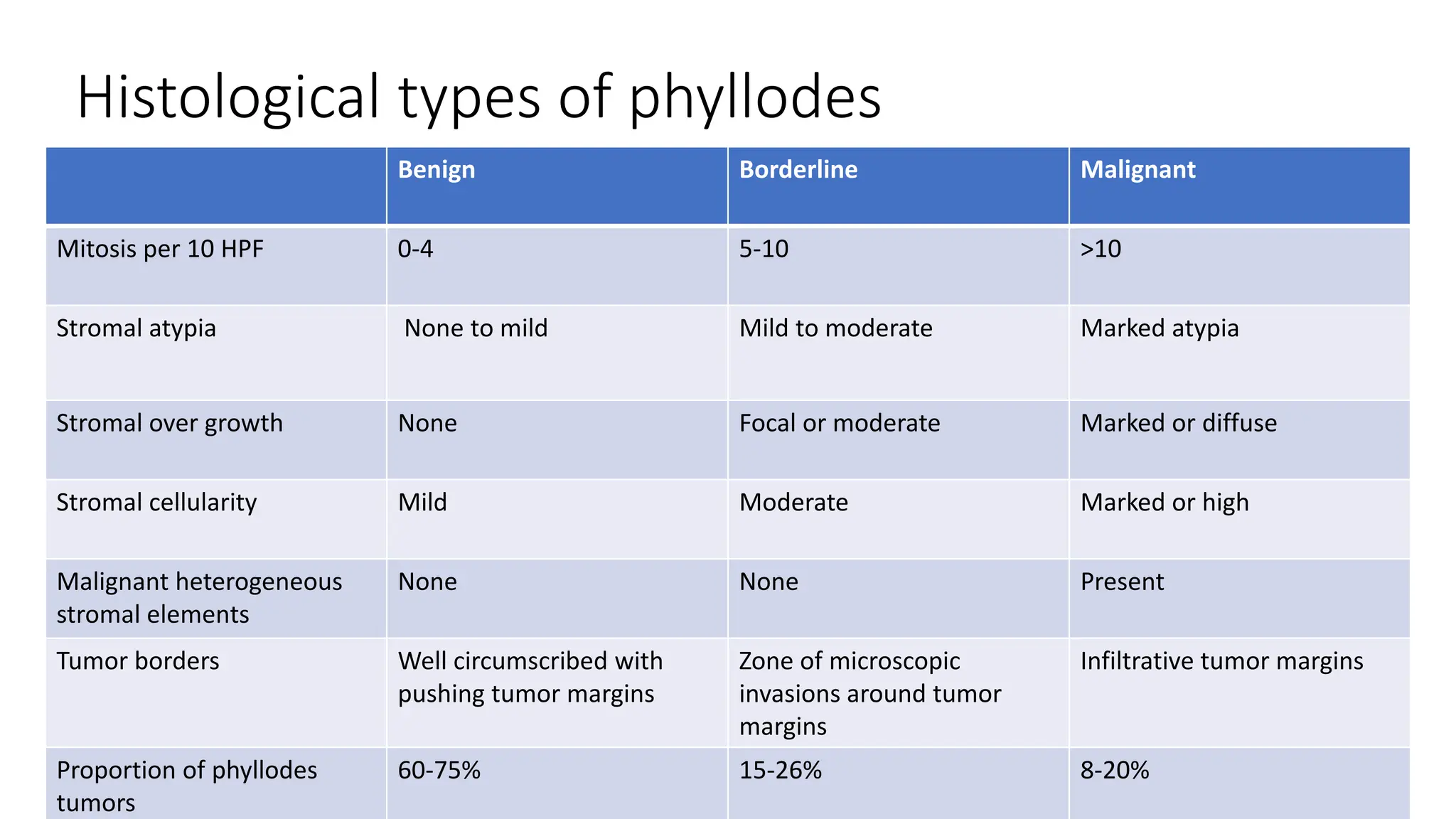
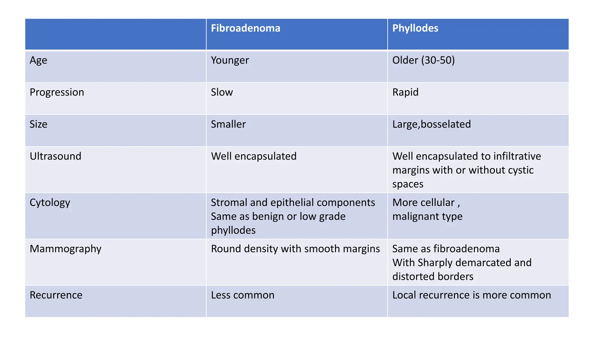
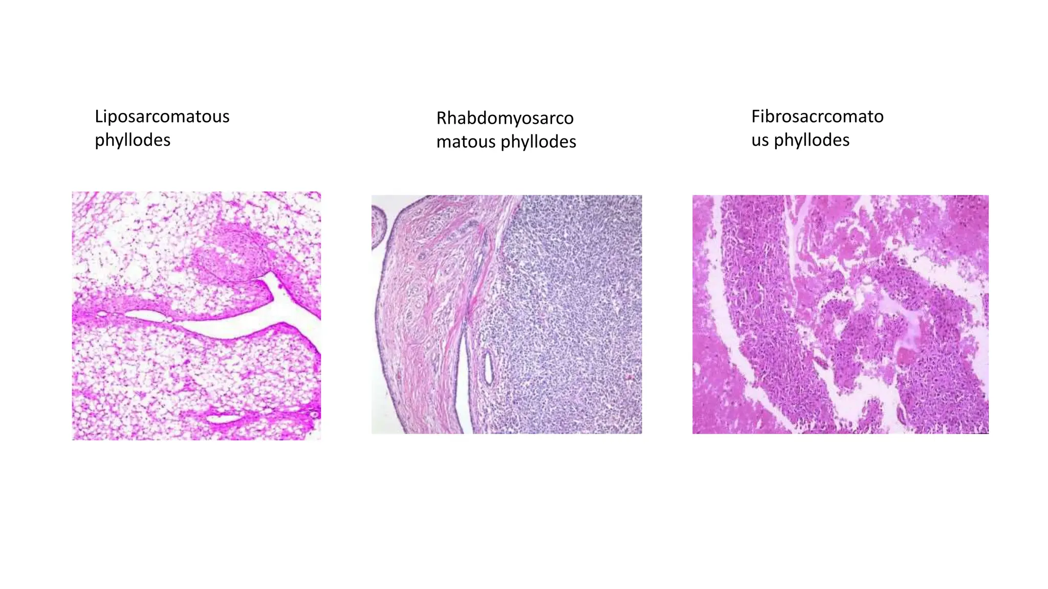
3. Anatomy
1.The mammary gland is located within the
superficial fascia of the anterior thoracic wall. It
consists of 15 to 20 lobes of glandular tissue of
the tubuloalveolar type.
2.Longitudinal fibrous connective stroma forms
a latticed framework that supports the lobes;
adipose tissue fills the space between the lobes
5. Arterial supply of breast
• Parenchyma and skin of the breast
receive their blood supply from:
• 1. perforating branches of the internal
mammary artery.
• 2. lateral branches of the posterior
intercostal arteries.
• 3. several branches from the axillary
artery, including highest thoracic, lateral
thoracic, and pectoral branches of the
thoracoacromial artery
6. Venous drainage of
breast
Superficial veins from base of nipple
drain into internal thoracic vein.
Deep veins from base of nipple drain
into internal thoracic, axillary and
posterior intercostal veins.
Breast tissue drains primarily into
axillary vein and internal mammary vein
Posterior intercostals with perforating
branches also have continuity with
Batsons Vertebral plexus.
7. Lymphatic drainage of
breast
1.75%of breast tissue to
axillary lymph nodes
2.20% into internal
mammary nodes
3.5% into posterior
intercostal nodes.
8. • Phyllodes tumors are most often unilateral, presenting as
mobile, palpable, and painless breast masses.
• Some are large and rapidly growing phyllodes tumors can
be symptomatic.
• They are on average larger than fibroadenomas, typically
measuring 4 to 5 cm and may be significantly larger and
accompanied by overlying skin ulceration and necrosis.
• Bilateral and multifocal lesions are rare.
• Metastasis to axillary lymph nodes is uncommon.
9. Epidemiology
• Previously known as Cystosarcoma Phyllodes
• Commonly seen in the age group 30-50 years
• About 90% of phyllodes tumors are benign and about 10% are malignant.
Although these tumors rarely metastasize, they can recur locally.
• Phyllodes tumors account for less than 1% of all primary breast
tumors and 2.5% of all fibroepithelial lesions in women
• Women with phyllodes tumors present at a slightly older age
(average 40–50 years) than women with fibroadenomas.
• Women developing malignant phyllodes tumors are typically older
than those presenting with benign tumors.
• Rarely, phyllodes tumors are diagnosed in young girls and
adolescents.
11. Clinical presentation
• Occurs in premenopausal women (30-50yrs)
• Usually unilateral
• Rapidly growing
• Huge swelling with bosselated surface
• Skin over breast is stretched,red and dilated veins over it with
necrosis over skin due to pressure
• Tumor is warmer, non tender,soft,fluctuating not fixed to skin,muscles
and chest wall with no axillary lymphadenopathy.
13. Macroscopic appearance
• Phyllodes tumors are circumscribed masses with tan, fleshy, and
bulging cut surfaces.
• demonstrate a whorled pattern, and owing to the degree of stromal
proliferation, display visible clefts or irregular protuberances.
• The borders are usually well defined and lobulated.
• an indistinct interface with the surrounding normal breast may
correspond to infiltrative growth, which often occurs with malignant
tumors.
• Cystic change, hemorrhage, and necrosis can be seen in larger
tumors.
14. • The classic architecture of phyllodes tumors features broad,
leaf-like, stromal projections that reside within large cleft-like
spaces.
• The proliferating stromal cells may compress the epithelial
component into slit-like cords.
• The epithelial component of phyllodes tumors is mostly benign,
enveloping the stromal projections and lining the clefts.
• The epithelial component consists of the usual mammary
bilayer of luminal epithelial cells and basal myoepithelial cells.
• Rarely, ductal carcinoma in situ or invasive carcinoma may also
be present.
Microscopic appearance
15. Types
• Based on cellularity, invasive margin, sarcomatous appearance it is
divided into
• 1.benign
• 2.borderline
• 3.malignant
16. Benign
• The stromal cellularity of benign phyllodes tumors is greater than
fibroadenomas with zones of stromal condensation present at the
epithelial-stromal interface.
• The stromal cells are bland with mild atypia and <5 mitoses per 10
HPF (<2.5 mitoses/mm 2 ).
• When compared with borderline and malignant phyllodes tumors,
the stroma of benign phyllodes tumors has a more homogeneous
distribution and a relatively consistent epithelium to stroma ratio.
• The borders of benign phyllodes tumors are typically well
demarcated and pushing, without evidence of infiltration into the
adjacent breast parenchyma and adipose tissue.
17. Benign
(A) Benign phyllodes tumor with broad,
leaf-like, stromal projections that
reside within large, cleft-like spaces
(B) The cellularity of the stromal leaves
is low in this region.
(C)In some regions the cellularity is
increased with condensation where
it borders the epithelium.
(D)The stromal nuclei show mild atypia
and occasional mitotic figures. The
epithelium surmounting the fronds
consists of the typical luminal
epithelial and myoepithelial bilayer.
18. Borderline
• the stromal component of borderline phyllodes tumors demonstrates
greater cellularity, moderate nuclear atypia, greater mitotic activity
(5–10 mitoses/10 HPF).
• Heterogeneous stromal expansion with marked distortion of the
epithelial component is typical.
• Stromal condensation at the epithelial-stromal interface may be
present and mitotic figures tend to be frequent in these areas .
• The interface of borderline phyllodes tumors with the surrounding
breast can be irregular, with foci of permeative growth into the
adjacent mammary parenchyma and adipose tissue.
• Periductal stromal tumors classified as a morphologic variant of
phyllodes tumor which are characterized by proliferative expansile
stroma, identical to phyllodes tumors, which surrounds dilated ducts,
forming an ill-defined multinodular mass.
19. Borderline
(A)Borderline phyllodes tumor with
marked stromal expansion that
does not meet the formal criteria
for stromal overgrowth (4× field
lacking epithelium
(B)The stroma is moderately to
highly cellular.
(C)Areas of increased mitotic
activity are present and
approximate the benign
epithelium.
20. Malignant
• The malignant phyllodes tumor demonstrates sarcomatous stromal
overgrowth (defined as a 4× field comprised of stroma only), marked
cellular pleomorphism, an increased mitotic rate (≥10 mitoses/10
HPF; ≥5 mitoses/mm 2 ), and infiltrative borders
• The characteristic leaf-like architecture is limited or nonevident and
the epithelial component can be very focal .
• Whereas the presence of malignant heterologous differentiation,
such as osteosarcoma, is diagnostic of a malignant phyllodes tumor,
even in the absence of the other criteria, a well-differentiated
liposarcoma component alone no longer qualifies a phyllodes tumor
as malignant.
21. Malignant
A) Malignant phyllodes tumor
characterized by heterogeneous
stromal expansion and highly
cellular broad, leaf-like stromal
projections.
B) (B) Although the degree of stromal
expansion does not fit the formal
definition of stromal overgrowth, the
marked atypia and abundant mitotic
activity, including atypical forms, is
diagnostic of a malignant phyllodes
tumor.
C) (C) The density of the stromal cells
varies throughout the tumor but the
high degree of atypia and mitotic
activity is consistent.
22. Histological types of phyllodes
Benign Borderline Malignant
Mitosis per 10 HPF 0-4 5-10 >10
Stromal atypia None to mild Mild to moderate Marked atypia
Stromal over growth None Focal or moderate Marked or diffuse
Stromal cellularity Mild Moderate Marked or high
Malignant heterogeneous
stromal elements
None None Present
Tumor borders Well circumscribed with
pushing tumor margins
Zone of microscopic
invasions around tumor
margins
Infiltrative tumor margins
Proportion of phyllodes
tumors
60-75% 15-26% 8-20%
23. Fibroadenoma Phyllodes
Age Younger Older (30-50)
Progression Slow Rapid
Size Smaller Large,bosselated
Ultrasound Well encapsulated Well encapsulated to infiltrative
margins with or without cystic
spaces
Cytology Stromal and epithelial components
Same as benign or low grade
phyllodes
More cellular ,
malignant type
Mammography Round density with smooth margins Same as fibroadenoma
With Sharply demarcated and
distorted borders
Recurrence Less common Local recurrence is more common
24.
25. Molecular alterations
• The cytogenetic abnormalities includes gains on chromosome 1q and
losses on chromosome 13, which have been associated with
malignant progression.
• Mutations of MED12 , more commonly seen in benign phyllodes
tumors and fibroadenomas, have also been identified in borderline
and malignant tumors.
• RARA , FLNA , SETD2 , and KMT2D mutations have also been
reported.
• Mutations in EGFR, RB1, TP53, c-KIT, PTEN, and TERT are
associated with malignant behavior.
• Increased incidence of phyllodes tumors has been found in patients
with Li-Fraumeni syndrome.
27. Ultrasound
• Ultrasound examination
characteristically shows a
hypoechoic mass with
striations that correlate with
the cleft-like spaces. Cystic
foci and posterior acoustic
enhancement reflect the more
complex cystic echogenicity
seen in borderline and
malignant phyllodes tumors
28. Mammogram
• phyllodes tumors are
mammographically
detected, appearing as
a round, oval, or
lobulated, well-
circumscribed mass
29. Cytology
• Core needle biopsy is more preferred than fine needle Cytology
• Stromal cells of fibroadenoma are polyclonal or monoclonal whereas
phyllodes are always monoclonal
• In malignant phyllodes, liposarcomatous and rhabdomyosarcomatous
elements are more common than fibrosacrcomatous elements
• Number of mitoses, presence or absence of invasive foci helps to
identify malignant type
31. Surgical management
• Benign and borderline: wide local excision with atleast 2cm margin
along with overlying skin and pectoralis major is removed to prevent
local recurrence.
• Malignant :simple or total mastectomy
32. • Treatment of phyllodes tumors requires complete excision with
negative margins.
• Breast conservation does not result in decreased survival but
better local control and improved disease-free intervals may be
achieved by mastectomy for large tumors.
• Sentinel lymph node evaluation is not typically performed due
to rare lymph node metastasis.
33. Wide local excision
• wide local excision with atleast
2cm margin along with
overlying skin and underlying
pectoralis major is removed to
prevent local recurrence.
34. Simple mastectomy
• Indications of mastectomy in phyllodes
1.Massive tumors
2.Recurrent tumors
3.Malignant type
• total mastectomy or simple mastectomy :removal of entire breast
tissue including skin over the tumor, nipple areolar complex and
axillary tail.
• total mastectomy was designed to treat local disease in the breast and
omit regional nodal dissection because nodal dissection has no
influence on survival and resulted in significant surgical morbidity.
• Sometimes adjacent lymph nodes are removed along with breast tissue
35. Position of patient
With the ipsilateral arm in a
relaxed, extended position
on the padded arm board.
36. • The choice of skin incision and the design of skin flaps are
carefully planned with respect to the quadrant in which
the primary neoplasm is located to have adequate
margins.
• The transverse mastectomy incision is now more widely
utilized. Skin incisions are made perpendicular to the
subcutaneous plane and elevates skin flaps to the
boundaries of the breast for adequate resection
38. • The breast tissue usually extends to a point where the anterior premammary fascia fuses
with the posterior pectoral fascia.
• We should remove the breast to the point of fusion between these two fasciae,which
usually extends to
superiorly to the level of the second rib
medially to the parasternal edge
Inferiorly to the inframammary crease
Laterally to the anterior border of latissimus dorsi.
• Dissection started superiorly and proceeded clockwise ending in the axillary region
• Care must be taken to ligate perforating branches of lateral thoracic and anterior
intercostal vessels
• Lateral branches of the medial pectoral neurovascular bundle is carefully dissected while
removing axillary tail.
• Romovac drains are placed
39. Postoperative care
• the patient should be instructed to limit the range of motion of
the shoulder and arm to augment flap adherence to the chest
wall.
• drains may be be removed when drainage becomes
predominantly serous and has decreased to a maximum of 20 to
30 mL during a 24-hour interval.
• Shoulder and arm range-of-motion exercises may be increased
the day after drains are removed.
40.
41.
42. Prognosis
• Risk of recurrence :
Benign phyllodes -10-15%
Borderline phyllodes-15-25%
Malignant phyllodes- 25-30%
• Margin status at the time of initial excision is the best predictor
of local recurrence for borderline and malignant tumors.
• Distant metastases typically occur 5 to 8 years after the initial
diagnosis. Large tumor size and the presence of malignant
heterologous elements are associated with increased metastatic
potential.
• When tumors metastasize, they may be in the form of a
malignant stromal component only.
• Most common metastatic sites are the lung and bones.
44. Radiotherapy and chemotherapy
• Indications of radiotherapy
1. Malignant phyllodes with tumor margin <1cm
2. Recurrent phyllodes
3. Metastatic phyllodes
• There is no currently accepted role for adjuvant radiation therapy,
especially for margins >1 cm.
• Indications of chemotherapy
1. Malignant phyllodes
• But there are very minimal success rates to chemotherapy.
• No role of hormonal therapy in phyllodes
45. Recurrence
• The grade of the tumor and margin status
determine the propensity for local recurrence and
metastasis.
• When phyllodes tumors recur, they present as
multiple nodules at the site of resection and in one-
fourth of cases are a higher histologic grade.
• Local recurrence is usually seen within first 2 years after
excision indicating need for follow up.
• Close follow-up with examination and imaging for early
detection of recurrence
• Patient should be followed up clinically for every 6
months in the first 2 years then annually with
mammography
• Treatment is reexcision with wide margins followed by
radiotherapy.
46.
47. References
• Bailey and love 28th edition
• Sabiston text book of surgery
• Schwartz’s principles of surgery
• Bland and Copeland’s The Breast