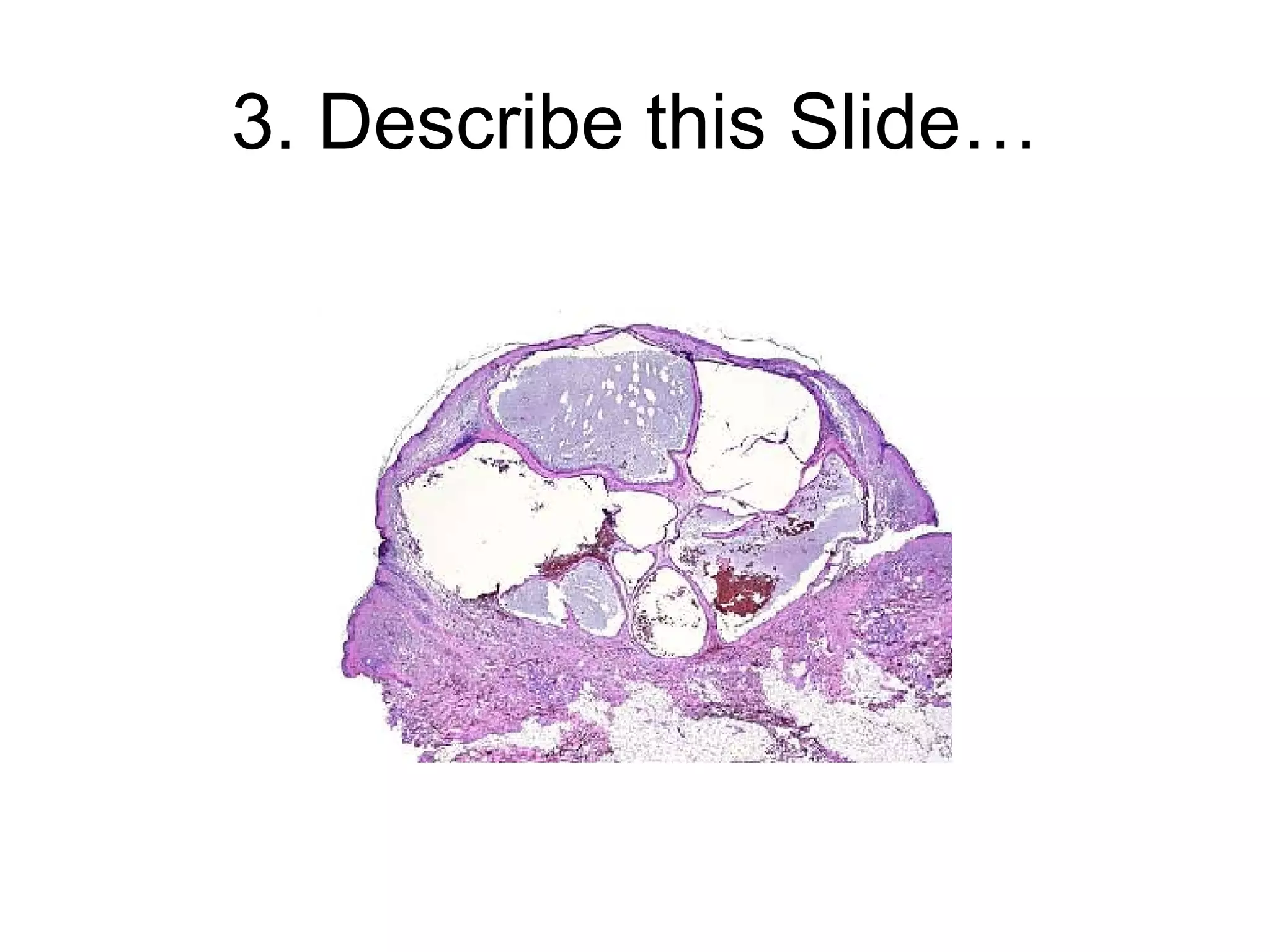
Grand Rounds Michael Rubin, MD Department of Ophthalmology and Visual Science The University of Chicago
Case Presentation 55 year old male presents for general ophthalmology evaluation.  States that he has no visual complaints, but that he has had a lesion on this left eye for the past few years that we wanted evaluated.
Medical History PMH: BPH, Borderline HTN MEDS: Finestride ALL: NKDA Ocular Hx: LUL lesion
Visual Examination BCVA: OD: 20/30 OS: 20/30 IOP: 16, 14 CVF: Full OU Pupils: 4mm to 2mm, No APD
Ocular Examination Adnexa: Right: MGD/Bleph   Left: See Photo Sclera/Conj: Clear OU Cornea: Clear OU AC: Deep and Quiet Iris: Normal OU Lens: 1 + NS and 1 + CC both eyes
LUL Lesion
Diagnosis Apocrine Hidrocystoma
Benign Adnexal Tumors Holocrine Tumors -- Glands of Zeis, Meibomian Glands Eccrine Tumors Apocrine Tumors --Sweat glands of Moll 4. Hair Follicle Tumors
Holocrine/Sebaceous Tumors Benign Lesions of Sebaceous Origin --Milia --Acquired Sebaceous Gland Hyperplasia --Sebaceous Adenoma
Benign Lessions of Eccrine Origin 1. Eccrine Hidrocystoma 2. Syringoma 3. Acrospiroma
Benign Lesions of Apocrine Origin Apocrine Hidrocystoma Cylindroma
Benign Lesions of Hair Follicle 1. Trichoepithelioma 2. Trichofolliculoma 3. Tricholemmoma 4. Pilomatrixoma
Background Apocrine hidrocystomas are benign cystic proliferations of the apocrine secretory glands.  These cysts most commonly appear as solitary, soft, dome-shaped, translucent papules or nodules and most frequently are located on the eyelids, especially the inner canthus.  Apocrine hidrocystomas grow slowly and usually persist indefinitely.
Pathophysiology The exact stimulus for the development of an apocrine hidrocystoma is unknown. Plausible causes of the closely related eccrine hidrocystoma include occlusion or blockage of the sweat duct apparatus, which results in the retention of sweat and a dilated cystic structure.
Predisposition Race:  No known predilection exists for race or geographic region.  Sex:  No known gender predilection exists.  Age:  Cysts occur in adulthood, although in no particular age group.  History:  Apocrine hidrocystomas usually are asymptomatic. No seasonal variation or familial tendencies have been identified. Cysts tend to appear during adulthood, grow slowly, and persist indefinitely.
Physical Characteristics The tumors usually occur as solitary translucent papules or nodules. Consistency is fluctuant cystic. Size varies from a few millimeters to approximately 1.5 cm. Tumors occasionally present as multiple lesions. Cysts often appear tense and shiny; coloration varies from flesh-colored to blue or black Tumors have a predilection for the eyelid, particularly the inner canthus. Tumors may arise on other areas of the head, neck, and trunk. Tumors also have been reported to occur on the penis, in the axillae, and in the anal region.
More Characteristics Walls, although translucent, are sufficiently thick that they seldom rupture spontaneously. When incised, the cyst collapses and a thin, clear, brownish or blackish fluid is released. Cysts are mobile with palpation and transilluminate. The fluid color of an apocrine hidrocystoma does not result from the presence of melanin or hemosiderin but may result either from the Tyndall phenomenon or the presence of lipofuscin pigment. Apocrine hidrocystomas are not affected by variation in temperature (unlike eccrine hidrocystomas).
Causes Although the origin of apocrine hidrocystomas is not known entirely, they are believed to be adenomatous cystic proliferations of the apocrine glands.
Need for Histology? The clinical appearance of a pea-sized cyst near the inner canthus of the eye, which contains a thin clear or pigmented fluid, suggests an apocrine hidrocystoma; however, histologic examination often is required to establish a specific and definitive diagnosis.
Histology On histologic exam, apocrine hidrocystomas show large unilocular or multilocular cystic spaces within the dermis. Apocrine hidrocystomas are more likely to be multilocular than the closely related eccrine hidrocystoma.
Slide One Scan power view showing multiple cystic areas. The fluid does not contain pigment granules though the fluid was dark.
Slide Two A high power view of the linings of two of the cysts. A few brown,  lipofuchsin pigment granules (PG) are in the basilar part of the epithelium. There is a suggestion of pigmented material in the apices of other cells. Apocrine snouts are prominent. 
Cyst Wall The cyst wall is lined by apocrine-type secretory epithelium.  The innermost layer of the wall is composed of a single (occasionally double) layer of cuboidal-to columnar-shaped cells. The nuclei of these cells are positioned basally.  The outer layer of cells composing the cyst wall is formed by myoepithelial cells in which the long axes run parallel to the cyst wall.
More Histology Well-organized fibrous tissue surrounds the cyst. Papillary projections extend from the secretory layer into the cyst cavity, depicting decapitation secretion.  The secretory cells are acid-Schiff–positive, and occasionally contain pigment granules, which provide the brown color of the cystic fluid. This pigment is neither melanin nor hemosiderin.  On electron microscopy, secretory cells have numerous, dense, lysosomal-type secretory granules typical of apocrine gland cells. They also have an increased number of annulate lamellae, which are unusual in normal apocrine cells.
Treatment Apocrine hidrocystomas can be incised and drained. Electrosurgical destruction of the cyst wall often is recommended to prevent recurrence. Punch, scissors, or elliptical excision also can remove tumors.
Prognosis Apocrine hidrocystomas grow gradually and persist indefinitely after attaining full size. They seldom recur after removal.
Pitfall If the diagnosis is made clinically without biopsy, a cystic basal cell carcinoma of the eyelid may be missed. Incision to observe the egress of fluid or biopsy of suspicious lesions excludes the possibility.
What’s This?
Syringoma Syringoma is a benign adnexal neoplasm formed by well-differentiated ductal elements.  Its name is derived from the Greek word  syrinx,  which means pipe or tube.
Appearance  Syringomas are skin-colored or yellowish, small, dermal papules. Sometimes, the lesions may appear translucent or cystic. The surface can be rounded or flat-topped. The lesions are usually smaller than 3 mm in diameter.
Pathophysiology Syringoma is a benign neoplasm that is traditionally considered to differentiate along eccrine lines.  Many tumors that were traditionally thought to be eccrine have recently been shown to have apocrine differentiation.
Que es eso?
Eccrine Hidrocystoma AKA Sudoriferous or Sweat gland Cysts Solitary or multiple small nodules on the eyelid Overlying skin is shiny and smooth Thought to be ductal retention cysts Tend to increase in size during hot and humid weather
Histology of Eccrine Hydrocystoma Eccrine Hydrocysomas are dermal cysts lined by double layered cuboidal epithelium. Cyst in usually translucent and fluid filled.
What the…
Cylindroma The cellular origin of cylindromas is debated by dermatopathologists. Cylindroma most likely is a very primitive sweat gland tumor differentiating toward either the eccrine line or the apocrine line.
Physical Characteristics of Cylindroma Solitary lesions are firm, rubbery nodules with pink, red, or sometimes blue coloring that range in size from a few millimeters to several centimeters. The multiple form has numerous masses of pink, red, or blue nodules, sometimes resembling bunches of grapes or small tomatoes (sometimes called a tomato tumor).
Histology of Cylindromas Note the focus of tubular differentiation. Tubules are usually few in number or absent. Small subunits are separated by glassy, eosinophilic basement membrane material. Some of the subunits have angulated outlines. The darker cells tend to be found around the periphery of the subunits.
Hair Follicle Lesions 1. Trichoepithelioma --Muliple, keratin-filled horn cysts 2. Trichofolliculoma --Well differentiated hamartomatous lesion --Solitary flesh-colored nodule with central umbilication 3. Trichilemmoma --Arises from outer hair sheath --Flesh colored, nodular, or papillomatous --Cowdens Disease: AD, Multiple lesions, acral keratosis, oral papillomas, increased risk for thyroid and breast CA. 4. Pilomatrixoma --Bening tumor of hair matrix origin --Solid or cystic, mobile, subcutaneous nodule, frim irregular, often reddish blue.
Lesion 1: Trichoepithelioma
Lesion 2: Trichofolliculoma
Image 3: Trichilemmoma
Image 4: Pilomatrixoma
Two Lesions, One disease
Basal Cell Carcinoma Basal cell carcinoma (BCC) is the most common malignancy in humans.  It typically occurs in areas of chronic sun exposure.  BCC is usually slow growing and rarely metastasizes, but it can cause significant local destruction and disfigurement if neglected or treated inadequately. Prognosis is excellent with proper therapy.
Features of Nodular BCC Nodular BCC: This is the most common variety of BCC. Nodular BCCs have some of the following features: Waxy papule(s) with central depression  Pearly appearance  Erosion or ulceration  Bleeding  Crusting  Rolled (raised) border  Translucency  Telangiectases over the surface  History of bleeding with minor trauma
Other forms of BCC Pigmented BCC : In addition to features seen in lesions of nodular BCC, lesions of pigmented BCC contain increased brown or black pigment and are seen more commonly in individuals with dark skin. Cystic BCC : Lesions of cystic BCC are translucent blue-gray cystic nodules that may mimic benign cystic lesions. Superficial BCC : This variety presents as scaly patches or papules that are pink to red-brown in color, often with central clearing. Micronodular BCC: This aggressive BCC subtype has the typical BCC distribution. It is less prone to ulceration, may appear yellow-white when stretched, and is firm to touch.  Morpheaform and infiltrating BCC : These are aggressive BCC subtypes with sclerotic (scarlike) plaques or papules. The border usually is not well defined and often extends well beyond clinical margins.
Resident Quiz
1. What is this?
2. What sydrome predisposing to thyroid and breast CA is associated with this lesion?  What is this lesion?
3. Describe this Slide…
Bibliography Alessi E, Gianotti R, Coggi A: Multiple apocrine hidrocystomas of the eyelids. Br J Dermatol 1997 Oct; 137(4): 642-5 Bures FA, Kotynek J: Differentiating between apocrine and eccrine hidrocystoma. Cutis 1982 Jun; 29(6): 616, 619-20 Combemale P, Kanitakis J, Dupin N: Multiple Moll's gland cysts (apocrine hidrocystomas) of the eyelids. Dermatology 1997; 194(2): 195-6 de Eusebio E, Lopez-Bran E, Rojo S: Multiple hidrocystomas. Dermatology 1996; 193(2): 152-3 Hashimoto K, Lever WF: Tumors of skin appendages. In: Freedberg IM, Eisen AZ, Wolff K, eds. Fitzpatrick's Dermatology in General Medicine. Vol 1. McGraw-Hill Professional Publishing; 1999: 899.  Mackie RM: Tumors of skin appendages. In: Champion RH, Wilkinson DS, Ebling FJG, eds. Rook/Wilkinson/Ebling: Textbook of Dermatology. Vol 2. Blackwell Science Inc; 1998: 1703-6.  Milum EA: A solitary pigmented tumor of the face. Apocrine hidrocystoma. Arch Dermatol 1991 Apr; 127(4): 572, 575 Schleicher SM: Multiple translucent facial papules. Apocrine hidrocystoma. Arch Dermatol 1998 Dec; 134(12): 1627-8, 1630-1  Shields JA, Eagle RC Jr, Shields CL: Apocrine hidrocystoma of the eyelid. Arch Ophthalmol 1993 Jun; 111(6): 866-7 Smith JD, Chernosky ME: Apocrine hidrocystoma (cystademnoma). Arch Dermatol 1974 May; 109(5): 700-2  Ter Poorten HJ: Apocrine hidrocystoma of the right scapula. Arch Dermatol 1977 Dec; 113(12): 1730

Apocrine Hidrocystoma

  • 1.
    Grand Rounds MichaelRubin, MD Department of Ophthalmology and Visual Science The University of Chicago
  • 2.
    Case Presentation 55year old male presents for general ophthalmology evaluation. States that he has no visual complaints, but that he has had a lesion on this left eye for the past few years that we wanted evaluated.
  • 3.
    Medical History PMH:BPH, Borderline HTN MEDS: Finestride ALL: NKDA Ocular Hx: LUL lesion
  • 4.
    Visual Examination BCVA:OD: 20/30 OS: 20/30 IOP: 16, 14 CVF: Full OU Pupils: 4mm to 2mm, No APD
  • 5.
    Ocular Examination Adnexa:Right: MGD/Bleph Left: See Photo Sclera/Conj: Clear OU Cornea: Clear OU AC: Deep and Quiet Iris: Normal OU Lens: 1 + NS and 1 + CC both eyes
  • 6.
  • 7.
  • 8.
    Benign Adnexal TumorsHolocrine Tumors -- Glands of Zeis, Meibomian Glands Eccrine Tumors Apocrine Tumors --Sweat glands of Moll 4. Hair Follicle Tumors
  • 9.
    Holocrine/Sebaceous Tumors BenignLesions of Sebaceous Origin --Milia --Acquired Sebaceous Gland Hyperplasia --Sebaceous Adenoma
  • 10.
    Benign Lessions ofEccrine Origin 1. Eccrine Hidrocystoma 2. Syringoma 3. Acrospiroma
  • 11.
    Benign Lesions ofApocrine Origin Apocrine Hidrocystoma Cylindroma
  • 12.
    Benign Lesions ofHair Follicle 1. Trichoepithelioma 2. Trichofolliculoma 3. Tricholemmoma 4. Pilomatrixoma
  • 13.
    Background Apocrine hidrocystomasare benign cystic proliferations of the apocrine secretory glands. These cysts most commonly appear as solitary, soft, dome-shaped, translucent papules or nodules and most frequently are located on the eyelids, especially the inner canthus. Apocrine hidrocystomas grow slowly and usually persist indefinitely.
  • 14.
    Pathophysiology The exactstimulus for the development of an apocrine hidrocystoma is unknown. Plausible causes of the closely related eccrine hidrocystoma include occlusion or blockage of the sweat duct apparatus, which results in the retention of sweat and a dilated cystic structure.
  • 15.
    Predisposition Race: No known predilection exists for race or geographic region. Sex: No known gender predilection exists. Age: Cysts occur in adulthood, although in no particular age group. History: Apocrine hidrocystomas usually are asymptomatic. No seasonal variation or familial tendencies have been identified. Cysts tend to appear during adulthood, grow slowly, and persist indefinitely.
  • 16.
    Physical Characteristics Thetumors usually occur as solitary translucent papules or nodules. Consistency is fluctuant cystic. Size varies from a few millimeters to approximately 1.5 cm. Tumors occasionally present as multiple lesions. Cysts often appear tense and shiny; coloration varies from flesh-colored to blue or black Tumors have a predilection for the eyelid, particularly the inner canthus. Tumors may arise on other areas of the head, neck, and trunk. Tumors also have been reported to occur on the penis, in the axillae, and in the anal region.
  • 17.
    More Characteristics Walls,although translucent, are sufficiently thick that they seldom rupture spontaneously. When incised, the cyst collapses and a thin, clear, brownish or blackish fluid is released. Cysts are mobile with palpation and transilluminate. The fluid color of an apocrine hidrocystoma does not result from the presence of melanin or hemosiderin but may result either from the Tyndall phenomenon or the presence of lipofuscin pigment. Apocrine hidrocystomas are not affected by variation in temperature (unlike eccrine hidrocystomas).
  • 18.
    Causes Although theorigin of apocrine hidrocystomas is not known entirely, they are believed to be adenomatous cystic proliferations of the apocrine glands.
  • 19.
    Need for Histology?The clinical appearance of a pea-sized cyst near the inner canthus of the eye, which contains a thin clear or pigmented fluid, suggests an apocrine hidrocystoma; however, histologic examination often is required to establish a specific and definitive diagnosis.
  • 20.
    Histology On histologicexam, apocrine hidrocystomas show large unilocular or multilocular cystic spaces within the dermis. Apocrine hidrocystomas are more likely to be multilocular than the closely related eccrine hidrocystoma.
  • 21.
    Slide One Scanpower view showing multiple cystic areas. The fluid does not contain pigment granules though the fluid was dark.
  • 22.
    Slide Two Ahigh power view of the linings of two of the cysts. A few brown,  lipofuchsin pigment granules (PG) are in the basilar part of the epithelium. There is a suggestion of pigmented material in the apices of other cells. Apocrine snouts are prominent. 
  • 23.
    Cyst Wall Thecyst wall is lined by apocrine-type secretory epithelium. The innermost layer of the wall is composed of a single (occasionally double) layer of cuboidal-to columnar-shaped cells. The nuclei of these cells are positioned basally. The outer layer of cells composing the cyst wall is formed by myoepithelial cells in which the long axes run parallel to the cyst wall.
  • 24.
    More Histology Well-organizedfibrous tissue surrounds the cyst. Papillary projections extend from the secretory layer into the cyst cavity, depicting decapitation secretion. The secretory cells are acid-Schiff–positive, and occasionally contain pigment granules, which provide the brown color of the cystic fluid. This pigment is neither melanin nor hemosiderin. On electron microscopy, secretory cells have numerous, dense, lysosomal-type secretory granules typical of apocrine gland cells. They also have an increased number of annulate lamellae, which are unusual in normal apocrine cells.
  • 25.
    Treatment Apocrine hidrocystomascan be incised and drained. Electrosurgical destruction of the cyst wall often is recommended to prevent recurrence. Punch, scissors, or elliptical excision also can remove tumors.
  • 26.
    Prognosis Apocrine hidrocystomasgrow gradually and persist indefinitely after attaining full size. They seldom recur after removal.
  • 27.
    Pitfall If thediagnosis is made clinically without biopsy, a cystic basal cell carcinoma of the eyelid may be missed. Incision to observe the egress of fluid or biopsy of suspicious lesions excludes the possibility.
  • 28.
  • 29.
    Syringoma Syringoma isa benign adnexal neoplasm formed by well-differentiated ductal elements. Its name is derived from the Greek word syrinx, which means pipe or tube.
  • 30.
    Appearance Syringomasare skin-colored or yellowish, small, dermal papules. Sometimes, the lesions may appear translucent or cystic. The surface can be rounded or flat-topped. The lesions are usually smaller than 3 mm in diameter.
  • 31.
    Pathophysiology Syringoma isa benign neoplasm that is traditionally considered to differentiate along eccrine lines. Many tumors that were traditionally thought to be eccrine have recently been shown to have apocrine differentiation.
  • 32.
  • 33.
    Eccrine Hidrocystoma AKASudoriferous or Sweat gland Cysts Solitary or multiple small nodules on the eyelid Overlying skin is shiny and smooth Thought to be ductal retention cysts Tend to increase in size during hot and humid weather
  • 34.
    Histology of EccrineHydrocystoma Eccrine Hydrocysomas are dermal cysts lined by double layered cuboidal epithelium. Cyst in usually translucent and fluid filled.
  • 35.
  • 36.
    Cylindroma The cellularorigin of cylindromas is debated by dermatopathologists. Cylindroma most likely is a very primitive sweat gland tumor differentiating toward either the eccrine line or the apocrine line.
  • 37.
    Physical Characteristics ofCylindroma Solitary lesions are firm, rubbery nodules with pink, red, or sometimes blue coloring that range in size from a few millimeters to several centimeters. The multiple form has numerous masses of pink, red, or blue nodules, sometimes resembling bunches of grapes or small tomatoes (sometimes called a tomato tumor).
  • 38.
    Histology of CylindromasNote the focus of tubular differentiation. Tubules are usually few in number or absent. Small subunits are separated by glassy, eosinophilic basement membrane material. Some of the subunits have angulated outlines. The darker cells tend to be found around the periphery of the subunits.
  • 39.
    Hair Follicle Lesions1. Trichoepithelioma --Muliple, keratin-filled horn cysts 2. Trichofolliculoma --Well differentiated hamartomatous lesion --Solitary flesh-colored nodule with central umbilication 3. Trichilemmoma --Arises from outer hair sheath --Flesh colored, nodular, or papillomatous --Cowdens Disease: AD, Multiple lesions, acral keratosis, oral papillomas, increased risk for thyroid and breast CA. 4. Pilomatrixoma --Bening tumor of hair matrix origin --Solid or cystic, mobile, subcutaneous nodule, frim irregular, often reddish blue.
  • 40.
  • 41.
  • 42.
  • 43.
  • 44.
  • 45.
    Basal Cell CarcinomaBasal cell carcinoma (BCC) is the most common malignancy in humans. It typically occurs in areas of chronic sun exposure. BCC is usually slow growing and rarely metastasizes, but it can cause significant local destruction and disfigurement if neglected or treated inadequately. Prognosis is excellent with proper therapy.
  • 46.
    Features of NodularBCC Nodular BCC: This is the most common variety of BCC. Nodular BCCs have some of the following features: Waxy papule(s) with central depression Pearly appearance Erosion or ulceration Bleeding Crusting Rolled (raised) border Translucency Telangiectases over the surface History of bleeding with minor trauma
  • 47.
    Other forms ofBCC Pigmented BCC : In addition to features seen in lesions of nodular BCC, lesions of pigmented BCC contain increased brown or black pigment and are seen more commonly in individuals with dark skin. Cystic BCC : Lesions of cystic BCC are translucent blue-gray cystic nodules that may mimic benign cystic lesions. Superficial BCC : This variety presents as scaly patches or papules that are pink to red-brown in color, often with central clearing. Micronodular BCC: This aggressive BCC subtype has the typical BCC distribution. It is less prone to ulceration, may appear yellow-white when stretched, and is firm to touch. Morpheaform and infiltrating BCC : These are aggressive BCC subtypes with sclerotic (scarlike) plaques or papules. The border usually is not well defined and often extends well beyond clinical margins.
  • 48.
  • 49.
  • 50.
    2. What sydromepredisposing to thyroid and breast CA is associated with this lesion? What is this lesion?
  • 51.
  • 52.
    Bibliography Alessi E,Gianotti R, Coggi A: Multiple apocrine hidrocystomas of the eyelids. Br J Dermatol 1997 Oct; 137(4): 642-5 Bures FA, Kotynek J: Differentiating between apocrine and eccrine hidrocystoma. Cutis 1982 Jun; 29(6): 616, 619-20 Combemale P, Kanitakis J, Dupin N: Multiple Moll's gland cysts (apocrine hidrocystomas) of the eyelids. Dermatology 1997; 194(2): 195-6 de Eusebio E, Lopez-Bran E, Rojo S: Multiple hidrocystomas. Dermatology 1996; 193(2): 152-3 Hashimoto K, Lever WF: Tumors of skin appendages. In: Freedberg IM, Eisen AZ, Wolff K, eds. Fitzpatrick's Dermatology in General Medicine. Vol 1. McGraw-Hill Professional Publishing; 1999: 899. Mackie RM: Tumors of skin appendages. In: Champion RH, Wilkinson DS, Ebling FJG, eds. Rook/Wilkinson/Ebling: Textbook of Dermatology. Vol 2. Blackwell Science Inc; 1998: 1703-6. Milum EA: A solitary pigmented tumor of the face. Apocrine hidrocystoma. Arch Dermatol 1991 Apr; 127(4): 572, 575 Schleicher SM: Multiple translucent facial papules. Apocrine hidrocystoma. Arch Dermatol 1998 Dec; 134(12): 1627-8, 1630-1 Shields JA, Eagle RC Jr, Shields CL: Apocrine hidrocystoma of the eyelid. Arch Ophthalmol 1993 Jun; 111(6): 866-7 Smith JD, Chernosky ME: Apocrine hidrocystoma (cystademnoma). Arch Dermatol 1974 May; 109(5): 700-2 Ter Poorten HJ: Apocrine hidrocystoma of the right scapula. Arch Dermatol 1977 Dec; 113(12): 1730