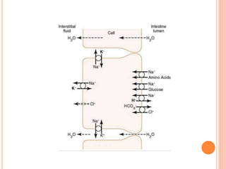
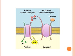
BASIC PRINCIPLES OF
GASTROINTESTINAL ABSORPTION
Prepared by FATIMA SUNDUS
Anatomical Basis of Absorption
The total quantity of fluid that must be absorbed each day by the
intestines is equal to the ingested fluid (about 1.5 liters) plus that
secreted in the various gastrointestinal secretions (about 7 liters). This
comes to a total of 8 to 9 liters..
The stomach is a poor absorptive area of the gastrointestinal
tract because it lacks the typical villus type of absorptive membrane,
and also because the junctions between the epithelial cells are tight
junctions. Only a few highly lipid-soluble substances, such as
alcohol and some drugs like aspirin, can be absorbed in small
quantities
the absorptive surface of the small intestinal mucosa, showing many
folds called valvulae conniventes (or folds of Kerckring), which
increase the surface area of the absorptive mucosa about
threefold.
These folds extend circularly most of the way around the intestine
and are especially well developed in the duodenum and jejunum,
where they often protrude up to 8 millimeters into the lumen
Also located on the epithelial surface of the small intestine
all the way down to the ileocecal valve are millions
of small villi.
These project about 1 millimeter from the surface of the mucosa
each intestinal epithelial cell on each villus is characterized by a
brush border, consisting of as many as 1000 microvilli 1
micrometer in length and 0.1 micrometer in diameter protruding
into the intestinal chyme
Thus, the combination of the folds of Kerckring, the villi, and the
microvilli increases the total absorptive area of the mucosa perhaps
1000-fold, making a tremendous total area of 250 or more square
meters for the entire small intestine—about the surface area of a
tennis court.
longitudinal section the general organization of the villus, emphasizing
(1) the advantageous arrangement of the vascular system for
absorption of fluid and dissolved material into the portal blood
(2) the arrangement of the “central lacteal” lymph vessel for
absorption into the lymph.
small pinocytic vesicles, which are pinched-off portions of infolded
enterocyte membrane forming vesicles of absorbed fluids that have been
entrapped.
Small amounts of substances are absorbed by this physical process of
pinocytosis.
Each microvillus of the brush border are multiple actin filaments
that contract rhythmically to cause continual movement of the
microvilli, keeping them constantly exposed to new quantities of
intestinal fluid
Absorption in the Small Intestine
Absorption from the small intestine each day consists of
 several hundred grams of carbohydrates,
 100 or more grams of fat,
 50 to 100 grams of amino acids,
 50 to 100 grams of ions,
 7 to 8 liters of water.
The absorptive capacity of the normal small intestine is far greater
than this: as much as
 several kilograms of carbohydrates per day,
 500 grams of fat per day,
 500 to 700 grams of proteins per day,
 20 or more liters of water per day.
Absorption of Water by Osmosis
Isosmotic Absorption.
laws of osmosis.
when the chyme is dilute enough, water is absorbed through the intestinal
mucosa into the blood of the villi almost entirely by osmosis.
when hyperosmotic solutions are discharged from the stomach into the
duodenum. Within minutes, sufficient water usually will be transferred by
osmosis to make the chyme isosmotic with the plasma.
Absorption of Ions
Sodium Is Actively Transported Through the Intestinal Membrane.
Twenty to 30 grams of sodium are secreted in the intestinal secretions each
day.
average person eats 5 to 8 grams of sodium each day.
Therefore, to prevent net loss of sodium into the feces, the intestines must
absorb 25 to 35 grams of sodium each day, which is equal to about one
seventh of all the sodium present in the body.
Whenever significant amounts of intestinal secretions are lost to the exterior,
as in extreme diarrhea, the sodium reserves of the body can sometimes be
depleted to lethal levels within hours.
Normally, however, less than 0.5 percent of the intestinal sodium is lost in
the feces each day because it is rapidly absorbed through the intestinal
mucosa.
Sodium absorption is powered by active transport of sodium
Active transport of sodium through the basolateral membranes of the
cell reduces the sodium concentration inside the cell.
Sodium is also co-transported through the brush border membrane by several
specific carrier proteins, including
(1) sodium-glucose co-transporter 1 (SGLT1)
(2) Sodium amino acid co-transporters,
(3) sodium-hydrogen exchanger.
At the same time they also provide secondary active absorption of glucose
and amino acids, powered by the active Na+-K+ ATPase pump on the
basolateral membrane.
Osmosis of the Water.
The next step in the transport process is osmosis of water by
transcellular and paracellular pathways.
Much of this osmosis occurs through the tight junctions between the
apical borders of the epithelial cells (paracellular pathway),
much also occurs through the cells themselves (transcellular pathway).
Aldosterone Greatly Enhances Sodium Absorption.
When a person becomes dehydrated, large amounts of aldosterone almost
always are secreted by the cortices of the adrenal glands.
Within 1 to 3 hours this aldosterone causes increased activation of the
enzyme and transport mechanisms for all aspects of sodium absorption
by the intestinal epithelium.
And the increased sodium absorption in turn causes secondary increases in
absorption of chloride ions, water, and some other substances.
This effect of aldosterone is especially important in the colon because it
allows virtually no loss of sodium chloride in the feces and also little
water loss.
Absorption of Chloride Ions in the Small Intestine.
In the upper part of the small intestine, chloride ion absorption is rapid and
occurs mainly by diffusion.
Then chloride ions move along this electrical gradient to “follow” the
sodium ions.
Chloride is also absorbed across the brush border membrane of parts of the
ileum and large intestine by a brush border membrane chloride
bicarbonate exchanger
chloride exits the cell on the basolateral membrane through chloride
channels.
Absorption of Bicarbonate Ions in the Duodenum and
Jejunum.
When sodium ions are absorbed, moderate amounts of hydrogen ions are
secreted into the lumen of the gut in exchange for some of the sodium.
These hydrogen ions in turn combine with the bicarbonate ions to form carbonic
acid (H2CO3), which then dissociates to form water and carbon dioxide.
The water remains as part of the chyme in the intestines, but the carbon dioxide is
readily absorbed into the blood and subsequently expired
through the lungs..
Secretion of Bicarbonate Ions and absorption of
chloride ions in the Ileum and Large Intestine
The epithelial cells on the surfaces of the villi in the ileum, as well as on all
surfaces of the large intestine, have a special capability of secreting
bicarbonate ions in exchange for absorption of chloride ions.
This is important because it provides alkaline bicarbonate ions that
neutralize acid products formed by bacteria in the large intestine.
Active Absorption of Calcium, Iron, Potassium,
Magnesium, and Phosphate.
Calcium ions are actively absorbed into the blood, especially from the
duodenum, and the amount of calcium ion absorption is exactly controlled
to supply the daily need of the body for calcium.
One important factor controlling calcium absorption is parathyroid
hormone secreted by the parathyroid glands, and another is vitamin D.
Parathyroid hormone activates vitamin D, and the activated vitamin D
in turn greatly enhances calcium absorption.
The principles of iron absorption and regulation of its absorption in
proportion to the body’s need for iron, especially for the formation of
hemoglobin
Potassium, magnesium, phosphate, and probably still other ions can also
be actively absorbed through the intestinal mucosa.
In general, the monovalent ions are absorbed with ease and in great
quantities. Conversely, bivalent ions are normally absorbed in only
small amounts; for example, maximum absorption of calcium ions is only
1/50 as great as the normal absorption of sodium ions.
Fortunately, only small quantities of the bivalent ions are normally required
daily by the body
Absorption of Nutrients
Carbohydrates Are Mainly Absorbed as Monosaccharides
Essentially all the carbohydrates in the food are absorbed in the form
of monosaccharides; only a small fraction is absorbed as disaccharides
and almost none as larger carbohydrate compounds.
By far the most abundant of the absorbed monosaccharides is
glucose, usually accounting for more than 80 percent of carbohydrate
calories absorbed.
The remaining 20 percent of absorbed monosaccharides is composed
almost entirely of galactose and fructose, the galactose derived from
milk and the fructose as one of the monosaccharides digested from
cane sugar.
Glucose Is Transported by a Sodium Co-Transport Mechanism.
In the absence of sodium transport through the intestinal membrane,
virtually no glucose can be absorbed. The reason is that glucose
absorption occurs in a co-transport mode with active transport of
sodium
There are two stages in the transport of sodium through the intestinal
membrane.
First is active transport of sodium ions through the basolateral
membranes of the intestinal epithelial cells into the blood, thereby
depleting sodium inside the epithelial cells.
Second, decrease of sodium inside the cells causes sodium from the
intestinal lumen to move through the brush border of the epithelial
cells to the cell interiors by a process of secondary active transport.
Sodium ion combines with a transport protein, SGLT1, which will not
transport sodium to the cell interior until SGLT1 also combines with
glucose.
Once inside the epithelial cell, another transport protein, glucose transporter
(GLUT2) facilitates diffusion of the glucose through the cell basolateral
membrane into the paracellular space and from there into the blood
Absorption of Other Monosaccharides.
Galactose is transported by almost exactly the same mechanism as glucose,
using the SGLT1 and SGLT2 transporters to cross the luminal and
basolateral membranes, respectively.
fructose transport does not occur by the sodium co-transport
mechanism.
Fructose is transported by facilitated diffusion all the way through the
intestinal epithelium and it is not coupled with sodium transport.
Transport of fructose from the intestinal lumen to the cell interior is facilitated
by GLUT5 and exit of fructose from the cell to the paracellular space is
facilitated by GLUT2
Much of the fructose, on entering the cell, becomes phosphorylated,
then converted to glucose, and finally transported in the form of
glucose the rest of the way into the blood.
Because fructose is not co-transported with sodium, its overall rate of
transport is only about one half that of glucose or galactose.
Absorption of Proteins as Dipeptides,
Tripeptides, or Amino Acids
most peptide or amino acid molecules bind in the cell’s
microvillus membrane with a specific transport protein that
requires sodium binding before transport can occur.
After binding, the sodium ion then moves down its electrochemical
gradient to the interior of the cell and pulls the amino acid or
peptide along with it.
This is called co-transport (or secondary active transport) of the
amino acids and peptides .
At least 10 different types of transport proteins for amino acids and
peptides have been found in the luminal membranes of intestinal epithelial
cells.
Absorption of Fats
When fats are digested to form monoglycerides and free fatty acids,
both of these digestive end products first become dissolved in the
central lipid portions of bile micelles.
Because the molecular dimensions of these micelles are only 3 to 6
nanometers in diameter, and because of their highly charged exterior,
they are soluble in chyme.
In this form, the monoglycerides and free fatty acids are carried to the
surfaces of the microvilli of the intestinal cell brush border and then
penetrate into the recesses among the moving, agitating microvilli.
Here, both the monoglycerides and fatty acids diffuse immediately out of
the micelles and into the interior of the epithelial cells, which is possible
because the lipids are also soluble in the epithelial cell membrane.
This process leaves the bile micelles still in the chyme, where they
function again and again to help absorb still more monoglycerides and
fatty acids.
Thus, the micelles perform a “ferrying” function that is highly
important for fat absorption.
In the presence of an abundance of bile micelles, about 97
percent of the fat is absorbed; in the absence of the bile micelles,
only 40 to 50 percent can be absorbed.
After entering the epithelial cell, the fatty acids and monoglycerides
are taken up by the cell’s smooth endoplasmic reticulum.
Direct Absorption of Fatty Acids into the Portal
Blood.
Small quantities of short- and medium-chain fatty acids, such as those from
butterfat, are absorbed directly into the portal blood rather than being converted
into triglycerides and absorbed by way of the lymphatics.
The cause of this difference between short- and long-chain fatty acid absorption
is that the short-chain fatty acids are more watersoluble and mostly are not
reconverted into triglycerides by the endoplasmic reticulum.
This allows direct diffusion of these short-chain fatty acids from the intestinal
epithelial cells directly into the capillary blood of the intestinal villi
Absorption in the Large Intestine: Formation of
Feces
About 1500 milliliters of chyme normally pass through the ileocecal
valve into the large intestine each day.
Most of the water and electrolytes in this chyme are absorbed in the
colon, usually leaving less than 100 milliliters of fluid to be excreted in
the feces.
all the ions are absorbed, leaving only 1 to 5 mEq each of sodium and
chloride ions to be lost in the feces.
Most of the absorption in the large intestine occurs in the proximal one
half of the colon, giving this portion the name absorbing colon,
whereas the distal colon functions principally for feces storage until a
propitious time for feces excretion and is therefore called the storage
colon.
Absorption and Secretion of Electrolytes and
Water.
The mucosa of the large intestine, like that of the small intestine, has a
high capability for active absorption of sodium, and the electrical
potential gradient created by absorption of the sodium causes chloride
absorption
The tight junctions between the epithelial cells of the large
intestinal epithelium are much tighter than those of the small intestine.
Absorption of sodium and chloride ions creates an osmotic gradient
across the large intestinal mucosa, which in turn causes absorption
of water.
Maximum Absorption Capacity of the Large
Intestine.
The large intestine can absorb a maximum of 5 to 8 liters of fluid
and electrolytes each day.
When the total quantity entering the large intestine through the
ileocecal valve or by way of large intestine secretion exceeds this
amount, the excess appears in the feces as diarrhea.
toxins from cholera or certain other bacterial infections often
cause the crypts in the terminal ileum and in the large intestine to
secrete 10 or more liters of fluid each day, leading to severe and
sometimes lethal diarrhea.
Composition of the Feces.
The feces normally are about three-fourths water and one-fourth
solid matter that is composed of about
 30 percent dead bacteria
 10 to 20 percent fat
 10 to 20 percent inorganic matter
 2 to 3 percent protein
 30 percent undigested roughage from the food and dried
constituents of digestive juices, such as bile pigment and sloughed
epithelial cells.
The brown color of feces is caused by stercobilin and urobilin,
derivatives of bilirubin.
The actual odoriferous products include
 indole
 skatole
 mercaptans
 hydrogen sulfide
The odor is caused principally by products of bacterial action; these
products vary from one person to another, depending on each
person’s colonic bacterial flora and on the type of food eaten.
Bacterial Action in the Colon.
Numerous bacteria, especially colon bacilli, are present even normally in the
absorbing colon.
They are capable of digesting small amounts of cellulose
Other substances formed as a result of bacterial activity are
 vitamin K
 vitamin B12
 thiamine
 riboflavin
 various gases that contribute to flatus in the colon, especially carbon
dioxide, hydrogen gas, and methane.
basic principle of absorption.pptx

basic principle of absorption.pptx

  • 1.
    BASIC PRINCIPLES OF GASTROINTESTINALABSORPTION Prepared by FATIMA SUNDUS
  • 2.
    Anatomical Basis ofAbsorption The total quantity of fluid that must be absorbed each day by the intestines is equal to the ingested fluid (about 1.5 liters) plus that secreted in the various gastrointestinal secretions (about 7 liters). This comes to a total of 8 to 9 liters..
  • 3.
    The stomach isa poor absorptive area of the gastrointestinal tract because it lacks the typical villus type of absorptive membrane, and also because the junctions between the epithelial cells are tight junctions. Only a few highly lipid-soluble substances, such as alcohol and some drugs like aspirin, can be absorbed in small quantities
  • 4.
    the absorptive surfaceof the small intestinal mucosa, showing many folds called valvulae conniventes (or folds of Kerckring), which increase the surface area of the absorptive mucosa about threefold. These folds extend circularly most of the way around the intestine and are especially well developed in the duodenum and jejunum, where they often protrude up to 8 millimeters into the lumen
  • 9.
    Also located onthe epithelial surface of the small intestine all the way down to the ileocecal valve are millions of small villi. These project about 1 millimeter from the surface of the mucosa each intestinal epithelial cell on each villus is characterized by a brush border, consisting of as many as 1000 microvilli 1 micrometer in length and 0.1 micrometer in diameter protruding into the intestinal chyme
  • 11.
    Thus, the combinationof the folds of Kerckring, the villi, and the microvilli increases the total absorptive area of the mucosa perhaps 1000-fold, making a tremendous total area of 250 or more square meters for the entire small intestine—about the surface area of a tennis court.
  • 12.
    longitudinal section thegeneral organization of the villus, emphasizing (1) the advantageous arrangement of the vascular system for absorption of fluid and dissolved material into the portal blood (2) the arrangement of the “central lacteal” lymph vessel for absorption into the lymph.
  • 14.
    small pinocytic vesicles,which are pinched-off portions of infolded enterocyte membrane forming vesicles of absorbed fluids that have been entrapped. Small amounts of substances are absorbed by this physical process of pinocytosis.
  • 16.
    Each microvillus ofthe brush border are multiple actin filaments that contract rhythmically to cause continual movement of the microvilli, keeping them constantly exposed to new quantities of intestinal fluid
  • 17.
    Absorption in theSmall Intestine Absorption from the small intestine each day consists of  several hundred grams of carbohydrates,  100 or more grams of fat,  50 to 100 grams of amino acids,  50 to 100 grams of ions,  7 to 8 liters of water. The absorptive capacity of the normal small intestine is far greater than this: as much as  several kilograms of carbohydrates per day,  500 grams of fat per day,  500 to 700 grams of proteins per day,  20 or more liters of water per day.
  • 18.
    Absorption of Waterby Osmosis Isosmotic Absorption. laws of osmosis. when the chyme is dilute enough, water is absorbed through the intestinal mucosa into the blood of the villi almost entirely by osmosis. when hyperosmotic solutions are discharged from the stomach into the duodenum. Within minutes, sufficient water usually will be transferred by osmosis to make the chyme isosmotic with the plasma.
  • 19.
    Absorption of Ions SodiumIs Actively Transported Through the Intestinal Membrane. Twenty to 30 grams of sodium are secreted in the intestinal secretions each day. average person eats 5 to 8 grams of sodium each day. Therefore, to prevent net loss of sodium into the feces, the intestines must absorb 25 to 35 grams of sodium each day, which is equal to about one seventh of all the sodium present in the body.
  • 20.
    Whenever significant amountsof intestinal secretions are lost to the exterior, as in extreme diarrhea, the sodium reserves of the body can sometimes be depleted to lethal levels within hours. Normally, however, less than 0.5 percent of the intestinal sodium is lost in the feces each day because it is rapidly absorbed through the intestinal mucosa. Sodium absorption is powered by active transport of sodium
  • 21.
    Active transport ofsodium through the basolateral membranes of the cell reduces the sodium concentration inside the cell. Sodium is also co-transported through the brush border membrane by several specific carrier proteins, including (1) sodium-glucose co-transporter 1 (SGLT1) (2) Sodium amino acid co-transporters, (3) sodium-hydrogen exchanger. At the same time they also provide secondary active absorption of glucose and amino acids, powered by the active Na+-K+ ATPase pump on the basolateral membrane.
  • 23.
    Osmosis of theWater. The next step in the transport process is osmosis of water by transcellular and paracellular pathways. Much of this osmosis occurs through the tight junctions between the apical borders of the epithelial cells (paracellular pathway), much also occurs through the cells themselves (transcellular pathway).
  • 25.
    Aldosterone Greatly EnhancesSodium Absorption. When a person becomes dehydrated, large amounts of aldosterone almost always are secreted by the cortices of the adrenal glands. Within 1 to 3 hours this aldosterone causes increased activation of the enzyme and transport mechanisms for all aspects of sodium absorption by the intestinal epithelium. And the increased sodium absorption in turn causes secondary increases in absorption of chloride ions, water, and some other substances. This effect of aldosterone is especially important in the colon because it allows virtually no loss of sodium chloride in the feces and also little water loss.
  • 27.
    Absorption of ChlorideIons in the Small Intestine. In the upper part of the small intestine, chloride ion absorption is rapid and occurs mainly by diffusion. Then chloride ions move along this electrical gradient to “follow” the sodium ions. Chloride is also absorbed across the brush border membrane of parts of the ileum and large intestine by a brush border membrane chloride bicarbonate exchanger chloride exits the cell on the basolateral membrane through chloride channels.
  • 29.
    Absorption of BicarbonateIons in the Duodenum and Jejunum. When sodium ions are absorbed, moderate amounts of hydrogen ions are secreted into the lumen of the gut in exchange for some of the sodium. These hydrogen ions in turn combine with the bicarbonate ions to form carbonic acid (H2CO3), which then dissociates to form water and carbon dioxide. The water remains as part of the chyme in the intestines, but the carbon dioxide is readily absorbed into the blood and subsequently expired through the lungs..
  • 30.
    Secretion of BicarbonateIons and absorption of chloride ions in the Ileum and Large Intestine The epithelial cells on the surfaces of the villi in the ileum, as well as on all surfaces of the large intestine, have a special capability of secreting bicarbonate ions in exchange for absorption of chloride ions. This is important because it provides alkaline bicarbonate ions that neutralize acid products formed by bacteria in the large intestine.
  • 31.
    Active Absorption ofCalcium, Iron, Potassium, Magnesium, and Phosphate. Calcium ions are actively absorbed into the blood, especially from the duodenum, and the amount of calcium ion absorption is exactly controlled to supply the daily need of the body for calcium. One important factor controlling calcium absorption is parathyroid hormone secreted by the parathyroid glands, and another is vitamin D. Parathyroid hormone activates vitamin D, and the activated vitamin D in turn greatly enhances calcium absorption.
  • 33.
    The principles ofiron absorption and regulation of its absorption in proportion to the body’s need for iron, especially for the formation of hemoglobin Potassium, magnesium, phosphate, and probably still other ions can also be actively absorbed through the intestinal mucosa. In general, the monovalent ions are absorbed with ease and in great quantities. Conversely, bivalent ions are normally absorbed in only small amounts; for example, maximum absorption of calcium ions is only 1/50 as great as the normal absorption of sodium ions. Fortunately, only small quantities of the bivalent ions are normally required daily by the body
  • 34.
    Absorption of Nutrients CarbohydratesAre Mainly Absorbed as Monosaccharides Essentially all the carbohydrates in the food are absorbed in the form of monosaccharides; only a small fraction is absorbed as disaccharides and almost none as larger carbohydrate compounds. By far the most abundant of the absorbed monosaccharides is glucose, usually accounting for more than 80 percent of carbohydrate calories absorbed. The remaining 20 percent of absorbed monosaccharides is composed almost entirely of galactose and fructose, the galactose derived from milk and the fructose as one of the monosaccharides digested from cane sugar.
  • 35.
    Glucose Is Transportedby a Sodium Co-Transport Mechanism. In the absence of sodium transport through the intestinal membrane, virtually no glucose can be absorbed. The reason is that glucose absorption occurs in a co-transport mode with active transport of sodium
  • 36.
    There are twostages in the transport of sodium through the intestinal membrane. First is active transport of sodium ions through the basolateral membranes of the intestinal epithelial cells into the blood, thereby depleting sodium inside the epithelial cells. Second, decrease of sodium inside the cells causes sodium from the intestinal lumen to move through the brush border of the epithelial cells to the cell interiors by a process of secondary active transport.
  • 39.
    Sodium ion combineswith a transport protein, SGLT1, which will not transport sodium to the cell interior until SGLT1 also combines with glucose. Once inside the epithelial cell, another transport protein, glucose transporter (GLUT2) facilitates diffusion of the glucose through the cell basolateral membrane into the paracellular space and from there into the blood
  • 41.
    Absorption of OtherMonosaccharides. Galactose is transported by almost exactly the same mechanism as glucose, using the SGLT1 and SGLT2 transporters to cross the luminal and basolateral membranes, respectively. fructose transport does not occur by the sodium co-transport mechanism. Fructose is transported by facilitated diffusion all the way through the intestinal epithelium and it is not coupled with sodium transport. Transport of fructose from the intestinal lumen to the cell interior is facilitated by GLUT5 and exit of fructose from the cell to the paracellular space is facilitated by GLUT2
  • 42.
    Much of thefructose, on entering the cell, becomes phosphorylated, then converted to glucose, and finally transported in the form of glucose the rest of the way into the blood. Because fructose is not co-transported with sodium, its overall rate of transport is only about one half that of glucose or galactose.
  • 43.
    Absorption of Proteinsas Dipeptides, Tripeptides, or Amino Acids most peptide or amino acid molecules bind in the cell’s microvillus membrane with a specific transport protein that requires sodium binding before transport can occur. After binding, the sodium ion then moves down its electrochemical gradient to the interior of the cell and pulls the amino acid or peptide along with it. This is called co-transport (or secondary active transport) of the amino acids and peptides .
  • 44.
    At least 10different types of transport proteins for amino acids and peptides have been found in the luminal membranes of intestinal epithelial cells.
  • 45.
    Absorption of Fats Whenfats are digested to form monoglycerides and free fatty acids, both of these digestive end products first become dissolved in the central lipid portions of bile micelles. Because the molecular dimensions of these micelles are only 3 to 6 nanometers in diameter, and because of their highly charged exterior, they are soluble in chyme. In this form, the monoglycerides and free fatty acids are carried to the surfaces of the microvilli of the intestinal cell brush border and then penetrate into the recesses among the moving, agitating microvilli. Here, both the monoglycerides and fatty acids diffuse immediately out of the micelles and into the interior of the epithelial cells, which is possible because the lipids are also soluble in the epithelial cell membrane. This process leaves the bile micelles still in the chyme, where they function again and again to help absorb still more monoglycerides and fatty acids.
  • 46.
    Thus, the micellesperform a “ferrying” function that is highly important for fat absorption. In the presence of an abundance of bile micelles, about 97 percent of the fat is absorbed; in the absence of the bile micelles, only 40 to 50 percent can be absorbed. After entering the epithelial cell, the fatty acids and monoglycerides are taken up by the cell’s smooth endoplasmic reticulum.
  • 47.
    Direct Absorption ofFatty Acids into the Portal Blood. Small quantities of short- and medium-chain fatty acids, such as those from butterfat, are absorbed directly into the portal blood rather than being converted into triglycerides and absorbed by way of the lymphatics. The cause of this difference between short- and long-chain fatty acid absorption is that the short-chain fatty acids are more watersoluble and mostly are not reconverted into triglycerides by the endoplasmic reticulum. This allows direct diffusion of these short-chain fatty acids from the intestinal epithelial cells directly into the capillary blood of the intestinal villi
  • 48.
    Absorption in theLarge Intestine: Formation of Feces About 1500 milliliters of chyme normally pass through the ileocecal valve into the large intestine each day. Most of the water and electrolytes in this chyme are absorbed in the colon, usually leaving less than 100 milliliters of fluid to be excreted in the feces. all the ions are absorbed, leaving only 1 to 5 mEq each of sodium and chloride ions to be lost in the feces. Most of the absorption in the large intestine occurs in the proximal one half of the colon, giving this portion the name absorbing colon, whereas the distal colon functions principally for feces storage until a propitious time for feces excretion and is therefore called the storage colon.
  • 50.
    Absorption and Secretionof Electrolytes and Water. The mucosa of the large intestine, like that of the small intestine, has a high capability for active absorption of sodium, and the electrical potential gradient created by absorption of the sodium causes chloride absorption The tight junctions between the epithelial cells of the large intestinal epithelium are much tighter than those of the small intestine. Absorption of sodium and chloride ions creates an osmotic gradient across the large intestinal mucosa, which in turn causes absorption of water.
  • 51.
    Maximum Absorption Capacityof the Large Intestine. The large intestine can absorb a maximum of 5 to 8 liters of fluid and electrolytes each day. When the total quantity entering the large intestine through the ileocecal valve or by way of large intestine secretion exceeds this amount, the excess appears in the feces as diarrhea. toxins from cholera or certain other bacterial infections often cause the crypts in the terminal ileum and in the large intestine to secrete 10 or more liters of fluid each day, leading to severe and sometimes lethal diarrhea.
  • 53.
    Composition of theFeces. The feces normally are about three-fourths water and one-fourth solid matter that is composed of about  30 percent dead bacteria  10 to 20 percent fat  10 to 20 percent inorganic matter  2 to 3 percent protein  30 percent undigested roughage from the food and dried constituents of digestive juices, such as bile pigment and sloughed epithelial cells. The brown color of feces is caused by stercobilin and urobilin, derivatives of bilirubin.
  • 55.
    The actual odoriferousproducts include  indole  skatole  mercaptans  hydrogen sulfide The odor is caused principally by products of bacterial action; these products vary from one person to another, depending on each person’s colonic bacterial flora and on the type of food eaten.
  • 56.
    Bacterial Action inthe Colon. Numerous bacteria, especially colon bacilli, are present even normally in the absorbing colon. They are capable of digesting small amounts of cellulose Other substances formed as a result of bacterial activity are  vitamin K  vitamin B12  thiamine  riboflavin  various gases that contribute to flatus in the colon, especially carbon dioxide, hydrogen gas, and methane.