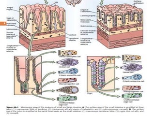
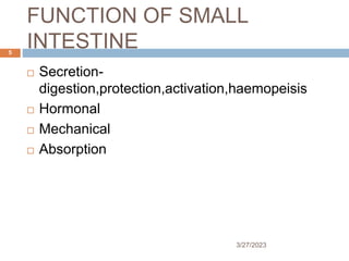
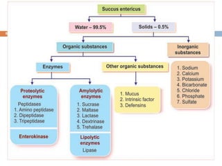
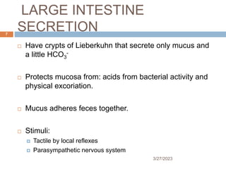
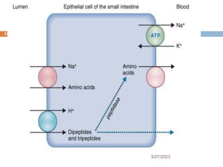
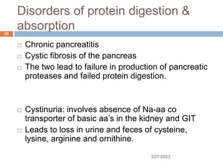
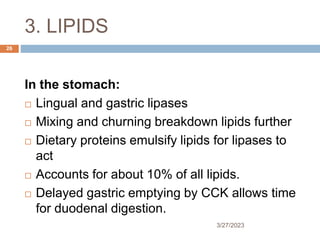
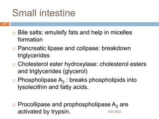
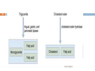
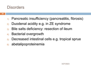
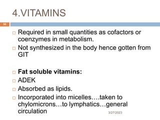
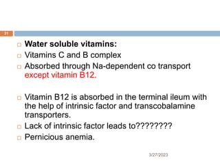
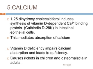
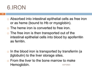
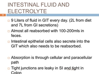
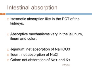
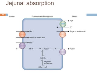
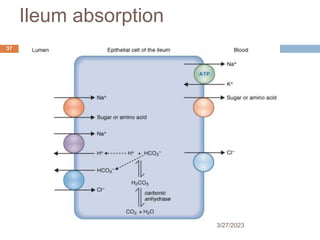
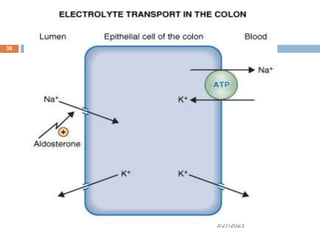
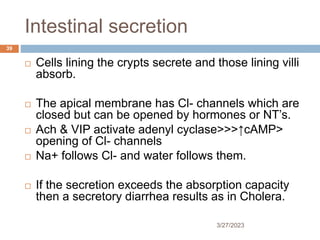
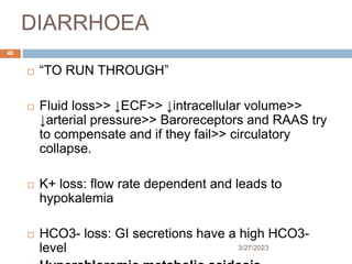
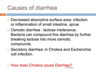
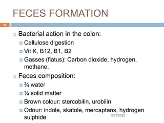
E STUDY 5-1 UNDER THE MICROSCOPE
FOCUSING IN
Members of the clinically significant group of
parasites located in blood and tissue that move
by means of flagella, known as the hemoflagel
lates, belong to the genera Leishmania and Try
panosoma. There are four morphologic forms of
clinical significance associated with these hemo
flagellates: amastigote, promastigote, epimasti
gote, and trypomastigote, all of which are defined
and described in detail in this chapter. Although
the specific life cycle may vary, all the organisms
in these two genera involve some combination of
the four morphologic forms. The transmission of
all hemoflagellates is via the bite of an arthropod
vector. The major difference between the two
genera is the primary diagnostic form found in
each; for Leishmania it is the amastigote and for
Trypanosoma it is the trypomastigote, with the
exception of Trypanosoma cruzi, in which amas
tigotes may also be found. Speciation within the
genera usually depends heavily on the patient
importance of this information, this text pro
vides a discussion of the geographic distribution
and symptomatology of each hemoflagellate.
Suspicions of hemoflagellate disease processes
are typically confirmed by more advanced diag
nostic techniques, such as serologic tests. Because
the initial diagnosis of hemoflagellate infections
relies primarily on the detection of the morpho
logic forms, this text will begin with a detailed
discussion of the morphologic forms.
MORPHOLOGY AND LIFE
CYCLE NOTES
Morphology
Amastigotes. The average roundish to oval
amastigote measures 5 by 3 μm in size (Figs. 51
and 5-2; Table 5-1). The amastigote contains a
nucleus, a basal body structure (called a blepha
roplast), and a small parabasal body. The large
single nucleus is typically located offcenter,
sometimes present more toward the edge of the
Nineyearold Chares, an African boy, recenty emigrated
to the United States from Kenya with his family. He began
complaining of chills and diarrhea 2 weeks prior to the
office vsit. After taking his temperature, whch revealed a
fever, hs mother took him into his pediatrician’s office.
During the examination, the doctor found a skn leson on
hs right arm and marked hepatosplenomegaly. A compete
bood count (CBC) was ordered, which revealed that
Charles was anemic. The doctor, afraid that the chid
was experiencing dum dum fever (kalaazar), ordered
a biopsy of the infected skin lesion and blood for
parasite study.
When the specimens were received in the laboratory,
the laboratory technician on duty made slides of the skn
lesion material and blood, stained them with Giemsa stan,
and carefully examined the slides. No parastes were found
in the bood slide The biopsy slide revealed an oval organ
ism (see diagram); it contained one nuceus, a parabasal
body, and an axonemelike structure.
Questions for Consideration
1. What parasite do you suspect? (Objective 5-10B)
2. Which morphologic form of the parasit
7. LARGE INTESTINE
SECRETION
3/27/2023
7
Have crypts of Lieberkuhn that secrete only mucus and
a little HCO3
-
Protects mucosa from: acids from bacterial activity and
physical excoriation.
Mucus adheres feces together.
Stimuli:
Tactile by local reflexes
Parasympathetic nervous system
10. DIGESTION AND
ABSORPTION
3/27/2023
10
OBJECTIVES:
To describe the site, digestive enzymes and
secretions involved in the digestion of:
CHO’s, Proteins, Lipids
To describe the absorbable units, transporters
involved and disorders in absorption of:
CHO’s, Proteins, Lipids, Fluid & Electrolytes.
11. Introduction
3/27/2023
11
Digestion and absorption is the ultimate
function of GIT.
Digestion is the breakdown of ingested food
into absorbable particles.
Absorption is movement of nutrients, water &
electrolytes from intestinal lumen into the
blood.
Occurs through cellular or paracellular paths
12. The small intestine
3/27/2023
12
Most of the absorption occurs here.
The intestinal mucosa has longitudinal folds of
Kerckring, villi & microvilli.
These increase absorptive surface area 600-1000
times.
They contain epithelial cells interspersed with
goblet cells.
Epithelial cells are shed every 3-6 days making
them vulnerable to irradiation and chemotherapy.
13. 1. CARBOHYDRATES
3/27/2023
13
Almost all ingested foods have >50% CHO’s
Contains polysaccharides (starch),
disaccharides (trehalase, maltose, sucrose &
lactose) and monosaccharide (glucose &
fructose).
Absorbed only as monosaccharide.
Digestion begins in the mouth and is as
follows:
16. Cho’s absorption
3/27/2023
16
Absorbed as monosaccharides.
Glucose and galactose: through Na-dependent 20 active
transport against the gradient on the apical membrane.
Fructose: by GLUT 5(fructose specific transporter) by
facilitated diffusion hence none against gradient.
Once in the cell all are absorbed across basolateral
membrane via GLUT 2 by facilitated diffusion.
18. Lactose intolerance
3/27/2023
18
A condition where there is absence of lactase and
one fails to breakdown lactose into glucose and
galactose.
As such lactose is not absorbed and remains in
the lumen
Being osmotic it retains water in the lumen and
leads to Diarrhea.
Treatment: avoid milk products or lactase
supplementation.
19. 2. PROTEINS
3/27/2023
19
Absorbable forms are amino acids, dipeptides and
tripeptides.
This is facilitated by the proteases in stomach and
small intestine.
In the stomach(pepsin), small intestines
(endopeptidases- trypsin, chymotrypsin, elastase
& exopeptidases- carboxypeptidase A&B)
Pancreatic secretions which are mostly proteins
are also absorbed in the same manner.
20. Digestion
3/27/2023
20
The pancreatic peptides are released in inactive
form.
Trypsinogen is activated by enterokinase and the
resulting trypsin catalyzes the activation of the
other proteases including trypsinogen.
Breakdown proteins to amino acids, dipeptides
and tripeptides.
Finally the proteases digest themselves.
23. Absorption
3/27/2023
23
Absorbable in larger molecules than CHO’s
Amino acids by the Na-aa co transporter each for
acidic, basic, neutral and imino amino acids.
Most proteins are absorbed as di/tri peptides
through the H+ dependent transporter
In the cells they are hydrolyzed by cytosolic
proteases to amino acids.
On the basolateral membrane, there is facilitated
diffusion of aa’s and remaining di/tri peptides.
25. Disorders of protein digestion &
absorption
3/27/2023
25
Chronic pancreatitis
Cystic fibrosis of the pancreas
The two lead to failure in production of pancreatic
proteases and failed protein digestion.
Cystinuria: involves absence of Na-aa co
transporter of basic aa’s in the kidney and GIT
Leads to loss in urine and feces of cysteine,
lysine, arginine and ornithine.
26. 3. LIPIDS
3/27/2023
26
In the stomach:
Lingual and gastric lipases
Mixing and churning breakdown lipids further
Dietary proteins emulsify lipids for lipases to
act
Accounts for about 10% of all lipids.
Delayed gastric emptying by CCK allows time
for duodenal digestion.
27. Small intestine
3/27/2023
27
Bile salts: emulsify fats and help in micelles
formation
Pancreatic lipase and colipase: breakdown
triglycerides
Cholesterol ester hydroxylase: cholesterol esters
and triglycerides (glycerol)
Phospholipase A2 : breaks phospholipids into
lysolecithin and fatty acids.
Procollipase and prophospholipase A2 are
activated by trypsin.
29. Disorders
3/27/2023
29
1) Pancreatic insufficiency (pancreatitis, fibrosis)
2) Duodenal acidity e.g. in ZE syndrome
3) Bile salts deficiency: resection of ileum
4) Bacterial overgrowth
5) Decreased intestinal cells e.g. tropical sprue
6) abetalipoproteinemia
30. 4.VITAMINS
3/27/2023
30
Required in small quantities as cofactors or
coenzymes in metabolism.
Not synthesized in the body hence gotten from
GIT
Fat soluble vitamins:
ADEK
Absorbed as lipids.
Incorporated into micelles….taken to
chylomicrons…to lymphatics…general
circulation
31. 3/27/2023
31
Water soluble vitamins:
Vitamins C and B complex
Absorbed through Na-dependent co transport
except vitamin B12.
Vitamin B12 is absorbed in the terminal ileum with
the help of intrinsic factor and transcobalamine
transporters.
Lack of intrinsic factor leads to????????
Pernicious anemia.
32. 5.CALCIUM
3/27/2023
32
1,25 dihydroxy cholecalciferol induces
synthesis of vitamin D-dependent Ca2+ binding
protein (Calbindin D-28K) in intestinal
epithelial cells.
This mediates absorption of calcium
Vitamin D deficiency impairs calcium
absorption and leads to deficiency.
Causes rickets in children and osteomalacia in
adults.
33. 6.IRON
3/27/2023
33
Absorbed into intestinal epithelial cells as free iron
or as heme (bound to Hb or myoglobin).
The heme iron is converted to free iron.
The free iron is then transported out of the
intestinal epithelial cells into blood by apoferritin
as ferritin.
In the blood iron is transported by transferrin (a
βglobulin) to the liver storage sites.
From the liver to the bone marrow to make
Hemoglobin.
34. INTESTINAL FLUID AND
ELECTROLYTE
3/27/2023
34
9 Liters of fluid in GIT every day. (2L from diet
and 7L from GI secretions)
Almost all reabsorbed with 100-200mls in
feces.
Intestinal epithelial cells also secrete into the
GIT which also needs to be reabsorbed.
Absorption is through cellular and paracellular
path
Tight junctions are leaky in SI and tight in
35. Intestinal absorption
3/27/2023
35
Isosmotic absorption like in the PCT of the
kidneys.
Absorptive mechanisms vary in the jejunum,
ileum and colon.
Jejunum: net absorption of NaHCO3
Ileum: net absorption of NaCl
Colon: net absorption of Na+ and K+
39. Intestinal secretion
3/27/2023
39
Cells lining the crypts secrete and those lining villi
absorb.
The apical membrane has Cl- channels which are
closed but can be opened by hormones or NT’s.
Ach & VIP activate adenyl cyclase>>>↑cAMP>
opening of Cl- channels
Na+ follows Cl- and water follows them.
If the secretion exceeds the absorption capacity
then a secretory diarrhea results as in Cholera.
40. DIARRHOEA
3/27/2023
40
“TO RUN THROUGH”
Fluid loss>> ↓ECF>> ↓intracellular volume>>
↓arterial pressure>> Baroreceptors and RAAS try
to compensate and if they fail>> circulatory
collapse.
K+ loss: flow rate dependent and leads to
hypokalemia
HCO3- loss: GI secretions have a high HCO3-
level
41. Causes of diarrhea
3/27/2023
41
Decreased absorptive surface area: infection
or inflammation of small intestine, sprue
Osmotic diarrhea : lactose intolerance.
Bacteria can compound this diarrhea by further
breaking lactose into more osmotic
compounds.
Secretory diarrhea: in Cholera and Escherichia
coli infection.
How does Cholera cause Diarrhea?
42. FECES FORMATION
3/27/2023
42
Bacterial action in the colon:
Cellulose digestion
Vit K, B12, B1, B2
Gasses (flatus): Carbon dioxide, hydrogen,
methane.
Feces composition:
¾ water
¼ solid matter
Brown colour: stercobilin, urobilin
Odour: indole, skatole, mercaptans, hydrogen
sulphide