1. Fertilization occurs when a sperm and egg fuse, forming a single diploid cell called a zygote containing genetic material from both parents.
2. The journey of sperm is difficult, with millions being overcome by acidity or blocked, and thousands destroyed before reaching the egg.
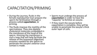
3. During its journey, sperm undergo capacitation to prepare for fertilization by improving motility and membrane changes.
4. Fertilization must occur in the uterine tube, where the egg is swept after ovulation. Hundreds of sperm help make a path for one to fuse with the egg's membrane.


![4-EMBRYOLOGICAL_DEVELOPMENT_OF_BODY_TISSUES,_ORGANS_AND_SYSTEMS.[1].pptx](https://cdn.slidesharecdn.com/ss_thumbnails/4-embryologicaldevelopmentofbodytissuesorgansandsystems-230811134542-e6d1c32e-thumbnail.jpg?width=640&height=640&fit=bounds)


