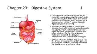
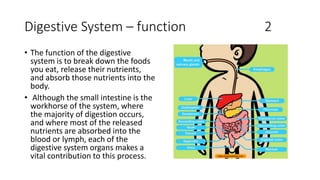
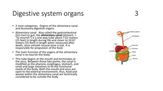
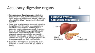
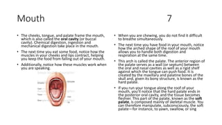
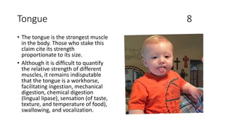
Chapter 23: Digestive System 1
• Consider what happens when you eat an
apple. Of course, you enjoy the apple’s taste
as you chew it, but in the hours that follow,
unless something goes amiss and you get a
stomachache, you don’t notice that your
digestive system is working.
• You may be taking a walk or studying or
sleeping, having forgotten all about the apple,
but your stomach and intestines are busy
digesting it and absorbing its vitamins and
other nutrients. By the time any waste
material is excreted, the body has
appropriated all it can use from the apple.
• In short, whether you pay attention or not,
the organs of the digestive system perform
their specific functions, allowing you to use
the food you eat to keep you going.
Digestive System – function 2
• The function of the digestive
system is to break down the foods
you eat, release their nutrients,
and absorb those nutrients into the
body.
• Although the small intestine is the
workhorse of the system, where
the majority of digestion occurs,
and where most of the released
nutrients are absorbed into the
blood or lymph, each of the
digestive system organs makes a
vital contribution to this process.
Digestive system organs 3
• 2 main categories: Organs of the alimentary canal
and Accessory digestive organs.
• Alimentary canal: Also called the gastrointestinal
(GI) tract or gut, the alimentary canal (aliment- =
“to nourish”) is a one-way tube about 7.62 meters
(25 feet) in length during life and closer to 10.67
meters (35 feet) in length when measured after
death, once smooth muscle tone is lost. It is
responsible for propulsion of the food.
• The main function of the organs of the alimentary
canal is to nourish the body.
• This tube begins at the mouth and terminates at
the anus. Between those two points, the canal is
modified as the pharynx, esophagus, stomach, and
small and large intestines to fit the functional
needs of the body. Both the mouth and anus are
open to the external environment; thus, food and
wastes within the alimentary canal are technically
considered to be outside the body.
Accessory digestive organs 4
• Each accessory digestive organ aids in the
breakdown of food. Within the mouth, the
teeth and tongue begin mechanical digestion,
whereas the salivary glands begin chemical
digestion.
• Once food products enter the small intestine,
the gallbladder, liver, and pancreas release
secretions—such as bile and enzymes—
essential for digestion to continue. Together,
these are called accessory organs because
they sprout from the lining cells of the
developing gut (mucosa) and augment its
function; indeed, you could not live without
their vital contributions, and many significant
diseases result from their malfunction.
• Even after development is complete, they
maintain a connection to the gut by way of
ducts.
Digestive process 5
• The processes of digestion include six
activities: ingestion, propulsion, mechanical
or physical digestion, chemical digestion,
absorption, and defecation.
• Ingestion: refers to the entry of food into the
alimentary canal through the mouth. There,
the food is chewed and mixed with saliva,
which contains enzymes that begin breaking
down the carbohydrates in the food plus
some lipid digestion via lingual lipase.
Chewing increases the surface area of the
food and allows an appropriately sized bolus
to be produced.
• Propulsion: Food leaves the mouth when the
tongue and pharyngeal muscles propel it into
the esophagus. This act of swallowing, the
last voluntary act until defecation, is an
example of propulsion, which refers to the
movement of food through the digestive
tract. It includes both the voluntary process of
swallowing and the involuntary process of
peristalsis.
• Peristalsis consists of sequential, alternating
waves of contraction and relaxation of
alimentary wall smooth muscles, which act to
propel food along. These waves also play a
role in mixing food with digestive juices.
• Peristalsis is so powerful that foods and
liquids you swallow enter your stomach even
if you are standing on your head.
Digestive process 6
• Digestion includes both mechanical and chemical
processes. Mechanical digestion is a purely
physical process that does not change the chemical
nature of the food. Instead, it makes the food
smaller to increase both surface area and mobility.
• It includes mastication, or chewing, as well as
tongue movements that help break food into
smaller bits and mix food with saliva.
• Although there may be a tendency to think that
mechanical digestion is limited to the first steps of
the digestive process, it occurs after the food
leaves the mouth, as well.
• The mechanical churning of food in the stomach
serves to further break it apart and expose more of
its surface area to digestive juices, creating an
acidic “soup” called chyme.
• In chemical digestion, starting in the mouth,
digestive secretions break down complex food
molecules into their chemical building blocks (for
example, proteins into separate amino acids).
These secretions vary in composition, but typically
contain water, various enzymes, acids, and salts.
The process is completed in the small intestine.
• Food that has been broken down is of no value to
the body unless it enters the bloodstream and its
nutrients are put to work. This occurs through the
process of absorption, which takes place primarily
within the small intestine. There, most nutrients
are absorbed from the lumen of the alimentary
canal into the bloodstream through the epithelial
cells that make up the mucosa.
• Defecation, the final step in digestion, undigested
materials are removed from the body as feces.
Mouth 7
• The cheeks, tongue, and palate frame the mouth,
which is also called the oral cavity (or buccal
cavity). Chemical digestion, ingestion and
mechanical digestion take place in the mouth.
• The next time you eat some food, notice how the
muscles in your cheeks and lips contract, helping
you keep the food from falling out of your mouth.
• Additionally, notice how these muscles work when
you are speaking.
• When you are chewing, you do not find it difficult
to breathe simultaneously.
• The next time you have food in your mouth, notice
how the arched shape of the roof of your mouth
allows you to handle both digestion and
respiration at the same time.
• This arch is called the palate. The anterior region of
the palate serves as a wall (or septum) between
the oral and nasal cavities as well as a rigid shelf
against which the tongue can push food. It is
created by the maxillary and palatine bones of the
skull and, given its bony structure, is known as the
hard palate.
• If you run your tongue along the roof of your
mouth, you’ll notice that the hard palate ends in
the posterior oral cavity, and the tissue becomes
fleshier. This part of the palate, known as the soft
palate, is composed mainly of skeletal muscle. You
can therefore manipulate, subconsciously, the soft
palate—for instance, to yawn, swallow, or sing
Tongue 8
• The tongue is the strongest muscle
in the body. Those who stake this
claim cite its strength
proportionate to its size.
• Although it is difficult to quantify
the relative strength of different
muscles, it remains indisputable
that the tongue is a workhorse,
facilitating ingestion, mechanical
digestion, chemical digestion
(lingual lipase), sensation (of taste,
texture, and temperature of food),
swallowing, and vocalization.
Pharynx 9
• The pharynx (throat) is involved in both
digestion and respiration. It receives food
and air from the mouth, and air from the
nasal cavities. When food enters the
pharynx, involuntary muscle contractions
close off the air passageways.
• The nasopharynx, is involved only in
breathing and speech. The other two
subdivisions, the oropharynx and the
laryngopharynx, are used for both
breathing and digestion.
• Usually during swallowing, the soft palate
and uvula rise reflexively to close off the
entrance to the nasopharynx. At the same
time, the larynx is pulled superiorly and
the cartilaginous epiglottis, its most
superior structure, folds inferiorly,
covering the glottis (the opening to the
larynx); this process effectively blocks
access to the trachea and bronchi.
• When the food “goes down the wrong
way,” it goes into the trachea. When food
enters the trachea, the reaction is to
cough, which usually forces the food up
and out of the trachea, and back into the
pharynx.
Esophagus 10
• The esophagus is a muscular tube that
connects the pharynx to the stomach.
• It is approximately 25.4 cm (10 in) in
length, located posterior to the trachea,
and remains in a collapsed form when not
engaged in swallowing.
• The upper esophageal sphincter, which is
continuous with the inferior pharyngeal
constrictor, controls the movement of
food from the pharynx into the
esophagus.
• Food passes from the esophagus into the
stomach at the lower esophageal
sphincter (also called the
gastroesophageal or cardiac sphincter).
Recall that sphincters are muscles that
surround tubes and serve as valves,
closing the tube when the sphincters
contract and opening it when they relax.
The lower esophageal sphincter relaxes to
let food pass into the stomach, and then
contracts to prevent stomach acids from
backing up into the esophagus.
• When the lower esophageal sphincter
does not completely close, the stomach’s
contents can reflux (that is, back up into
the esophagus), causing heartburn or
gastroesophageal reflux disease (GERD).
Deglutition 3 phases 11
• Deglutition is another word for
swallowing—the movement of
food from the mouth to the
stomach. The entire process takes
about 4 to 8 seconds for solid or
semisolid food, and about 1 second
for very soft food and liquids.
• Although this sounds quick and
effortless, deglutition is, in fact, a
complex process that involves both
the skeletal muscle of the tongue
and the muscles of the pharynx
and esophagus. It is aided by the
presence of mucus and saliva.
• There are three stages in
deglutition: the voluntary phase,
the pharyngeal phase, and the
esophageal phase.
• The voluntary phase of deglutition
(also known as the oral or buccal
phase) is so called because you can
control when you swallow food.
Deglutition: 3 phases 12
• In the pharyngeal phase, stimulation of
receptors in the oropharynx sends impulses
to the deglutition center (a collection of
neurons that controls swallowing) in the
medulla oblongata. Impulses are then sent
back to the uvula and soft palate, causing
them to move upward and close off the
nasopharynx.
• The laryngeal muscles also constrict to
prevent aspiration of food into the trachea. At
this point, deglutition apnea takes place,
which means that breathing ceases for a very
brief time.
• Contractions of the pharyngeal constrictor
muscles move the bolus through the
oropharynx and laryngopharynx. Relaxation of
the upper esophageal sphincter then allows
food to enter the esophagus.
• The entry of food into the esophagus marks
the beginning of the esophageal phase of
deglutition and the initiation of peristalsis.
Peristalsis propels the bolus through the
esophagus and toward the stomach.
• During the esophageal phase, esophageal
glands secrete mucus that lubricates the
bolus and minimizes friction.
Stomach 13
• Chemical digestion really gets underway in the
stomach. The empty stomach is only about the size
of your fist, but can stretch to hold as much as 4
liters of food and fluid, or more than 75 times its
empty volume, and then return to its resting size
when empty.
• The stomach participates in virtually all the
digestive activities with the exception of ingestion
and defecation.
• Although almost all absorption takes place in the
small intestine, the stomach does absorb some
nonpolar substances, such as alcohol and aspirin.
• Popular culture tends to refer to the stomach as
the location where all digestion takes place. Of
course, this is not true.
• An important function of the stomach is to serve as
a temporary holding chamber. You can ingest a
meal far more quickly than it can be digested and
absorbed by the small intestine.
• Thus, the stomach holds food and parses only
small amounts into the small intestine at a time.
Foods are not processed in the order they are
eaten; rather, they are mixed together with
digestive juices in the stomach until they are
converted into chyme, which is released into the
small intestine.
• In a process called gastric emptying, rhythmic
mixing waves force about 3 mL of chyme at a time
through the pyloric sphincter and into the
duodenum.
Small Intestine Large Intestine 14
• Chyme released from the stomach
enters the small intestine, which is
the primary digestive organ in the
body. Not only is this where most
digestion occurs, it is also where
practically all absorption occurs. The
longest part of the alimentary canal,
the small intestine is about 3.05
meters (10 feet) long in a living person
(but about twice as long in a cadaver
due to the loss of muscle tone).
• The large intestine is the terminal
part of the alimentary canal. The
primary function of this organ is to
finish absorption of nutrients and
water, synthesize certain vitamins,
form feces, and eliminate feces from
the body.
• The large intestine runs from the
appendix to the anus. It frames the
small intestine on three sides. Despite
its being about one-half as long as the
small intestine, it is called large
because it is more than twice the
diameter of the small intestine, about
3 inches.
Absorption, Feces Formation, and Defecation 15
• The small intestine absorbs about 90 percent of
the water you ingest (either as liquid or within
solid food).
• The large intestine absorbs most of the remaining
water, a process that converts the liquid chyme
residue into semisolid feces (“stool”). Feces is
composed of undigested food residues,
unabsorbed digested substances, millions of
bacteria, old epithelial cells from the GI mucosa,
inorganic salts, and enough water to let it pass
smoothly out of the body.
• Of every 500 mL (17 ounces) of food residue that
enters the cecum each day, about 150 mL (5
ounces) become feces.
• Valsalva’s maneuver voluntary contraction of the
diaphragm and abdominal wall muscles and closing
of the glottis, which increases intra-abdominal
pressure and facilitates defecation
• If defecation is delayed for an extended time,
additional water is absorbed, making the feces
firmer and potentially leading to constipation.
• On the other hand, if the waste matter moves too
quickly through the intestines, not enough water is
absorbed, and diarrhea can result. This can be
caused by the ingestion of foodborne pathogens.
• In general, diet, health, and stress determine the
frequency of bowel movements. The number of
bowel movements varies greatly between
individuals, ranging from two or three per day to
three or four per week.
Accessory Organs 16
• Chemical digestion in the small intestine
relies on the activities of three accessory
digestive organs: the liver, pancreas, and
gallbladder.
• The digestive role of the liver is to produce
bile and export it to the duodenum.
• The gallbladder primarily stores,
concentrates, and releases bile.
• The pancreas produces pancreatic juice,
which contains digestive enzymes and
bicarbonate ions, and delivers it to the
duodenum.
• The appendix (or vermiform appendix) is a
winding tube that attaches to the cecum.
• Although the 7.6-cm (3-in) long appendix
contains lymphoid tissue, suggesting an
immunologic function, this organ is generally
considered vestigial.
• However, at least one recent report
postulates a survival advantage conferred by
the appendix: In diarrheal illness, the
appendix may serve as a bacterial reservoir to
repopulate the enteric bacteria for those
surviving the initial phases of the illness.
• Moreover, its twisted anatomy provides a
haven for the accumulation and multiplication
of enteric bacteria.

Basic ap chapter 23 powerpoint 2017

  • 1.
    Chapter 23: DigestiveSystem 1 • Consider what happens when you eat an apple. Of course, you enjoy the apple’s taste as you chew it, but in the hours that follow, unless something goes amiss and you get a stomachache, you don’t notice that your digestive system is working. • You may be taking a walk or studying or sleeping, having forgotten all about the apple, but your stomach and intestines are busy digesting it and absorbing its vitamins and other nutrients. By the time any waste material is excreted, the body has appropriated all it can use from the apple. • In short, whether you pay attention or not, the organs of the digestive system perform their specific functions, allowing you to use the food you eat to keep you going.
  • 2.
    Digestive System –function 2 • The function of the digestive system is to break down the foods you eat, release their nutrients, and absorb those nutrients into the body. • Although the small intestine is the workhorse of the system, where the majority of digestion occurs, and where most of the released nutrients are absorbed into the blood or lymph, each of the digestive system organs makes a vital contribution to this process.
  • 3.
    Digestive system organs3 • 2 main categories: Organs of the alimentary canal and Accessory digestive organs. • Alimentary canal: Also called the gastrointestinal (GI) tract or gut, the alimentary canal (aliment- = “to nourish”) is a one-way tube about 7.62 meters (25 feet) in length during life and closer to 10.67 meters (35 feet) in length when measured after death, once smooth muscle tone is lost. It is responsible for propulsion of the food. • The main function of the organs of the alimentary canal is to nourish the body. • This tube begins at the mouth and terminates at the anus. Between those two points, the canal is modified as the pharynx, esophagus, stomach, and small and large intestines to fit the functional needs of the body. Both the mouth and anus are open to the external environment; thus, food and wastes within the alimentary canal are technically considered to be outside the body.
  • 4.
    Accessory digestive organs4 • Each accessory digestive organ aids in the breakdown of food. Within the mouth, the teeth and tongue begin mechanical digestion, whereas the salivary glands begin chemical digestion. • Once food products enter the small intestine, the gallbladder, liver, and pancreas release secretions—such as bile and enzymes— essential for digestion to continue. Together, these are called accessory organs because they sprout from the lining cells of the developing gut (mucosa) and augment its function; indeed, you could not live without their vital contributions, and many significant diseases result from their malfunction. • Even after development is complete, they maintain a connection to the gut by way of ducts.
  • 5.
    Digestive process 5 •The processes of digestion include six activities: ingestion, propulsion, mechanical or physical digestion, chemical digestion, absorption, and defecation. • Ingestion: refers to the entry of food into the alimentary canal through the mouth. There, the food is chewed and mixed with saliva, which contains enzymes that begin breaking down the carbohydrates in the food plus some lipid digestion via lingual lipase. Chewing increases the surface area of the food and allows an appropriately sized bolus to be produced. • Propulsion: Food leaves the mouth when the tongue and pharyngeal muscles propel it into the esophagus. This act of swallowing, the last voluntary act until defecation, is an example of propulsion, which refers to the movement of food through the digestive tract. It includes both the voluntary process of swallowing and the involuntary process of peristalsis. • Peristalsis consists of sequential, alternating waves of contraction and relaxation of alimentary wall smooth muscles, which act to propel food along. These waves also play a role in mixing food with digestive juices. • Peristalsis is so powerful that foods and liquids you swallow enter your stomach even if you are standing on your head.
  • 6.
    Digestive process 6 •Digestion includes both mechanical and chemical processes. Mechanical digestion is a purely physical process that does not change the chemical nature of the food. Instead, it makes the food smaller to increase both surface area and mobility. • It includes mastication, or chewing, as well as tongue movements that help break food into smaller bits and mix food with saliva. • Although there may be a tendency to think that mechanical digestion is limited to the first steps of the digestive process, it occurs after the food leaves the mouth, as well. • The mechanical churning of food in the stomach serves to further break it apart and expose more of its surface area to digestive juices, creating an acidic “soup” called chyme. • In chemical digestion, starting in the mouth, digestive secretions break down complex food molecules into their chemical building blocks (for example, proteins into separate amino acids). These secretions vary in composition, but typically contain water, various enzymes, acids, and salts. The process is completed in the small intestine. • Food that has been broken down is of no value to the body unless it enters the bloodstream and its nutrients are put to work. This occurs through the process of absorption, which takes place primarily within the small intestine. There, most nutrients are absorbed from the lumen of the alimentary canal into the bloodstream through the epithelial cells that make up the mucosa. • Defecation, the final step in digestion, undigested materials are removed from the body as feces.
  • 7.
    Mouth 7 • Thecheeks, tongue, and palate frame the mouth, which is also called the oral cavity (or buccal cavity). Chemical digestion, ingestion and mechanical digestion take place in the mouth. • The next time you eat some food, notice how the muscles in your cheeks and lips contract, helping you keep the food from falling out of your mouth. • Additionally, notice how these muscles work when you are speaking. • When you are chewing, you do not find it difficult to breathe simultaneously. • The next time you have food in your mouth, notice how the arched shape of the roof of your mouth allows you to handle both digestion and respiration at the same time. • This arch is called the palate. The anterior region of the palate serves as a wall (or septum) between the oral and nasal cavities as well as a rigid shelf against which the tongue can push food. It is created by the maxillary and palatine bones of the skull and, given its bony structure, is known as the hard palate. • If you run your tongue along the roof of your mouth, you’ll notice that the hard palate ends in the posterior oral cavity, and the tissue becomes fleshier. This part of the palate, known as the soft palate, is composed mainly of skeletal muscle. You can therefore manipulate, subconsciously, the soft palate—for instance, to yawn, swallow, or sing
  • 8.
    Tongue 8 • Thetongue is the strongest muscle in the body. Those who stake this claim cite its strength proportionate to its size. • Although it is difficult to quantify the relative strength of different muscles, it remains indisputable that the tongue is a workhorse, facilitating ingestion, mechanical digestion, chemical digestion (lingual lipase), sensation (of taste, texture, and temperature of food), swallowing, and vocalization.
  • 9.
    Pharynx 9 • Thepharynx (throat) is involved in both digestion and respiration. It receives food and air from the mouth, and air from the nasal cavities. When food enters the pharynx, involuntary muscle contractions close off the air passageways. • The nasopharynx, is involved only in breathing and speech. The other two subdivisions, the oropharynx and the laryngopharynx, are used for both breathing and digestion. • Usually during swallowing, the soft palate and uvula rise reflexively to close off the entrance to the nasopharynx. At the same time, the larynx is pulled superiorly and the cartilaginous epiglottis, its most superior structure, folds inferiorly, covering the glottis (the opening to the larynx); this process effectively blocks access to the trachea and bronchi. • When the food “goes down the wrong way,” it goes into the trachea. When food enters the trachea, the reaction is to cough, which usually forces the food up and out of the trachea, and back into the pharynx.
  • 10.
    Esophagus 10 • Theesophagus is a muscular tube that connects the pharynx to the stomach. • It is approximately 25.4 cm (10 in) in length, located posterior to the trachea, and remains in a collapsed form when not engaged in swallowing. • The upper esophageal sphincter, which is continuous with the inferior pharyngeal constrictor, controls the movement of food from the pharynx into the esophagus. • Food passes from the esophagus into the stomach at the lower esophageal sphincter (also called the gastroesophageal or cardiac sphincter). Recall that sphincters are muscles that surround tubes and serve as valves, closing the tube when the sphincters contract and opening it when they relax. The lower esophageal sphincter relaxes to let food pass into the stomach, and then contracts to prevent stomach acids from backing up into the esophagus. • When the lower esophageal sphincter does not completely close, the stomach’s contents can reflux (that is, back up into the esophagus), causing heartburn or gastroesophageal reflux disease (GERD).
  • 11.
    Deglutition 3 phases11 • Deglutition is another word for swallowing—the movement of food from the mouth to the stomach. The entire process takes about 4 to 8 seconds for solid or semisolid food, and about 1 second for very soft food and liquids. • Although this sounds quick and effortless, deglutition is, in fact, a complex process that involves both the skeletal muscle of the tongue and the muscles of the pharynx and esophagus. It is aided by the presence of mucus and saliva. • There are three stages in deglutition: the voluntary phase, the pharyngeal phase, and the esophageal phase. • The voluntary phase of deglutition (also known as the oral or buccal phase) is so called because you can control when you swallow food.
  • 12.
    Deglutition: 3 phases12 • In the pharyngeal phase, stimulation of receptors in the oropharynx sends impulses to the deglutition center (a collection of neurons that controls swallowing) in the medulla oblongata. Impulses are then sent back to the uvula and soft palate, causing them to move upward and close off the nasopharynx. • The laryngeal muscles also constrict to prevent aspiration of food into the trachea. At this point, deglutition apnea takes place, which means that breathing ceases for a very brief time. • Contractions of the pharyngeal constrictor muscles move the bolus through the oropharynx and laryngopharynx. Relaxation of the upper esophageal sphincter then allows food to enter the esophagus. • The entry of food into the esophagus marks the beginning of the esophageal phase of deglutition and the initiation of peristalsis. Peristalsis propels the bolus through the esophagus and toward the stomach. • During the esophageal phase, esophageal glands secrete mucus that lubricates the bolus and minimizes friction.
  • 13.
    Stomach 13 • Chemicaldigestion really gets underway in the stomach. The empty stomach is only about the size of your fist, but can stretch to hold as much as 4 liters of food and fluid, or more than 75 times its empty volume, and then return to its resting size when empty. • The stomach participates in virtually all the digestive activities with the exception of ingestion and defecation. • Although almost all absorption takes place in the small intestine, the stomach does absorb some nonpolar substances, such as alcohol and aspirin. • Popular culture tends to refer to the stomach as the location where all digestion takes place. Of course, this is not true. • An important function of the stomach is to serve as a temporary holding chamber. You can ingest a meal far more quickly than it can be digested and absorbed by the small intestine. • Thus, the stomach holds food and parses only small amounts into the small intestine at a time. Foods are not processed in the order they are eaten; rather, they are mixed together with digestive juices in the stomach until they are converted into chyme, which is released into the small intestine. • In a process called gastric emptying, rhythmic mixing waves force about 3 mL of chyme at a time through the pyloric sphincter and into the duodenum.
  • 14.
    Small Intestine LargeIntestine 14 • Chyme released from the stomach enters the small intestine, which is the primary digestive organ in the body. Not only is this where most digestion occurs, it is also where practically all absorption occurs. The longest part of the alimentary canal, the small intestine is about 3.05 meters (10 feet) long in a living person (but about twice as long in a cadaver due to the loss of muscle tone). • The large intestine is the terminal part of the alimentary canal. The primary function of this organ is to finish absorption of nutrients and water, synthesize certain vitamins, form feces, and eliminate feces from the body. • The large intestine runs from the appendix to the anus. It frames the small intestine on three sides. Despite its being about one-half as long as the small intestine, it is called large because it is more than twice the diameter of the small intestine, about 3 inches.
  • 15.
    Absorption, Feces Formation,and Defecation 15 • The small intestine absorbs about 90 percent of the water you ingest (either as liquid or within solid food). • The large intestine absorbs most of the remaining water, a process that converts the liquid chyme residue into semisolid feces (“stool”). Feces is composed of undigested food residues, unabsorbed digested substances, millions of bacteria, old epithelial cells from the GI mucosa, inorganic salts, and enough water to let it pass smoothly out of the body. • Of every 500 mL (17 ounces) of food residue that enters the cecum each day, about 150 mL (5 ounces) become feces. • Valsalva’s maneuver voluntary contraction of the diaphragm and abdominal wall muscles and closing of the glottis, which increases intra-abdominal pressure and facilitates defecation • If defecation is delayed for an extended time, additional water is absorbed, making the feces firmer and potentially leading to constipation. • On the other hand, if the waste matter moves too quickly through the intestines, not enough water is absorbed, and diarrhea can result. This can be caused by the ingestion of foodborne pathogens. • In general, diet, health, and stress determine the frequency of bowel movements. The number of bowel movements varies greatly between individuals, ranging from two or three per day to three or four per week.
  • 16.
    Accessory Organs 16 •Chemical digestion in the small intestine relies on the activities of three accessory digestive organs: the liver, pancreas, and gallbladder. • The digestive role of the liver is to produce bile and export it to the duodenum. • The gallbladder primarily stores, concentrates, and releases bile. • The pancreas produces pancreatic juice, which contains digestive enzymes and bicarbonate ions, and delivers it to the duodenum. • The appendix (or vermiform appendix) is a winding tube that attaches to the cecum. • Although the 7.6-cm (3-in) long appendix contains lymphoid tissue, suggesting an immunologic function, this organ is generally considered vestigial. • However, at least one recent report postulates a survival advantage conferred by the appendix: In diarrheal illness, the appendix may serve as a bacterial reservoir to repopulate the enteric bacteria for those surviving the initial phases of the illness. • Moreover, its twisted anatomy provides a haven for the accumulation and multiplication of enteric bacteria.