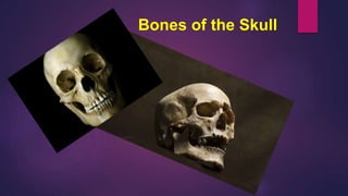
SlideShare a Scribd company logo
1 of 147
Bones of the Skull
The skull:
is the bony casing ( a box ) of
the head of humans and other
vertebrates.
The human skull consists primarily of two parts :
A- the cranium (the protective casing of the brain), and
B- the bones of the face, which include the maxilla
(upper jaw bone), mandible (lower jaw bone),
zygomatic(cheekbones), and the nasal bones. A
B
Closely associated with, but not
strictly part of, the skull are the hyoid
(a small bone at the back of the
tongue) and the auditory ossicles (three
tiny bones in each middle ear).
The 22 skull bones are made up of external and internal
tables of compact bone separated by a layer of spongy
bone called the diploe. The internal table is thinner and
more brittle than the external table. The bones are
covered on the outer surface with periosteum and inner
surfaces with endosteum. These bones are connected
togather by strong fibrous joint called sutures.
A- The cranium consists of the
following 8 bones, two of which
are paired (Figs. ):
 Frontal bone: 1
 Parietal bones: 2
 Occipital bone: 1
 Temporal bones: 2
 Sphenoid bone: 1
 Ethmoid bone: 1
B- The facial bones are 14 in number consist of
the following, two of which are single:
 Zygomatic bones: 2
 Maxillae: 2
 Nasal bones: 2
 Lacrimal bones: 2
 Vomer: 1
 Palatine bones: 2
 Inferior conchae: 2
 Mandible: 1
It is unnecessary for students of medicine to
know the detailed structure of each individual
skull bone. However, students should be
familiar with the skull as a whole and should
have a dried skull available for reference as they
read the following description.
Thank you
Lecture two
Paranasal Air-Sinuses
Paranasal air-sinuses are air-filled
spaces, communicating with the
nasal cavity, within cranial, and the
facial bones of the skull. Humans
possess a number of paranasal air-
sinuses, divided into subgroups.
The subgroups of the paranasal
air sinuses
1- the maxillary air sinuses, also called the
maxillary antra (or Antrum of Highmore). They are
the largest of the paranasal sinuses, are under the
eyes, in the maxillary bones (cheek bones).
2- the frontal air-sinus
over the eyes, in the frontal
bone, which forms the hard part
of the forehead.
Frontal air-SINUSES are absent at
birth, they are generally fairly well
developed between the seventh and
eighth years, but only reach their full
size after puberty.
The frontal air sinuses:
Are situated behind the superciliary arches, are
rarely symmetrical, and the septum between them
frequently deviates to one or other side of the
middle line.
Each opens into the anterior part of the
corresponding middle meatus of the nose through
the frontonasal duct which traverses the anterior
part of the labyrinth of the ethmoid. These
structures then open into the hiatus semilunaris in
the middle meatus.
Their average measurements are as
follows:
1- height, 3 cm.;
2- breadth, 2.5 cm.;
3- depth from before backward, 2.5
cm.
3- the ethmoid air- sinus,
which are formed from several
discrete air cells within the
ethmoid bonee between the
nose and the eyes.
4- the sphenoid air-sinus:
found within the sphenoid
bone at the center of the skull
base under the pituitary gland
Biological function of the
paranasal air-sinuses :
The biological role of the sinuses is
debated, but a number of possible
functions have been proposed:
1- Decreasing the relative weight of the
front of the skull, and especially the bones
of the face. The shape of the facial bones is
important, as a point of origin and insertion
for the muscles of facial expression.
2- Increasing resonance of the
voice.
3- Providing a buffer against blows
to the face.
4- Insulating sensitive structures like
dental roots and eyes from rapid
temperature fluctuations in the nasal
cavity.
5- Humidifying and heating of
inhaled air because of slow air
turnover in this region.
6- Regulation of intranasal
and serum gas pressures.
7-Immunological defense.
8-Regulating the temperature
of the C.S.F. ( cerebrospinal
fluid ) contacting the inner
layer of the frontal air sinus.
Thank you
Lecture Three
The scalp
The scalp is the part of the head that
extends from the superciliary arches
anteriorly to the external occipital
protuberance and superior nuchal lines
posteriorly. Laterally it continues inferiorly to
the zygomatic arch.
The scalp is a multilayered structure with
layers that can be defined by the word
itself:
S-skin;
C-connective tissue (dense);
A-aponeurotic layer;
L-loose connective tissue;
P-pericranium (Fig. ).
Examining the layers of the scalp
reveals that the first three layers are
tightly held together, forming a single
unit . This unit is sometimes referred
to as the scalp proper and is the
tissue torn away during serious
'scalping' injuries.
1- The skin :
is the outer layer of the scalp (Figs.
and ). It is similar structurally to skin
throughout the body with the
exception that hair is present on a
large amount of it.
2- Connective tissue (dense) :
Deep to the skin is dense connective tissue.
This layer anchors the skin to the third layer
and contains the arteries, veins, and nerves
supplying the scalp. When the scalp is cut,
the dense connective tissue surrounding the
vessels tends to hold cut vessels open. This
results in profuse bleeding.
3-Aponeurotic layer :
The deepest layer of the first three layers is the
aponeurotic layer. Firmly attached to the skin by
the dense connective tissue of the second layer,
this layer consists of the occipitofrontalis muscle,
which has a frontal belly anteriorly, an occipital
belly posteriorly, and an aponeurotic tendon-the
epicranial aponeurosis (galea aponeurotica)-connecting the
two (Fig. ).
The frontal belly of
occipitofrontalis begins
anteriorly where it is attached
to the skin of the eyebrows. It
passes upward, across the
forehead, to become
continuous with the
aponeurotic tendon.
The occipitofrontalis muscles move the
scalp, wrinkle the forehead, and raise the
eyebrows. The frontal belly is innervated
by temporal branches of the facial nerve
[VII] and the posterior belly by the
posterior auricular branch.
Posteriorly, each occipital belly of
occipitofrontalis arises from the
lateral part of the superior nuchal
line of the occipital bone and the
mastoid process of the temporal
bone. It also passes superiorly to
attach to the aponeurotic tendon.
A layer of loose connective tissue separates the
aponeurotic layer from the pericranium and
facilitates movement of the scalp proper over the
calvaria (Figs. And ). Because of its
consistency, infections tend to localize and
spread through the loose connective tissue.
4- Loose connective tissue
The pericranium is the deepest layer of
the scalp and is the periosteum on the
outer surface of the calvaria. It is
attached to the bones of the calvaria,
but is removable, except in the area of
the sutures.
5- Pericranium
Lecture Four
The Innervation of the scalp
Arterial Supply
Venous and Lymphatic Drainage
The Innervation of the scalp
1- Sensory innervation of the scalp is
from two major sources, 1- cranial
nerves or
2- cervical nerves,depending on
whether it is anterior or posterior to the
ears and the vertex of the head (Fig. ),
2- Motor supply
A- The frontal branch of the facial nerve
supplies the frontal bellies of the
occipitofrontalis muscle, and
B- the auricular branch of the facial nerve
supplies the occipital bellies of the muscle.
1-Supratrochlear nerve - A branch of the ophthalmic
division of the trigeminal nerve; this nerve supplies
the scalp in the medial plane at the frontal region, up
to the vertex
2-Supraorbital nerve - Also a branch of the
ophthalmic division of the trigeminal nerve; this
nerve supplies the scalp at the front, lateral to the
supratrochlear nerve distribution, up to the vertex
3-Zygomaticotemporal nerve - A branch of the
maxillary division of the trigeminal nerve; it supplies
the scalp over the temple region
4-Auriculotemporal nerve - A branch of the
mandibular division of the trigeminal nerve; it
supplies the skin over the temporal region of the
scalp
Posterior to the ears and vertex, sensory
innervation of the scalp is by cervical nerves,
specifically branches from spinal cord levels C2
and C3 (Fig. ). These branches are
1- the great auricular,
2- the lesser occipital,
3- the greater occipital, and
4- the third occipital nerves.
Arterial Supply
The scalp has a rich vascular supply. The blood
vessels traverse the connective tissue layer,
which receives vascular contribution from the
internal and external carotid arteries. The blood
vessels anastomose freely in the scalp. From
the midline anteriorly, the arteries present as
follows:
1- Supratrochlear artery
2- Supraorbital artery
3- Superficial temporal
artery
4- Posterior auricular
artery
5- Occipital artery
1 & 2 :
The supratrochlear and supraorbital arteries
are 2 branches of the ophthalmic artery,
which, in turn, is a branch of the internal
carotid artery. These arteries accompany the
corresponding nerves.
3- The superficial temporal artery is a terminal
branch of the external carotid artery that ascends
in front of the auricle. This artery, which supplies
the scalp over the temporal region, travels with the
auriculotemporal nerve and divides into anterior
and posterior branches.
4- The posterior auricular artery is a
branch of the external carotid artery
that ascends posterior to the auricle.
5- The occipital artery is a branch of the
external carotid artery; it is
accompanied by the greater occipital
nerve.
The veins of the scalp freely
anastomose with one another
and are connected to the
diploic veins of the skull
bones and the intracranial
dural sinuses through several
emissary veins. The emissary
veins are valveless. The scalp
veins, which are as follows,
accompany the arteries and
have similar names (see the
image ))
Venous and Lymphatic
Drainage
Lymphatic drainage
The part of the scalp that is
anterior to the auricles is
drained to the
1-parotid,
2-submandibular, and
3-deep cervical lymph nodes.
The posterior part of the scalp
is drained to
1- the posterior auricular
(mastoid) and
2- occipital lymph nodes.
Applied Anatomy
1-Wounds in the scalp bleed profusely, because the fibrous
fascia prevents vasoconstriction. However, wounds superficial
to the aponeurosis gap much less than do wounds that cut
through it, because aponeurosis holds the skin tight.
During a difficult birth, bleeding may occur between the
neonate's pericranium and calvaria, usually over 1 parietal bone,
2-The emissary veins do not have valves and open
in the loose areolar tissue; therefore, infection can
be transmitted from the scalp to the cranial cavity.
The layer of loose areolar tissue is known as the
dangerous area of the scalp.
Metastatic spread of malignant lesions in front of
the auricle is to the parotid and cervical groups of
lymph nodes.
3-Anastomosis exists at the medial angle of the
eye, between the facial branch of the external
carotid artery and the cutaneous branch of the
internal carotid artery. During old age, if the
internal carotid artery undergoes atherosclerotic
changes, the intracranial structures can receive
blood from the connection of the facial artery to
the dorsal nasal branch of the ophthalmic artery.
4-Because it contains numerous
sebaceous glands, the scalp is one of
the most common sites for sebaceous
cysts.
Thank you
Lecture five
& Six
are like elastic sheets that are stretched in
layers over the cranium, facial bones, the
openings they form, and the cartilage, fat, and
other tissues of the head. These are the
muscles of facial expression, acting singly and
in combination.
The facial muscles
face-to-face meeting is an important initial
contact between individuals. Part of this
exchange is the use of facial expressions to
convey emotions. In fact, a Physician can
gain important information about an
individual's general health by observing a
patient's face.
These muscles control expressions of
the face so they are sometimes
referred to as muscles of 'facial
expression'. They also act as
sphincters and dilators of the orifices
of the face (i.e. the orbits, nose, and
mouth).
1- The orbital group :
Two muscles are associated with the orbital
group-
A- the orbicularis oculi and
B- the corrugator supercilii.
1- Orbicularis oculi
-Palpebral part Medial palpebral
ligament
Lateral palpebral
raphe
Facial nerve [VII] Closes the
eyelids gently
-Orbital part Nasal part of
frontal bone;
frontal process of
maxilla; medial
palpebral
ligament
Fibers form an
uninterrupted
ellipse around
orbit
Facial nerve [VII] Closes the
eyelids forcefully
2- Corrugator
supercilii
Medial end of the
superciliary arch
Skin of the medial
half of eye-brow
Facial nerve [VII] Draws the
eyebrows
medially and
downward
1- The orbital group :
1-Nasalis
Nerve supply Action
-Transverse part Maxilla just lateral to
nose
Aponeurosis across
dorsum of nose with
muscle fibers from
the other side
Facial nerve [VII] Compresses nasal
aperture
-Alar part Maxilla over lateral
incisor
Alar cartilage of
nose
Facial nerve [VII] Draws cartilage
downward and
laterally opening
nostril
2-Procerus Nasal bone and
upper part of lateral
nasal cartilage
Skin of lower
forehead between
eyebrows
Facial nerve [VII] Draws down medial
angle of eyebrows
producing
transverse wrinkles
over bridge of nose
3- Depressor
septi
Maxilla above medial
incisor
Mobile part of the
nasal septum
Facial nerve [VII] Pulls nose inferiorly
2- The Nasal group
3- The Oral group
1-Depressor
anguli oris
Oblique line of
mandible below
canine, premolar and
first molar teeth
Skin at the corner of
mouth and blending
with orbicularis oris
Facial nerve [VII] Draws corner of
mouth down and
laterally
2-Depressor labii
inferioris
Anterior part of
oblique line of
mandible
Lower lip at midline;
blends with muscle
from opposite side
Facial nerve [VII] Draws lower lip
downward and
laterally
3-Mentalis Mandible inferior to
incisor teeth
Skin of chin Facial nerve [VII] Raises and protrudes
lower lip as it wrinkles
skin on chin
4-Risorius Fascia over masseter
muscle
Skin at the corner of
the mouth
Facial nerve [VII] Retracts corner of
mouth
1-Zygomaticus major Posterior part of lateral
surface of zygomatic
bone
Skin at the corner
of the mouth
Facial nerve [VII] Draws the corner of
the mouth upward
and laterally
2-Zygomaticus minor Anterior part of lateral
surface of zygomatic
bone
Upper lip just
medial to corner of
mouth
Facial nerve [VII] Draws the upper lip
upward
3-Levator labii
superioris
Infra-orbital margin of
maxilla
Skin of upper
lateral half of
upper lip
Facial nerve [VII] Raises upper lip;
helps form nasolabial
furrow
4-Levator labii
superioris alaeque
nasi
Frontal process of
maxilla
Alar cartilage of
nose and upper lip
Facial nerve [VII] Raises upper lip and
opens nostril
5-Levator anguli oris Maxilla below infra-
orbital foramen
Skin at the corner
of mouth
Facial nerve [VII] Raises corner of
mouth; helps form
nasolabial furrow
Other muscles or groups
Anterior auricular Anterior part of
temporal fascia
Into helix of ear Facial nerve [VII] Draws ear upward
and forward
Superior auricular Epicranial
aponeurosis on side
of head
Upper part of auricle Facial nerve [VII] Elevates ear
Posterior auricular Mastoid process of
temporal bone
Convexity of concha
of ear
Facial nerve [VII] Draws ear upward
and backward
Occipitofrontalis
-Frontal belly Skin of eyebrows Into galea
aponeurotica
Facial nerve [VII] Wrinkles forehead;
raises eyebrows
-Occipital belly Lateral part of
superior nuchal
line of occipital
bone and mastoid
process of
temporal bone
Into galea
aponeurotica
Facial nerve [VII] Draws scalp
backward
Thank you
Lecture seven
The oral cavity
Part one
The oral cavity, also known as the buccal cavity or
the mouth, is the orifice through which an individual
takes in food and water.it extends from the vermilion
(red) border of the lips to the junction of the hard and
soft palates in the roof of the mouth, and to the
circumvallate papillae on the tongue.
The oral cavity
X
The Oral Cavity is designed for:
1-articulation in speech and
2- mastication of food,
3-the oral cavity also functions as an
alternate airway passage.
The oral cavity is divided in:
1-The vestibule: is the space between
the teeth and the inner mucosal lining
of the lips and checks.
2-The oral cavity proper: is the space
contained within the upper and lower
dental arches.
The entire oral cavity is lined by :
a stratified squamous epithelium. The epithelial
lining is divided into two broad types:
1-Masticatory epithelium covers the surfaces involved in the
processing of food (tongue, gingivae and hard palate). The
epithelium is keratinized to different degrees depending on the
extent of physical forces exerted on it.
2-Lining epithelium, i.e. non-keratinised stratified squamous
epithelium, covers the remaining surfaces of the oral cavity.
A. Boundaries
1- Anterior - the lips
2- Posterior - the anterior tonsillar pillars
3-Roof - hard and soft palate Floor –
4- mucosa overlying sublingual and submandibular
glands.
5- Walls - buccal mucosa
Pharynx
A. Nasopharynx - extends from posterior choanae of the nose to the soft palate. Related
posteriorly to the base of the skull. Contains adenoid tissue and the orifices of the eustachian
tubes. This area is not accessible to direct inspection and must be examined by mirrors or
optical instruments.
B. Oropharynx - that portion which is visible via
the mouth. Extends from soft palate superiorly to
vallecula inferiorly. Posterior and lateral walls are
formed by the superior and middle pharyngeal
constrictors
D. Hypopharynx - Is the portion of the pharynx that lies inferior to the
tip of epiglottis. The posterior and lateral walls are formed by middle
and inferior pharyngeal constrictors. It extends inferiorly to the
cricopharyngeus, where the pharynx empties into the cervical
esophagus. Anteriorly, it extends from the valleculae and contains
the epiglottis and the larynx. Lateral to the larynx are the pyriform
sinuses, two mucosal pouches whose medial borders are the lateral
walls of the larynx. The posterior aspect of the hypopharynx contains
the posterior pharyngeal wall and post cricoid mucosa.
B. Contents
1- Alveolar processes and teeth
2- Anterior tongue to circumvallate papilla
3- Orifice of parotid gland (Stenson's duct) in
buccal mucosa opposite upper second
molars
4-Orifice of submandibular duct (Wharton's
duct) in anterior floor of mouth
5-Orifices of sublingual glands
The oral cavity has numerous functions:
1- One function is called oral competence,
which is the ability to hold food and saliva in
the mouth without drooling.
The specialized lining of the mouth as well as
the many saliva glands provide lubrication
which aide in speech, swallowing and in the
digestion of food.
2-The grinding and crushing of food, which
occurs in the oral cavity, is also important for
digestion. Once foods are prepared for
swallowing, the oral cavity helps in swallowing
as the tongue and the mouth push the food
backward towards the swallowing tube - the
oesophagus.
3-The grinding and crushing of food, which
occurs in the oral cavity, is also important for
digestion. Once foods are prepared for
swallowing, the oral cavity helps in swallowing
as the tongue and the mouth push the food
backward towards the swallowing tube - the
oesophagus.
4- Finally, our highly coordinated and
specialized speech, which is so important to
communication, would not be possible
without the structures of the oral cavity.
The oral cavity consists of:
1-the lips,
2-commissures,
3-all surfaces (anterior 2/3 or oral, dorsal, ventral,
border) of the tongue except the base of tongue,
4-lingual tonsils,
5-gums (alveolar ridge),
6-floor of mouth,
7-hard palate,
8-buccal mucosa, and
9-retromolar trigone.
Lecture Eight
The oral cavity
Part two
The tongue
13th,Dec. 2015
The tongue is a muscular organ in the mouth. The
tongue is covered with moist, pink tissue called
mucosa. Tiny bumps called papillae give the tongue
its rough texture. Thousands of taste buds cover the
surfaces of the papillae. Taste buds are collections
of nerve-like cells that connect to nerves running
into the brain.
The tongue
The tongue
The tongue consists of a buccal and a pharyngeal
portion separated by a V-shaped groove on its
dorsal surface, the sulcus terminalis.
At the apex of this groove is a shallow
depression, the foramen caecum,embryological
origin of the thyroid gland. Immediately in front of
the sulcus lie a row of large vallate papilliae.
The under aspect of the tongue bears the 1-
median frenulum linguae;
2-the mucosa is thin on this surface and
3-the lingual veins can thus be seen on either
side of the frenulum. The lingual nerve and the
lingual artery are medial to the vein but not
visible.
4-On either side of the base of the frenulum can
be seen the orifice of the submandibular duct on
its papilla. Inspect this in a mirror and note the
discharge of saliva when you press on your
submandibular gland just below the angle of the
jaw.
The four common tastes
are
1-sweet,
2-sour,
3- bitter, and
4-salty.
5-A fifth taste,
called umami, results from tasting
glutamate (present in MSG). The tongue
has many nerves that help detect and
transmit taste signals to the brain.
Because of this, all parts of the tongue
can detect these four common tastes;
the commonly described “taste map” of
the tongue doesn’t really exist.
Lecture Nine
The oral cavity
Part Three
The tongue II
20th,Dec. 2015
Blood supply
Blood is supplied from the lingual branch of
the external carotid artery. There is little
cross-circulation across the median raphe,
which is therefore a relatively avascular
plane
Branches of the lingual artery:
Supply blood to the tongue
A- Dorsal lingual arteries: Supply the posterior
part of the tongue
B- Deep lingual artery: Supplies the anterior part
of the tongue and communicates with the dorsal
arteries at the apex
C- Sublingual artery: Supplies the sublingual
gland and the floor of the oral cavity
Venous drainage:
Veins of the tongue are arranged in two sets,
superficial and deep. Superficial vein drains the tip
and under surface of the tongue, passes superficial
to the hyoglossus accompanying the hypoglossal
nerve and ends into internal jugular vein.
Deep vein drains the dorsum of tongue
accompanying the lingual artery, passes deep to the
hyoglossus and terminates into the internal jugular
vein either directly or after joining the superficial
vein.
Lymph drainage
The drainage zones of the mucosa of the tongue can be grouped
into three:
the tip drains to the submental nodes;
the anterior two-thirds drains to the submental and
submandibular
nodes and thence to the lower nodes of the deep cervical
chain along the carotid sheath;
the posterior one-third drains to the upper nodes of the deep
cervical chain.
There is a rich anastomosis across the midline between the
lymphatics
of the posterior one-third of the tongue so that a tumour on one
side readily metastasizes to contralateral nodes. In contrast,
there is little crosscommunication in the anterior two-thirds,
where growths more than 0.5in (12mm) from the midline do not
metastasize to the opposite side of the neck till late in the
disease.
Diagram of the lymph drainage of the tongue. Note two
points.
The anterior part of the tongue tends to drain to the nodes
farthest
Down the deep cervical chain, whereas the posterior part
drains to the upper
chain.
(ii) The anterior two-thirds of the tongue drain unilaterally,
the posterior one-third bilaterally.
Lymphatic drainage:
Lymphatics of the tongue consist of intramuscular and submucous plexuses,
and are arranged in four sets—apical, marginal, central and dorsal (Fig ).
Apical set:
It drains the tip and frenulum linguae, descends with or without decussation and terminates
as follows:
(a) Some vessels pierce the mylohyoid and drain into submental lymph nodes; a few
vessels pass downwards in front of hyoid bond and drain directly into jugulo-omohyoid
lymph nodes.
(b) Some vessels drain into submandibular nodes after piercing the mylohyoid.
(c) A few vessels pass deep to the mylohyoid and terminate into jugulo-digastric or jugulo-
omohyoid lymph nodes.
B- The Marginal set:
The vessels drain the side of the tongue in front of sulcus
terminalis and terminate as follows:
(a) Some vessels drain into submandibular nodes after
piercing the mylohyoid;
(b) Some vessels pass deep to the mylohyoid and drain into
jugulo-digastric and jugulo- omohyoid nodes.
C- The Central set:
The vessels drain the dorsal surface of anterior two-thirds of tongue in
front of vallate papillae. They descend between the two genioglossi with or
without decussations and terminate as follows:
(a) Most of the vessels drain into jugulo digastric or jugulo-omohyoid
nodes without piercing the mylohyoid.
(b) A few vessels pierce the mylohyoid and drain into submandibular
nodes.
Dorsal (or basal) Set:
It drains the posterior one-third of the tongue including
vallate papillae.
(a) Most of the vessels drain bilaterally into jugulo-
digastric nodes after piercing the pharyngeal wall.
(b) One vessel passes downward behind the tongue and
hyoid bone, pierces the thyrohyoid membrane and
drains directly into jugulo-omohyoid nodes.
Peculiarities of lymphatics:
1. Lymphatics do not accompany the blood
vessels.
2. In the middle line of the tongue a free
decussation takes place and the lymphatics pass
bilaterally.
3. Tip of the tongue presents richest lymph
drainage. A cancer affecting the tip spreads to all
cervical lymph nodes of both sides.
4. A group of lymph nodes situated at the
bifurcation of common carotid artery is known as
the principal lymph nodes of the tongue.
The lymphatics of the mouth
Lymph from the upper lip, teeth, lateral parts of the anterior
part of the tongue, and gingivae drains into the
submandibular lymph nodes.
Lymph from the lower lip and apex of the tongue drains
into the submental lymph nodes.
Lymph from the medial anterior portion of the tongue
drains into the inferior deep cervical lymph nodes, and the
posterior portion of the tongue drains into the superior
deep cervical lymph nodes.
The parotid glands drain their lymph into the superficial
and deep cervical lymph nodes. The submandibular glands
drain lymph into the deep cervical lymph nodes.
Thank You
Lecture Ten
The Parotid Gland
The parotid gland
The parotid gland:
They are Paired unilobular glands. Pyramidal in shape
divided non anatomically by the facial nerve into deep
and superficial lobes•
Accessory parotid tissue may extend along parotid duct
into buccal space•
1- is the largest of the three main salivary glands in the
head and numerous structures pass through it.
2-It is anterior to and below the lower half of the ear,
superficial, posterior, and deep to the ramus of the
mandible .
3- It extends down to the lower border of the mandible
and up to the zygomatic arch.
4-Posteriorly it covers the anterior part of the
sternocleidomastoid muscle and continues anteriorly to
halfway across the masseter muscle.
The facial nerve divides the
gland into superficial and deep
lobes and numerous structures
pass through it.
The parotid duct emerges from
the anterior border of the gland
and passes forward over the
lateral surface of the masseter
The duct is about 2 in. (5 cm)
long and passes forward across
the masseter about a
fingerbreadth below the
zygomatic arch. turns deeply into
the buccal fat pad and pierces
the buccinator muscle to enter
the vestibule of the mouth
opposite the upper second molar
tooth..
Important relationships
Several major structures enter and pass
through or pass just deep to the parotid
gland. These include :
1- the facial nerve [VII],
2- the external carotid artery and its
branches, and
3- the retromandibular vein and its
tributaries
1- the facial nerve [VII] :
The facial nerve [VII] exits the skull through the
stylomastoid foramen and passes into the deep
substance of the parotid gland, where it usually
divides into upper and lower trunks. These pass
through the substance of the parotid gland, where
there may be further branching and anastomosing
of the nerves.
Five terminal groups of branches of
the facial nerve [VII] branches-emerge
from the upper, anterior, and lower
borders of the parotid gland:
1- the temporal,
2- zygomatic,
3- buccal,
4- marginal mandibular, and
5- cervical
1
2
3
4
5
2- the external carotid artery and its
branches :
The external carotid artery enters into or
passes deep to the inferior border of the
parotid gland . As it continues in a superior
direction it gives off
1-the posterior auricular artery
2- before dividing into its two terminal
branches
A- the maxillary and
B- superficial temporal arteries emerges
from the upper border of the gland after
giving off the transverse facial artery.
3- the retromandibular vein and its
tributaries :
The retromandibular vein is formed
in the substance of the parotid
gland when the superficial temporal
and maxillary veins join together
(and passes inferiorly in the
substance of the parotid gland. It
usually divides into anterior and
posterior branches just below the
inferior border of the gland.
Arterial supply of the parotid
gland :
The parotid gland receives its
arterial supply from the numerous
arteries that pass through its
substance.
Sensory innervation of the parotid
gland is provided by the
auriculotemporal nerve, which is a
branch of the mandibular nerve [V3].
This division of the trigeminal nerve
exits the skull through the foramen
ovale.
The auriculotemporal nerve also
carries secretomotor fibers to the
parotid gland. the mandibular nerve
[V3] and are just inferior to the
foramen ovale.
Thank you
الاسراء 2015
الاسراء 2015
الاسراء 2015

More Related Content

What's hot

Ppt.infra temporal fossa
Ppt.infra temporal fossaPpt.infra temporal fossa
Ppt.infra temporal fossadocnviv
 
Blood supply of face /certified fixed orthodontic courses by Indian dental a...
Blood supply of face  /certified fixed orthodontic courses by Indian dental a...Blood supply of face  /certified fixed orthodontic courses by Indian dental a...
Blood supply of face /certified fixed orthodontic courses by Indian dental a...Indian dental academy
 
Anatomy of the Scalp
Anatomy of the ScalpAnatomy of the Scalp
Anatomy of the ScalpRafid Rashid
 
Facial nerve & TRIGEMINAL NERVES
Facial nerve & TRIGEMINAL NERVESFacial nerve & TRIGEMINAL NERVES
Facial nerve & TRIGEMINAL NERVESDr. Vishal Gohil
 
Nerve supply of head & neck by Dr. Amit T. Suryawanshi, Oral Surgeon, Pune
Nerve supply of head & neck  by Dr. Amit T. Suryawanshi,  Oral Surgeon, Pune Nerve supply of head & neck  by Dr. Amit T. Suryawanshi,  Oral Surgeon, Pune
Nerve supply of head & neck by Dr. Amit T. Suryawanshi, Oral Surgeon, Pune All Good Things
 
trigeminal nerve and pathology/rotary endodontic courses by indian dental aca...
trigeminal nerve and pathology/rotary endodontic courses by indian dental aca...trigeminal nerve and pathology/rotary endodontic courses by indian dental aca...
trigeminal nerve and pathology/rotary endodontic courses by indian dental aca...Indian dental academy
 
Trigeminal Nerve and its applied aspects
Trigeminal Nerve and its applied aspectsTrigeminal Nerve and its applied aspects
Trigeminal Nerve and its applied aspectsAMBARKHAN4
 
Nerve supply of face 1
Nerve supply of face 1Nerve supply of face 1
Nerve supply of face 1RenukaAjay
 
Trigeminal nerve and its applied anatomy
Trigeminal nerve and its applied anatomyTrigeminal nerve and its applied anatomy
Trigeminal nerve and its applied anatomyILA YADAV
 
Anatomy of Cranial Nerves 5,7,8
Anatomy of Cranial Nerves 5,7,8Anatomy of Cranial Nerves 5,7,8
Anatomy of Cranial Nerves 5,7,8RushdanZakariah
 
Trigeminal nerve / orthodontic courses by Indian dental academy
Trigeminal nerve   / orthodontic courses by Indian dental academyTrigeminal nerve   / orthodontic courses by Indian dental academy
Trigeminal nerve / orthodontic courses by Indian dental academyIndian dental academy
 

What's hot (19)

Facial artery
Facial arteryFacial artery
Facial artery
 
Ppt.infra temporal fossa
Ppt.infra temporal fossaPpt.infra temporal fossa
Ppt.infra temporal fossa
 
Blood supply of face /certified fixed orthodontic courses by Indian dental a...
Blood supply of face  /certified fixed orthodontic courses by Indian dental a...Blood supply of face  /certified fixed orthodontic courses by Indian dental a...
Blood supply of face /certified fixed orthodontic courses by Indian dental a...
 
Trigeminal nerve ppt
Trigeminal nerve  pptTrigeminal nerve  ppt
Trigeminal nerve ppt
 
Maxillary nerve
Maxillary nerveMaxillary nerve
Maxillary nerve
 
Anatomy of the Scalp
Anatomy of the ScalpAnatomy of the Scalp
Anatomy of the Scalp
 
Facial nerve & TRIGEMINAL NERVES
Facial nerve & TRIGEMINAL NERVESFacial nerve & TRIGEMINAL NERVES
Facial nerve & TRIGEMINAL NERVES
 
Nerve supply of head & neck by Dr. Amit T. Suryawanshi, Oral Surgeon, Pune
Nerve supply of head & neck  by Dr. Amit T. Suryawanshi,  Oral Surgeon, Pune Nerve supply of head & neck  by Dr. Amit T. Suryawanshi,  Oral Surgeon, Pune
Nerve supply of head & neck by Dr. Amit T. Suryawanshi, Oral Surgeon, Pune
 
trigeminal nerve and pathology/rotary endodontic courses by indian dental aca...
trigeminal nerve and pathology/rotary endodontic courses by indian dental aca...trigeminal nerve and pathology/rotary endodontic courses by indian dental aca...
trigeminal nerve and pathology/rotary endodontic courses by indian dental aca...
 
Trigeminal Nerve and its applied aspects
Trigeminal Nerve and its applied aspectsTrigeminal Nerve and its applied aspects
Trigeminal Nerve and its applied aspects
 
Anatomy of Facial Nerve
Anatomy of Facial NerveAnatomy of Facial Nerve
Anatomy of Facial Nerve
 
Trigeminal nerve
Trigeminal nerveTrigeminal nerve
Trigeminal nerve
 
Nerve supply of face 1
Nerve supply of face 1Nerve supply of face 1
Nerve supply of face 1
 
Trigeminal Nerve
Trigeminal NerveTrigeminal Nerve
Trigeminal Nerve
 
Trigeminal nerve and its applied anatomy
Trigeminal nerve and its applied anatomyTrigeminal nerve and its applied anatomy
Trigeminal nerve and its applied anatomy
 
Anatomy of Cranial Nerves 5,7,8
Anatomy of Cranial Nerves 5,7,8Anatomy of Cranial Nerves 5,7,8
Anatomy of Cranial Nerves 5,7,8
 
the scalp by dr. haydar muneer salih
the scalp by dr. haydar muneer salihthe scalp by dr. haydar muneer salih
the scalp by dr. haydar muneer salih
 
Trigeminal nerve / orthodontic courses by Indian dental academy
Trigeminal nerve   / orthodontic courses by Indian dental academyTrigeminal nerve   / orthodontic courses by Indian dental academy
Trigeminal nerve / orthodontic courses by Indian dental academy
 
Trigeminal nerve
Trigeminal nerveTrigeminal nerve
Trigeminal nerve
 

Viewers also liked

Focus group 1
Focus group 1Focus group 1
Focus group 1toyee
 
Rapport administation systèmes et supervision réseaux tp1 diabang master1 tr
Rapport administation systèmes et supervision réseaux tp1 diabang master1 trRapport administation systèmes et supervision réseaux tp1 diabang master1 tr
Rapport administation systèmes et supervision réseaux tp1 diabang master1 trCheikh Tidiane DIABANG
 
Porivnyalna tablitsya
Porivnyalna tablitsyaPorivnyalna tablitsya
Porivnyalna tablitsyaMarina Vogel
 
Informe Misionero Adultos Nº11 para el 06 de Diciembre 2014
Informe Misionero Adultos  Nº11 para el 06 de Diciembre 2014Informe Misionero Adultos  Nº11 para el 06 de Diciembre 2014
Informe Misionero Adultos Nº11 para el 06 de Diciembre 2014Escuela Sabatica
 
Inside Panel Text
Inside Panel Text Inside Panel Text
Inside Panel Text toyee
 
Rapport tp2 diabang ,pape moussa fall et maguette mbacké
Rapport tp2 diabang ,pape moussa fall et maguette mbackéRapport tp2 diabang ,pape moussa fall et maguette mbacké
Rapport tp2 diabang ,pape moussa fall et maguette mbackéCheikh Tidiane DIABANG
 

Viewers also liked (7)

Focus group 1
Focus group 1Focus group 1
Focus group 1
 
Internet
InternetInternet
Internet
 
Rapport administation systèmes et supervision réseaux tp1 diabang master1 tr
Rapport administation systèmes et supervision réseaux tp1 diabang master1 trRapport administation systèmes et supervision réseaux tp1 diabang master1 tr
Rapport administation systèmes et supervision réseaux tp1 diabang master1 tr
 
Porivnyalna tablitsya
Porivnyalna tablitsyaPorivnyalna tablitsya
Porivnyalna tablitsya
 
Informe Misionero Adultos Nº11 para el 06 de Diciembre 2014
Informe Misionero Adultos  Nº11 para el 06 de Diciembre 2014Informe Misionero Adultos  Nº11 para el 06 de Diciembre 2014
Informe Misionero Adultos Nº11 para el 06 de Diciembre 2014
 
Inside Panel Text
Inside Panel Text Inside Panel Text
Inside Panel Text
 
Rapport tp2 diabang ,pape moussa fall et maguette mbacké
Rapport tp2 diabang ,pape moussa fall et maguette mbackéRapport tp2 diabang ,pape moussa fall et maguette mbacké
Rapport tp2 diabang ,pape moussa fall et maguette mbacké
 

Similar to الاسراء 2015

The cranial bones
The cranial bonesThe cranial bones
The cranial bonesDoc Lorie B
 
anatomy - Scalp + face
anatomy - Scalp + faceanatomy - Scalp + face
anatomy - Scalp + faceMBBS IMS MSU
 
Spinal cord Gross anatomy with Clinical Anatomy.pptx
Spinal cord Gross anatomy with Clinical Anatomy.pptxSpinal cord Gross anatomy with Clinical Anatomy.pptx
Spinal cord Gross anatomy with Clinical Anatomy.pptxsiddharthroy26587
 
Osteology of head and face
Osteology of head and faceOsteology of head and face
Osteology of head and facesweetdil
 
Base of skull by dr kifayat
Base of skull by dr kifayatBase of skull by dr kifayat
Base of skull by dr kifayatKifayat Khan
 
Anatomy(2) The skull and face The temporal region Infratemporal and pterygopa...
Anatomy(2) The skull and face The temporal region Infratemporal and pterygopa...Anatomy(2) The skull and face The temporal region Infratemporal and pterygopa...
Anatomy(2) The skull and face The temporal region Infratemporal and pterygopa...Hamzeh AlBattikhi
 
fetal head development(week 12)
fetal head development(week 12)fetal head development(week 12)
fetal head development(week 12)warkaa ali
 
Abhijith head and neck bs
Abhijith head and neck bsAbhijith head and neck bs
Abhijith head and neck bsAbhijith George
 
Skull base anatomy by Dr. Aditya Tiwari
Skull base anatomy by Dr. Aditya TiwariSkull base anatomy by Dr. Aditya Tiwari
Skull base anatomy by Dr. Aditya TiwariAditya Tiwari
 
Nasal cavity and Paranasal Air Sinuses
Nasal cavity and Paranasal Air Sinuses Nasal cavity and Paranasal Air Sinuses
Nasal cavity and Paranasal Air Sinuses Prabhakar Yadav
 
9. Anatomy of the Nose and Paranasal sinuses, Dr. Maxprince.pptx
9. Anatomy of the Nose and Paranasal sinuses, Dr. Maxprince.pptx9. Anatomy of the Nose and Paranasal sinuses, Dr. Maxprince.pptx
9. Anatomy of the Nose and Paranasal sinuses, Dr. Maxprince.pptxIvanMorgan2
 
VEMP Dr.dr. HR Yusa Herwanto, MKed(ORL-HNS), Sp.THTKL(K)
VEMP Dr.dr. HR Yusa Herwanto, MKed(ORL-HNS), Sp.THTKL(K)VEMP Dr.dr. HR Yusa Herwanto, MKed(ORL-HNS), Sp.THTKL(K)
VEMP Dr.dr. HR Yusa Herwanto, MKed(ORL-HNS), Sp.THTKL(K)florensiapratiwi
 
Blood supply of head and neck
Blood supply of head and neckBlood supply of head and neck
Blood supply of head and neckDr.Vamsi Reddy
 
Meninges of brain & spinal cord
Meninges of brain & spinal cordMeninges of brain & spinal cord
Meninges of brain & spinal cordABDUL QADEER MEMON
 
Anatomy Of Nose And Paranasal Sinuses - Copy.pptx
Anatomy Of Nose And Paranasal Sinuses - Copy.pptxAnatomy Of Nose And Paranasal Sinuses - Copy.pptx
Anatomy Of Nose And Paranasal Sinuses - Copy.pptxHtet Ko
 

Similar to الاسراء 2015 (20)

The Scalp.pptx
The Scalp.pptxThe Scalp.pptx
The Scalp.pptx
 
The cranial bones
The cranial bonesThe cranial bones
The cranial bones
 
anatomy - Scalp + face
anatomy - Scalp + faceanatomy - Scalp + face
anatomy - Scalp + face
 
Scalp + face
Scalp + faceScalp + face
Scalp + face
 
Trigeminal nerve.pdf
Trigeminal nerve.pdfTrigeminal nerve.pdf
Trigeminal nerve.pdf
 
Spinal cord Gross anatomy with Clinical Anatomy.pptx
Spinal cord Gross anatomy with Clinical Anatomy.pptxSpinal cord Gross anatomy with Clinical Anatomy.pptx
Spinal cord Gross anatomy with Clinical Anatomy.pptx
 
Osteology of head and face
Osteology of head and faceOsteology of head and face
Osteology of head and face
 
Skull
SkullSkull
Skull
 
Base of skull by dr kifayat
Base of skull by dr kifayatBase of skull by dr kifayat
Base of skull by dr kifayat
 
Anatomy(2) The skull and face The temporal region Infratemporal and pterygopa...
Anatomy(2) The skull and face The temporal region Infratemporal and pterygopa...Anatomy(2) The skull and face The temporal region Infratemporal and pterygopa...
Anatomy(2) The skull and face The temporal region Infratemporal and pterygopa...
 
fetal head development(week 12)
fetal head development(week 12)fetal head development(week 12)
fetal head development(week 12)
 
Abhijith head and neck bs
Abhijith head and neck bsAbhijith head and neck bs
Abhijith head and neck bs
 
Skull base anatomy by Dr. Aditya Tiwari
Skull base anatomy by Dr. Aditya TiwariSkull base anatomy by Dr. Aditya Tiwari
Skull base anatomy by Dr. Aditya Tiwari
 
Nasal cavity and Paranasal Air Sinuses
Nasal cavity and Paranasal Air Sinuses Nasal cavity and Paranasal Air Sinuses
Nasal cavity and Paranasal Air Sinuses
 
orofacial muscles.pptx
orofacial muscles.pptxorofacial muscles.pptx
orofacial muscles.pptx
 
9. Anatomy of the Nose and Paranasal sinuses, Dr. Maxprince.pptx
9. Anatomy of the Nose and Paranasal sinuses, Dr. Maxprince.pptx9. Anatomy of the Nose and Paranasal sinuses, Dr. Maxprince.pptx
9. Anatomy of the Nose and Paranasal sinuses, Dr. Maxprince.pptx
 
VEMP Dr.dr. HR Yusa Herwanto, MKed(ORL-HNS), Sp.THTKL(K)
VEMP Dr.dr. HR Yusa Herwanto, MKed(ORL-HNS), Sp.THTKL(K)VEMP Dr.dr. HR Yusa Herwanto, MKed(ORL-HNS), Sp.THTKL(K)
VEMP Dr.dr. HR Yusa Herwanto, MKed(ORL-HNS), Sp.THTKL(K)
 
Blood supply of head and neck
Blood supply of head and neckBlood supply of head and neck
Blood supply of head and neck
 
Meninges of brain & spinal cord
Meninges of brain & spinal cordMeninges of brain & spinal cord
Meninges of brain & spinal cord
 
Anatomy Of Nose And Paranasal Sinuses - Copy.pptx
Anatomy Of Nose And Paranasal Sinuses - Copy.pptxAnatomy Of Nose And Paranasal Sinuses - Copy.pptx
Anatomy Of Nose And Paranasal Sinuses - Copy.pptx
 

More from Ahmed S.Ibrahim (20)

Oral Hygiene
Oral HygieneOral Hygiene
Oral Hygiene
 
Oral Hygiene
Oral HygieneOral Hygiene
Oral Hygiene
 
Oral Hygiene
Oral HygieneOral Hygiene
Oral Hygiene
 
Periodontal ligament
Periodontal ligamentPeriodontal ligament
Periodontal ligament
 
Presentation1
Presentation1Presentation1
Presentation1
 
Hist 12 lab
Hist 12 labHist 12 lab
Hist 12 lab
 
عرض اجنة
عرض اجنةعرض اجنة
عرض اجنة
 
Lec 11 polymer and polymerization part ii
Lec 11 polymer and polymerization part iiLec 11 polymer and polymerization part ii
Lec 11 polymer and polymerization part ii
 
Lec 10 part i polymer and polymerization
Lec 10 part i polymer and polymerizationLec 10 part i polymer and polymerization
Lec 10 part i polymer and polymerization
 
General anatomy fierst year 2016
General anatomy  fierst year 2016General anatomy  fierst year 2016
General anatomy fierst year 2016
 
Hist 9
Hist 9Hist 9
Hist 9
 
Hist 7
Hist 7Hist 7
Hist 7
 
Hist 6
Hist 6Hist 6
Hist 6
 
Hist 5
Hist 5Hist 5
Hist 5
 
Hist 4 pract
Hist 4 practHist 4 pract
Hist 4 pract
 
Hist 3
Hist 3Hist 3
Hist 3
 
Hist 2
Hist 2Hist 2
Hist 2
 
Hist 1
Hist 1Hist 1
Hist 1
 
First trimester-in-oral-histology-projections-slides-dr.-nada
First trimester-in-oral-histology-projections-slides-dr.-nadaFirst trimester-in-oral-histology-projections-slides-dr.-nada
First trimester-in-oral-histology-projections-slides-dr.-nada
 
Oral histology lab 3,4
Oral histology lab 3,4Oral histology lab 3,4
Oral histology lab 3,4
 

Recently uploaded

Call Girls Horamavu WhatsApp Number 7001035870 Meeting With Bangalore Escorts
Call Girls Horamavu WhatsApp Number 7001035870 Meeting With Bangalore EscortsCall Girls Horamavu WhatsApp Number 7001035870 Meeting With Bangalore Escorts
Call Girls Horamavu WhatsApp Number 7001035870 Meeting With Bangalore Escortsvidya singh
 
VIP Call Girls Indore Kirti 💚😋 9256729539 🚀 Indore Escorts
VIP Call Girls Indore Kirti 💚😋  9256729539 🚀 Indore EscortsVIP Call Girls Indore Kirti 💚😋  9256729539 🚀 Indore Escorts
VIP Call Girls Indore Kirti 💚😋 9256729539 🚀 Indore Escortsaditipandeya
 
Top Rated Bangalore Call Girls Richmond Circle ⟟ 8250192130 ⟟ Call Me For Gen...
Top Rated Bangalore Call Girls Richmond Circle ⟟ 8250192130 ⟟ Call Me For Gen...Top Rated Bangalore Call Girls Richmond Circle ⟟ 8250192130 ⟟ Call Me For Gen...
Top Rated Bangalore Call Girls Richmond Circle ⟟ 8250192130 ⟟ Call Me For Gen...narwatsonia7
 
Kesar Bagh Call Girl Price 9548273370 , Lucknow Call Girls Service
Kesar Bagh Call Girl Price 9548273370 , Lucknow Call Girls ServiceKesar Bagh Call Girl Price 9548273370 , Lucknow Call Girls Service
Kesar Bagh Call Girl Price 9548273370 , Lucknow Call Girls Servicemakika9823
 
Call Girls Bhubaneswar Just Call 9907093804 Top Class Call Girl Service Avail...
Call Girls Bhubaneswar Just Call 9907093804 Top Class Call Girl Service Avail...Call Girls Bhubaneswar Just Call 9907093804 Top Class Call Girl Service Avail...
Call Girls Bhubaneswar Just Call 9907093804 Top Class Call Girl Service Avail...Dipal Arora
 
Call Girl Number in Vashi Mumbai📲 9833363713 💞 Full Night Enjoy
Call Girl Number in Vashi Mumbai📲 9833363713 💞 Full Night EnjoyCall Girl Number in Vashi Mumbai📲 9833363713 💞 Full Night Enjoy
Call Girl Number in Vashi Mumbai📲 9833363713 💞 Full Night Enjoybabeytanya
 
💎VVIP Kolkata Call Girls Parganas🩱7001035870🩱Independent Girl ( Ac Rooms Avai...
💎VVIP Kolkata Call Girls Parganas🩱7001035870🩱Independent Girl ( Ac Rooms Avai...💎VVIP Kolkata Call Girls Parganas🩱7001035870🩱Independent Girl ( Ac Rooms Avai...
💎VVIP Kolkata Call Girls Parganas🩱7001035870🩱Independent Girl ( Ac Rooms Avai...Taniya Sharma
 
Low Rate Call Girls Kochi Anika 8250192130 Independent Escort Service Kochi
Low Rate Call Girls Kochi Anika 8250192130 Independent Escort Service KochiLow Rate Call Girls Kochi Anika 8250192130 Independent Escort Service Kochi
Low Rate Call Girls Kochi Anika 8250192130 Independent Escort Service KochiSuhani Kapoor
 
Call Girl Number in Panvel Mumbai📲 9833363713 💞 Full Night Enjoy
Call Girl Number in Panvel Mumbai📲 9833363713 💞 Full Night EnjoyCall Girl Number in Panvel Mumbai📲 9833363713 💞 Full Night Enjoy
Call Girl Number in Panvel Mumbai📲 9833363713 💞 Full Night Enjoybabeytanya
 
Call Girls Service Jaipur Grishma WhatsApp ❤8445551418 VIP Call Girls Jaipur
Call Girls Service Jaipur Grishma WhatsApp ❤8445551418 VIP Call Girls JaipurCall Girls Service Jaipur Grishma WhatsApp ❤8445551418 VIP Call Girls Jaipur
Call Girls Service Jaipur Grishma WhatsApp ❤8445551418 VIP Call Girls Jaipurparulsinha
 
Call Girls Colaba Mumbai ❤️ 9920874524 👈 Cash on Delivery
Call Girls Colaba Mumbai ❤️ 9920874524 👈 Cash on DeliveryCall Girls Colaba Mumbai ❤️ 9920874524 👈 Cash on Delivery
Call Girls Colaba Mumbai ❤️ 9920874524 👈 Cash on Deliverynehamumbai
 
Chandrapur Call girls 8617370543 Provides all area service COD available
Chandrapur Call girls 8617370543 Provides all area service COD availableChandrapur Call girls 8617370543 Provides all area service COD available
Chandrapur Call girls 8617370543 Provides all area service COD availableDipal Arora
 
Call Girls Kochi Just Call 9907093804 Top Class Call Girl Service Available
Call Girls Kochi Just Call 9907093804 Top Class Call Girl Service AvailableCall Girls Kochi Just Call 9907093804 Top Class Call Girl Service Available
Call Girls Kochi Just Call 9907093804 Top Class Call Girl Service AvailableDipal Arora
 
Call Girls Ludhiana Just Call 9907093804 Top Class Call Girl Service Available
Call Girls Ludhiana Just Call 9907093804 Top Class Call Girl Service AvailableCall Girls Ludhiana Just Call 9907093804 Top Class Call Girl Service Available
Call Girls Ludhiana Just Call 9907093804 Top Class Call Girl Service AvailableDipal Arora
 
Best Rate (Hyderabad) Call Girls Jahanuma ⟟ 8250192130 ⟟ High Class Call Girl...
Best Rate (Hyderabad) Call Girls Jahanuma ⟟ 8250192130 ⟟ High Class Call Girl...Best Rate (Hyderabad) Call Girls Jahanuma ⟟ 8250192130 ⟟ High Class Call Girl...
Best Rate (Hyderabad) Call Girls Jahanuma ⟟ 8250192130 ⟟ High Class Call Girl...astropune
 
VIP Russian Call Girls in Varanasi Samaira 8250192130 Independent Escort Serv...
VIP Russian Call Girls in Varanasi Samaira 8250192130 Independent Escort Serv...VIP Russian Call Girls in Varanasi Samaira 8250192130 Independent Escort Serv...
VIP Russian Call Girls in Varanasi Samaira 8250192130 Independent Escort Serv...Neha Kaur
 
Lucknow Call girls - 8800925952 - 24x7 service with hotel room
Lucknow Call girls - 8800925952 - 24x7 service with hotel roomLucknow Call girls - 8800925952 - 24x7 service with hotel room
Lucknow Call girls - 8800925952 - 24x7 service with hotel roomdiscovermytutordmt
 
(Rocky) Jaipur Call Girl - 9521753030 Escorts Service 50% Off with Cash ON De...
(Rocky) Jaipur Call Girl - 9521753030 Escorts Service 50% Off with Cash ON De...(Rocky) Jaipur Call Girl - 9521753030 Escorts Service 50% Off with Cash ON De...
(Rocky) Jaipur Call Girl - 9521753030 Escorts Service 50% Off with Cash ON De...indiancallgirl4rent
 
Premium Call Girls Cottonpet Whatsapp 7001035870 Independent Escort Service
Premium Call Girls Cottonpet Whatsapp 7001035870 Independent Escort ServicePremium Call Girls Cottonpet Whatsapp 7001035870 Independent Escort Service
Premium Call Girls Cottonpet Whatsapp 7001035870 Independent Escort Servicevidya singh
 
Call Girls Ooty Just Call 9907093804 Top Class Call Girl Service Available
Call Girls Ooty Just Call 9907093804 Top Class Call Girl Service AvailableCall Girls Ooty Just Call 9907093804 Top Class Call Girl Service Available
Call Girls Ooty Just Call 9907093804 Top Class Call Girl Service AvailableDipal Arora
 

Recently uploaded (20)

Call Girls Horamavu WhatsApp Number 7001035870 Meeting With Bangalore Escorts
Call Girls Horamavu WhatsApp Number 7001035870 Meeting With Bangalore EscortsCall Girls Horamavu WhatsApp Number 7001035870 Meeting With Bangalore Escorts
Call Girls Horamavu WhatsApp Number 7001035870 Meeting With Bangalore Escorts
 
VIP Call Girls Indore Kirti 💚😋 9256729539 🚀 Indore Escorts
VIP Call Girls Indore Kirti 💚😋  9256729539 🚀 Indore EscortsVIP Call Girls Indore Kirti 💚😋  9256729539 🚀 Indore Escorts
VIP Call Girls Indore Kirti 💚😋 9256729539 🚀 Indore Escorts
 
Top Rated Bangalore Call Girls Richmond Circle ⟟ 8250192130 ⟟ Call Me For Gen...
Top Rated Bangalore Call Girls Richmond Circle ⟟ 8250192130 ⟟ Call Me For Gen...Top Rated Bangalore Call Girls Richmond Circle ⟟ 8250192130 ⟟ Call Me For Gen...
Top Rated Bangalore Call Girls Richmond Circle ⟟ 8250192130 ⟟ Call Me For Gen...
 
Kesar Bagh Call Girl Price 9548273370 , Lucknow Call Girls Service
Kesar Bagh Call Girl Price 9548273370 , Lucknow Call Girls ServiceKesar Bagh Call Girl Price 9548273370 , Lucknow Call Girls Service
Kesar Bagh Call Girl Price 9548273370 , Lucknow Call Girls Service
 
Call Girls Bhubaneswar Just Call 9907093804 Top Class Call Girl Service Avail...
Call Girls Bhubaneswar Just Call 9907093804 Top Class Call Girl Service Avail...Call Girls Bhubaneswar Just Call 9907093804 Top Class Call Girl Service Avail...
Call Girls Bhubaneswar Just Call 9907093804 Top Class Call Girl Service Avail...
 
Call Girl Number in Vashi Mumbai📲 9833363713 💞 Full Night Enjoy
Call Girl Number in Vashi Mumbai📲 9833363713 💞 Full Night EnjoyCall Girl Number in Vashi Mumbai📲 9833363713 💞 Full Night Enjoy
Call Girl Number in Vashi Mumbai📲 9833363713 💞 Full Night Enjoy
 
💎VVIP Kolkata Call Girls Parganas🩱7001035870🩱Independent Girl ( Ac Rooms Avai...
💎VVIP Kolkata Call Girls Parganas🩱7001035870🩱Independent Girl ( Ac Rooms Avai...💎VVIP Kolkata Call Girls Parganas🩱7001035870🩱Independent Girl ( Ac Rooms Avai...
💎VVIP Kolkata Call Girls Parganas🩱7001035870🩱Independent Girl ( Ac Rooms Avai...
 
Low Rate Call Girls Kochi Anika 8250192130 Independent Escort Service Kochi
Low Rate Call Girls Kochi Anika 8250192130 Independent Escort Service KochiLow Rate Call Girls Kochi Anika 8250192130 Independent Escort Service Kochi
Low Rate Call Girls Kochi Anika 8250192130 Independent Escort Service Kochi
 
Call Girl Number in Panvel Mumbai📲 9833363713 💞 Full Night Enjoy
Call Girl Number in Panvel Mumbai📲 9833363713 💞 Full Night EnjoyCall Girl Number in Panvel Mumbai📲 9833363713 💞 Full Night Enjoy
Call Girl Number in Panvel Mumbai📲 9833363713 💞 Full Night Enjoy
 
Call Girls Service Jaipur Grishma WhatsApp ❤8445551418 VIP Call Girls Jaipur
Call Girls Service Jaipur Grishma WhatsApp ❤8445551418 VIP Call Girls JaipurCall Girls Service Jaipur Grishma WhatsApp ❤8445551418 VIP Call Girls Jaipur
Call Girls Service Jaipur Grishma WhatsApp ❤8445551418 VIP Call Girls Jaipur
 
Call Girls Colaba Mumbai ❤️ 9920874524 👈 Cash on Delivery
Call Girls Colaba Mumbai ❤️ 9920874524 👈 Cash on DeliveryCall Girls Colaba Mumbai ❤️ 9920874524 👈 Cash on Delivery
Call Girls Colaba Mumbai ❤️ 9920874524 👈 Cash on Delivery
 
Chandrapur Call girls 8617370543 Provides all area service COD available
Chandrapur Call girls 8617370543 Provides all area service COD availableChandrapur Call girls 8617370543 Provides all area service COD available
Chandrapur Call girls 8617370543 Provides all area service COD available
 
Call Girls Kochi Just Call 9907093804 Top Class Call Girl Service Available
Call Girls Kochi Just Call 9907093804 Top Class Call Girl Service AvailableCall Girls Kochi Just Call 9907093804 Top Class Call Girl Service Available
Call Girls Kochi Just Call 9907093804 Top Class Call Girl Service Available
 
Call Girls Ludhiana Just Call 9907093804 Top Class Call Girl Service Available
Call Girls Ludhiana Just Call 9907093804 Top Class Call Girl Service AvailableCall Girls Ludhiana Just Call 9907093804 Top Class Call Girl Service Available
Call Girls Ludhiana Just Call 9907093804 Top Class Call Girl Service Available
 
Best Rate (Hyderabad) Call Girls Jahanuma ⟟ 8250192130 ⟟ High Class Call Girl...
Best Rate (Hyderabad) Call Girls Jahanuma ⟟ 8250192130 ⟟ High Class Call Girl...Best Rate (Hyderabad) Call Girls Jahanuma ⟟ 8250192130 ⟟ High Class Call Girl...
Best Rate (Hyderabad) Call Girls Jahanuma ⟟ 8250192130 ⟟ High Class Call Girl...
 
VIP Russian Call Girls in Varanasi Samaira 8250192130 Independent Escort Serv...
VIP Russian Call Girls in Varanasi Samaira 8250192130 Independent Escort Serv...VIP Russian Call Girls in Varanasi Samaira 8250192130 Independent Escort Serv...
VIP Russian Call Girls in Varanasi Samaira 8250192130 Independent Escort Serv...
 
Lucknow Call girls - 8800925952 - 24x7 service with hotel room
Lucknow Call girls - 8800925952 - 24x7 service with hotel roomLucknow Call girls - 8800925952 - 24x7 service with hotel room
Lucknow Call girls - 8800925952 - 24x7 service with hotel room
 
(Rocky) Jaipur Call Girl - 9521753030 Escorts Service 50% Off with Cash ON De...
(Rocky) Jaipur Call Girl - 9521753030 Escorts Service 50% Off with Cash ON De...(Rocky) Jaipur Call Girl - 9521753030 Escorts Service 50% Off with Cash ON De...
(Rocky) Jaipur Call Girl - 9521753030 Escorts Service 50% Off with Cash ON De...
 
Premium Call Girls Cottonpet Whatsapp 7001035870 Independent Escort Service
Premium Call Girls Cottonpet Whatsapp 7001035870 Independent Escort ServicePremium Call Girls Cottonpet Whatsapp 7001035870 Independent Escort Service
Premium Call Girls Cottonpet Whatsapp 7001035870 Independent Escort Service
 
Call Girls Ooty Just Call 9907093804 Top Class Call Girl Service Available
Call Girls Ooty Just Call 9907093804 Top Class Call Girl Service AvailableCall Girls Ooty Just Call 9907093804 Top Class Call Girl Service Available
Call Girls Ooty Just Call 9907093804 Top Class Call Girl Service Available
 

الاسراء 2015

  • 1. Bones of the Skull
  • 2.
  • 3.
  • 4. The skull: is the bony casing ( a box ) of the head of humans and other vertebrates.
  • 5. The human skull consists primarily of two parts : A- the cranium (the protective casing of the brain), and B- the bones of the face, which include the maxilla (upper jaw bone), mandible (lower jaw bone), zygomatic(cheekbones), and the nasal bones. A B
  • 6. Closely associated with, but not strictly part of, the skull are the hyoid (a small bone at the back of the tongue) and the auditory ossicles (three tiny bones in each middle ear).
  • 7. The 22 skull bones are made up of external and internal tables of compact bone separated by a layer of spongy bone called the diploe. The internal table is thinner and more brittle than the external table. The bones are covered on the outer surface with periosteum and inner surfaces with endosteum. These bones are connected togather by strong fibrous joint called sutures.
  • 8. A- The cranium consists of the following 8 bones, two of which are paired (Figs. ):  Frontal bone: 1  Parietal bones: 2  Occipital bone: 1  Temporal bones: 2  Sphenoid bone: 1  Ethmoid bone: 1
  • 9. B- The facial bones are 14 in number consist of the following, two of which are single:  Zygomatic bones: 2  Maxillae: 2  Nasal bones: 2  Lacrimal bones: 2  Vomer: 1  Palatine bones: 2  Inferior conchae: 2  Mandible: 1
  • 10. It is unnecessary for students of medicine to know the detailed structure of each individual skull bone. However, students should be familiar with the skull as a whole and should have a dried skull available for reference as they read the following description.
  • 11.
  • 12.
  • 15. Paranasal Air-Sinuses Paranasal air-sinuses are air-filled spaces, communicating with the nasal cavity, within cranial, and the facial bones of the skull. Humans possess a number of paranasal air- sinuses, divided into subgroups.
  • 16.
  • 17. The subgroups of the paranasal air sinuses 1- the maxillary air sinuses, also called the maxillary antra (or Antrum of Highmore). They are the largest of the paranasal sinuses, are under the eyes, in the maxillary bones (cheek bones).
  • 18.
  • 19. 2- the frontal air-sinus over the eyes, in the frontal bone, which forms the hard part of the forehead.
  • 20. Frontal air-SINUSES are absent at birth, they are generally fairly well developed between the seventh and eighth years, but only reach their full size after puberty.
  • 21. The frontal air sinuses: Are situated behind the superciliary arches, are rarely symmetrical, and the septum between them frequently deviates to one or other side of the middle line. Each opens into the anterior part of the corresponding middle meatus of the nose through the frontonasal duct which traverses the anterior part of the labyrinth of the ethmoid. These structures then open into the hiatus semilunaris in the middle meatus.
  • 22. Their average measurements are as follows: 1- height, 3 cm.; 2- breadth, 2.5 cm.; 3- depth from before backward, 2.5 cm.
  • 23. 3- the ethmoid air- sinus, which are formed from several discrete air cells within the ethmoid bonee between the nose and the eyes.
  • 24. 4- the sphenoid air-sinus: found within the sphenoid bone at the center of the skull base under the pituitary gland
  • 25.
  • 26.
  • 27.
  • 28.
  • 29. Biological function of the paranasal air-sinuses : The biological role of the sinuses is debated, but a number of possible functions have been proposed:
  • 30. 1- Decreasing the relative weight of the front of the skull, and especially the bones of the face. The shape of the facial bones is important, as a point of origin and insertion for the muscles of facial expression.
  • 31. 2- Increasing resonance of the voice.
  • 32. 3- Providing a buffer against blows to the face.
  • 33. 4- Insulating sensitive structures like dental roots and eyes from rapid temperature fluctuations in the nasal cavity.
  • 34. 5- Humidifying and heating of inhaled air because of slow air turnover in this region.
  • 35. 6- Regulation of intranasal and serum gas pressures.
  • 37. 8-Regulating the temperature of the C.S.F. ( cerebrospinal fluid ) contacting the inner layer of the frontal air sinus.
  • 41. The scalp is the part of the head that extends from the superciliary arches anteriorly to the external occipital protuberance and superior nuchal lines posteriorly. Laterally it continues inferiorly to the zygomatic arch.
  • 42. The scalp is a multilayered structure with layers that can be defined by the word itself: S-skin; C-connective tissue (dense); A-aponeurotic layer; L-loose connective tissue; P-pericranium (Fig. ).
  • 43. Examining the layers of the scalp reveals that the first three layers are tightly held together, forming a single unit . This unit is sometimes referred to as the scalp proper and is the tissue torn away during serious 'scalping' injuries.
  • 44. 1- The skin : is the outer layer of the scalp (Figs. and ). It is similar structurally to skin throughout the body with the exception that hair is present on a large amount of it.
  • 45. 2- Connective tissue (dense) : Deep to the skin is dense connective tissue. This layer anchors the skin to the third layer and contains the arteries, veins, and nerves supplying the scalp. When the scalp is cut, the dense connective tissue surrounding the vessels tends to hold cut vessels open. This results in profuse bleeding.
  • 46. 3-Aponeurotic layer : The deepest layer of the first three layers is the aponeurotic layer. Firmly attached to the skin by the dense connective tissue of the second layer, this layer consists of the occipitofrontalis muscle, which has a frontal belly anteriorly, an occipital belly posteriorly, and an aponeurotic tendon-the epicranial aponeurosis (galea aponeurotica)-connecting the two (Fig. ).
  • 47. The frontal belly of occipitofrontalis begins anteriorly where it is attached to the skin of the eyebrows. It passes upward, across the forehead, to become continuous with the aponeurotic tendon.
  • 48. The occipitofrontalis muscles move the scalp, wrinkle the forehead, and raise the eyebrows. The frontal belly is innervated by temporal branches of the facial nerve [VII] and the posterior belly by the posterior auricular branch.
  • 49. Posteriorly, each occipital belly of occipitofrontalis arises from the lateral part of the superior nuchal line of the occipital bone and the mastoid process of the temporal bone. It also passes superiorly to attach to the aponeurotic tendon.
  • 50. A layer of loose connective tissue separates the aponeurotic layer from the pericranium and facilitates movement of the scalp proper over the calvaria (Figs. And ). Because of its consistency, infections tend to localize and spread through the loose connective tissue. 4- Loose connective tissue
  • 51. The pericranium is the deepest layer of the scalp and is the periosteum on the outer surface of the calvaria. It is attached to the bones of the calvaria, but is removable, except in the area of the sutures. 5- Pericranium
  • 52. Lecture Four The Innervation of the scalp Arterial Supply Venous and Lymphatic Drainage
  • 53. The Innervation of the scalp 1- Sensory innervation of the scalp is from two major sources, 1- cranial nerves or 2- cervical nerves,depending on whether it is anterior or posterior to the ears and the vertex of the head (Fig. ),
  • 54.
  • 55. 2- Motor supply A- The frontal branch of the facial nerve supplies the frontal bellies of the occipitofrontalis muscle, and B- the auricular branch of the facial nerve supplies the occipital bellies of the muscle.
  • 56. 1-Supratrochlear nerve - A branch of the ophthalmic division of the trigeminal nerve; this nerve supplies the scalp in the medial plane at the frontal region, up to the vertex 2-Supraorbital nerve - Also a branch of the ophthalmic division of the trigeminal nerve; this nerve supplies the scalp at the front, lateral to the supratrochlear nerve distribution, up to the vertex 3-Zygomaticotemporal nerve - A branch of the maxillary division of the trigeminal nerve; it supplies the scalp over the temple region 4-Auriculotemporal nerve - A branch of the mandibular division of the trigeminal nerve; it supplies the skin over the temporal region of the scalp
  • 57. Posterior to the ears and vertex, sensory innervation of the scalp is by cervical nerves, specifically branches from spinal cord levels C2 and C3 (Fig. ). These branches are 1- the great auricular, 2- the lesser occipital, 3- the greater occipital, and 4- the third occipital nerves.
  • 58. Arterial Supply The scalp has a rich vascular supply. The blood vessels traverse the connective tissue layer, which receives vascular contribution from the internal and external carotid arteries. The blood vessels anastomose freely in the scalp. From the midline anteriorly, the arteries present as follows:
  • 59. 1- Supratrochlear artery 2- Supraorbital artery 3- Superficial temporal artery 4- Posterior auricular artery 5- Occipital artery
  • 60. 1 & 2 : The supratrochlear and supraorbital arteries are 2 branches of the ophthalmic artery, which, in turn, is a branch of the internal carotid artery. These arteries accompany the corresponding nerves.
  • 61. 3- The superficial temporal artery is a terminal branch of the external carotid artery that ascends in front of the auricle. This artery, which supplies the scalp over the temporal region, travels with the auriculotemporal nerve and divides into anterior and posterior branches.
  • 62. 4- The posterior auricular artery is a branch of the external carotid artery that ascends posterior to the auricle.
  • 63. 5- The occipital artery is a branch of the external carotid artery; it is accompanied by the greater occipital nerve.
  • 64. The veins of the scalp freely anastomose with one another and are connected to the diploic veins of the skull bones and the intracranial dural sinuses through several emissary veins. The emissary veins are valveless. The scalp veins, which are as follows, accompany the arteries and have similar names (see the image )) Venous and Lymphatic Drainage
  • 65. Lymphatic drainage The part of the scalp that is anterior to the auricles is drained to the 1-parotid, 2-submandibular, and 3-deep cervical lymph nodes. The posterior part of the scalp is drained to 1- the posterior auricular (mastoid) and 2- occipital lymph nodes.
  • 66. Applied Anatomy 1-Wounds in the scalp bleed profusely, because the fibrous fascia prevents vasoconstriction. However, wounds superficial to the aponeurosis gap much less than do wounds that cut through it, because aponeurosis holds the skin tight. During a difficult birth, bleeding may occur between the neonate's pericranium and calvaria, usually over 1 parietal bone,
  • 67. 2-The emissary veins do not have valves and open in the loose areolar tissue; therefore, infection can be transmitted from the scalp to the cranial cavity. The layer of loose areolar tissue is known as the dangerous area of the scalp. Metastatic spread of malignant lesions in front of the auricle is to the parotid and cervical groups of lymph nodes.
  • 68. 3-Anastomosis exists at the medial angle of the eye, between the facial branch of the external carotid artery and the cutaneous branch of the internal carotid artery. During old age, if the internal carotid artery undergoes atherosclerotic changes, the intracranial structures can receive blood from the connection of the facial artery to the dorsal nasal branch of the ophthalmic artery.
  • 69. 4-Because it contains numerous sebaceous glands, the scalp is one of the most common sites for sebaceous cysts.
  • 72. are like elastic sheets that are stretched in layers over the cranium, facial bones, the openings they form, and the cartilage, fat, and other tissues of the head. These are the muscles of facial expression, acting singly and in combination. The facial muscles
  • 73. face-to-face meeting is an important initial contact between individuals. Part of this exchange is the use of facial expressions to convey emotions. In fact, a Physician can gain important information about an individual's general health by observing a patient's face.
  • 74. These muscles control expressions of the face so they are sometimes referred to as muscles of 'facial expression'. They also act as sphincters and dilators of the orifices of the face (i.e. the orbits, nose, and mouth).
  • 75. 1- The orbital group : Two muscles are associated with the orbital group- A- the orbicularis oculi and B- the corrugator supercilii.
  • 76. 1- Orbicularis oculi -Palpebral part Medial palpebral ligament Lateral palpebral raphe Facial nerve [VII] Closes the eyelids gently -Orbital part Nasal part of frontal bone; frontal process of maxilla; medial palpebral ligament Fibers form an uninterrupted ellipse around orbit Facial nerve [VII] Closes the eyelids forcefully 2- Corrugator supercilii Medial end of the superciliary arch Skin of the medial half of eye-brow Facial nerve [VII] Draws the eyebrows medially and downward 1- The orbital group :
  • 77. 1-Nasalis Nerve supply Action -Transverse part Maxilla just lateral to nose Aponeurosis across dorsum of nose with muscle fibers from the other side Facial nerve [VII] Compresses nasal aperture -Alar part Maxilla over lateral incisor Alar cartilage of nose Facial nerve [VII] Draws cartilage downward and laterally opening nostril 2-Procerus Nasal bone and upper part of lateral nasal cartilage Skin of lower forehead between eyebrows Facial nerve [VII] Draws down medial angle of eyebrows producing transverse wrinkles over bridge of nose 3- Depressor septi Maxilla above medial incisor Mobile part of the nasal septum Facial nerve [VII] Pulls nose inferiorly 2- The Nasal group
  • 78.
  • 79. 3- The Oral group
  • 80. 1-Depressor anguli oris Oblique line of mandible below canine, premolar and first molar teeth Skin at the corner of mouth and blending with orbicularis oris Facial nerve [VII] Draws corner of mouth down and laterally 2-Depressor labii inferioris Anterior part of oblique line of mandible Lower lip at midline; blends with muscle from opposite side Facial nerve [VII] Draws lower lip downward and laterally 3-Mentalis Mandible inferior to incisor teeth Skin of chin Facial nerve [VII] Raises and protrudes lower lip as it wrinkles skin on chin 4-Risorius Fascia over masseter muscle Skin at the corner of the mouth Facial nerve [VII] Retracts corner of mouth
  • 81. 1-Zygomaticus major Posterior part of lateral surface of zygomatic bone Skin at the corner of the mouth Facial nerve [VII] Draws the corner of the mouth upward and laterally 2-Zygomaticus minor Anterior part of lateral surface of zygomatic bone Upper lip just medial to corner of mouth Facial nerve [VII] Draws the upper lip upward 3-Levator labii superioris Infra-orbital margin of maxilla Skin of upper lateral half of upper lip Facial nerve [VII] Raises upper lip; helps form nasolabial furrow 4-Levator labii superioris alaeque nasi Frontal process of maxilla Alar cartilage of nose and upper lip Facial nerve [VII] Raises upper lip and opens nostril 5-Levator anguli oris Maxilla below infra- orbital foramen Skin at the corner of mouth Facial nerve [VII] Raises corner of mouth; helps form nasolabial furrow
  • 82.
  • 83.
  • 84.
  • 85.
  • 86. Other muscles or groups Anterior auricular Anterior part of temporal fascia Into helix of ear Facial nerve [VII] Draws ear upward and forward Superior auricular Epicranial aponeurosis on side of head Upper part of auricle Facial nerve [VII] Elevates ear Posterior auricular Mastoid process of temporal bone Convexity of concha of ear Facial nerve [VII] Draws ear upward and backward
  • 87. Occipitofrontalis -Frontal belly Skin of eyebrows Into galea aponeurotica Facial nerve [VII] Wrinkles forehead; raises eyebrows -Occipital belly Lateral part of superior nuchal line of occipital bone and mastoid process of temporal bone Into galea aponeurotica Facial nerve [VII] Draws scalp backward
  • 89. Lecture seven The oral cavity Part one
  • 90. The oral cavity, also known as the buccal cavity or the mouth, is the orifice through which an individual takes in food and water.it extends from the vermilion (red) border of the lips to the junction of the hard and soft palates in the roof of the mouth, and to the circumvallate papillae on the tongue. The oral cavity
  • 91. X
  • 92. The Oral Cavity is designed for: 1-articulation in speech and 2- mastication of food, 3-the oral cavity also functions as an alternate airway passage.
  • 93. The oral cavity is divided in: 1-The vestibule: is the space between the teeth and the inner mucosal lining of the lips and checks. 2-The oral cavity proper: is the space contained within the upper and lower dental arches.
  • 94. The entire oral cavity is lined by : a stratified squamous epithelium. The epithelial lining is divided into two broad types: 1-Masticatory epithelium covers the surfaces involved in the processing of food (tongue, gingivae and hard palate). The epithelium is keratinized to different degrees depending on the extent of physical forces exerted on it. 2-Lining epithelium, i.e. non-keratinised stratified squamous epithelium, covers the remaining surfaces of the oral cavity.
  • 95. A. Boundaries 1- Anterior - the lips 2- Posterior - the anterior tonsillar pillars 3-Roof - hard and soft palate Floor – 4- mucosa overlying sublingual and submandibular glands. 5- Walls - buccal mucosa
  • 96. Pharynx A. Nasopharynx - extends from posterior choanae of the nose to the soft palate. Related posteriorly to the base of the skull. Contains adenoid tissue and the orifices of the eustachian tubes. This area is not accessible to direct inspection and must be examined by mirrors or optical instruments.
  • 97. B. Oropharynx - that portion which is visible via the mouth. Extends from soft palate superiorly to vallecula inferiorly. Posterior and lateral walls are formed by the superior and middle pharyngeal constrictors
  • 98. D. Hypopharynx - Is the portion of the pharynx that lies inferior to the tip of epiglottis. The posterior and lateral walls are formed by middle and inferior pharyngeal constrictors. It extends inferiorly to the cricopharyngeus, where the pharynx empties into the cervical esophagus. Anteriorly, it extends from the valleculae and contains the epiglottis and the larynx. Lateral to the larynx are the pyriform sinuses, two mucosal pouches whose medial borders are the lateral walls of the larynx. The posterior aspect of the hypopharynx contains the posterior pharyngeal wall and post cricoid mucosa.
  • 99.
  • 100. B. Contents 1- Alveolar processes and teeth 2- Anterior tongue to circumvallate papilla 3- Orifice of parotid gland (Stenson's duct) in buccal mucosa opposite upper second molars 4-Orifice of submandibular duct (Wharton's duct) in anterior floor of mouth 5-Orifices of sublingual glands
  • 101. The oral cavity has numerous functions: 1- One function is called oral competence, which is the ability to hold food and saliva in the mouth without drooling. The specialized lining of the mouth as well as the many saliva glands provide lubrication which aide in speech, swallowing and in the digestion of food.
  • 102. 2-The grinding and crushing of food, which occurs in the oral cavity, is also important for digestion. Once foods are prepared for swallowing, the oral cavity helps in swallowing as the tongue and the mouth push the food backward towards the swallowing tube - the oesophagus.
  • 103. 3-The grinding and crushing of food, which occurs in the oral cavity, is also important for digestion. Once foods are prepared for swallowing, the oral cavity helps in swallowing as the tongue and the mouth push the food backward towards the swallowing tube - the oesophagus.
  • 104. 4- Finally, our highly coordinated and specialized speech, which is so important to communication, would not be possible without the structures of the oral cavity.
  • 105. The oral cavity consists of: 1-the lips, 2-commissures, 3-all surfaces (anterior 2/3 or oral, dorsal, ventral, border) of the tongue except the base of tongue, 4-lingual tonsils, 5-gums (alveolar ridge), 6-floor of mouth, 7-hard palate, 8-buccal mucosa, and 9-retromolar trigone.
  • 106. Lecture Eight The oral cavity Part two The tongue 13th,Dec. 2015
  • 107. The tongue is a muscular organ in the mouth. The tongue is covered with moist, pink tissue called mucosa. Tiny bumps called papillae give the tongue its rough texture. Thousands of taste buds cover the surfaces of the papillae. Taste buds are collections of nerve-like cells that connect to nerves running into the brain. The tongue
  • 108. The tongue The tongue consists of a buccal and a pharyngeal portion separated by a V-shaped groove on its dorsal surface, the sulcus terminalis. At the apex of this groove is a shallow depression, the foramen caecum,embryological origin of the thyroid gland. Immediately in front of the sulcus lie a row of large vallate papilliae.
  • 109. The under aspect of the tongue bears the 1- median frenulum linguae; 2-the mucosa is thin on this surface and 3-the lingual veins can thus be seen on either side of the frenulum. The lingual nerve and the lingual artery are medial to the vein but not visible. 4-On either side of the base of the frenulum can be seen the orifice of the submandibular duct on its papilla. Inspect this in a mirror and note the discharge of saliva when you press on your submandibular gland just below the angle of the jaw.
  • 110.
  • 111.
  • 112.
  • 113.
  • 114.
  • 115. The four common tastes are 1-sweet, 2-sour, 3- bitter, and 4-salty. 5-A fifth taste, called umami, results from tasting glutamate (present in MSG). The tongue has many nerves that help detect and transmit taste signals to the brain. Because of this, all parts of the tongue can detect these four common tastes; the commonly described “taste map” of the tongue doesn’t really exist.
  • 116.
  • 117. Lecture Nine The oral cavity Part Three The tongue II 20th,Dec. 2015
  • 118. Blood supply Blood is supplied from the lingual branch of the external carotid artery. There is little cross-circulation across the median raphe, which is therefore a relatively avascular plane
  • 119. Branches of the lingual artery: Supply blood to the tongue A- Dorsal lingual arteries: Supply the posterior part of the tongue B- Deep lingual artery: Supplies the anterior part of the tongue and communicates with the dorsal arteries at the apex C- Sublingual artery: Supplies the sublingual gland and the floor of the oral cavity
  • 120. Venous drainage: Veins of the tongue are arranged in two sets, superficial and deep. Superficial vein drains the tip and under surface of the tongue, passes superficial to the hyoglossus accompanying the hypoglossal nerve and ends into internal jugular vein. Deep vein drains the dorsum of tongue accompanying the lingual artery, passes deep to the hyoglossus and terminates into the internal jugular vein either directly or after joining the superficial vein.
  • 121. Lymph drainage The drainage zones of the mucosa of the tongue can be grouped into three: the tip drains to the submental nodes; the anterior two-thirds drains to the submental and submandibular nodes and thence to the lower nodes of the deep cervical chain along the carotid sheath; the posterior one-third drains to the upper nodes of the deep cervical chain. There is a rich anastomosis across the midline between the lymphatics of the posterior one-third of the tongue so that a tumour on one side readily metastasizes to contralateral nodes. In contrast, there is little crosscommunication in the anterior two-thirds, where growths more than 0.5in (12mm) from the midline do not metastasize to the opposite side of the neck till late in the disease.
  • 122.
  • 123. Diagram of the lymph drainage of the tongue. Note two points. The anterior part of the tongue tends to drain to the nodes farthest Down the deep cervical chain, whereas the posterior part drains to the upper chain. (ii) The anterior two-thirds of the tongue drain unilaterally, the posterior one-third bilaterally.
  • 124.
  • 125.
  • 126. Lymphatic drainage: Lymphatics of the tongue consist of intramuscular and submucous plexuses, and are arranged in four sets—apical, marginal, central and dorsal (Fig ). Apical set: It drains the tip and frenulum linguae, descends with or without decussation and terminates as follows: (a) Some vessels pierce the mylohyoid and drain into submental lymph nodes; a few vessels pass downwards in front of hyoid bond and drain directly into jugulo-omohyoid lymph nodes. (b) Some vessels drain into submandibular nodes after piercing the mylohyoid. (c) A few vessels pass deep to the mylohyoid and terminate into jugulo-digastric or jugulo- omohyoid lymph nodes.
  • 127. B- The Marginal set: The vessels drain the side of the tongue in front of sulcus terminalis and terminate as follows: (a) Some vessels drain into submandibular nodes after piercing the mylohyoid; (b) Some vessels pass deep to the mylohyoid and drain into jugulo-digastric and jugulo- omohyoid nodes.
  • 128. C- The Central set: The vessels drain the dorsal surface of anterior two-thirds of tongue in front of vallate papillae. They descend between the two genioglossi with or without decussations and terminate as follows: (a) Most of the vessels drain into jugulo digastric or jugulo-omohyoid nodes without piercing the mylohyoid. (b) A few vessels pierce the mylohyoid and drain into submandibular nodes.
  • 129. Dorsal (or basal) Set: It drains the posterior one-third of the tongue including vallate papillae. (a) Most of the vessels drain bilaterally into jugulo- digastric nodes after piercing the pharyngeal wall. (b) One vessel passes downward behind the tongue and hyoid bone, pierces the thyrohyoid membrane and drains directly into jugulo-omohyoid nodes.
  • 130. Peculiarities of lymphatics: 1. Lymphatics do not accompany the blood vessels. 2. In the middle line of the tongue a free decussation takes place and the lymphatics pass bilaterally. 3. Tip of the tongue presents richest lymph drainage. A cancer affecting the tip spreads to all cervical lymph nodes of both sides. 4. A group of lymph nodes situated at the bifurcation of common carotid artery is known as the principal lymph nodes of the tongue.
  • 131. The lymphatics of the mouth Lymph from the upper lip, teeth, lateral parts of the anterior part of the tongue, and gingivae drains into the submandibular lymph nodes. Lymph from the lower lip and apex of the tongue drains into the submental lymph nodes. Lymph from the medial anterior portion of the tongue drains into the inferior deep cervical lymph nodes, and the posterior portion of the tongue drains into the superior deep cervical lymph nodes. The parotid glands drain their lymph into the superficial and deep cervical lymph nodes. The submandibular glands drain lymph into the deep cervical lymph nodes.
  • 134. The parotid gland The parotid gland: They are Paired unilobular glands. Pyramidal in shape divided non anatomically by the facial nerve into deep and superficial lobes• Accessory parotid tissue may extend along parotid duct into buccal space• 1- is the largest of the three main salivary glands in the head and numerous structures pass through it. 2-It is anterior to and below the lower half of the ear, superficial, posterior, and deep to the ramus of the mandible . 3- It extends down to the lower border of the mandible and up to the zygomatic arch. 4-Posteriorly it covers the anterior part of the sternocleidomastoid muscle and continues anteriorly to halfway across the masseter muscle.
  • 135. The facial nerve divides the gland into superficial and deep lobes and numerous structures pass through it.
  • 136. The parotid duct emerges from the anterior border of the gland and passes forward over the lateral surface of the masseter The duct is about 2 in. (5 cm) long and passes forward across the masseter about a fingerbreadth below the zygomatic arch. turns deeply into the buccal fat pad and pierces the buccinator muscle to enter the vestibule of the mouth opposite the upper second molar tooth..
  • 137. Important relationships Several major structures enter and pass through or pass just deep to the parotid gland. These include : 1- the facial nerve [VII], 2- the external carotid artery and its branches, and 3- the retromandibular vein and its tributaries
  • 138. 1- the facial nerve [VII] : The facial nerve [VII] exits the skull through the stylomastoid foramen and passes into the deep substance of the parotid gland, where it usually divides into upper and lower trunks. These pass through the substance of the parotid gland, where there may be further branching and anastomosing of the nerves.
  • 139. Five terminal groups of branches of the facial nerve [VII] branches-emerge from the upper, anterior, and lower borders of the parotid gland: 1- the temporal, 2- zygomatic, 3- buccal, 4- marginal mandibular, and 5- cervical 1 2 3 4 5
  • 140. 2- the external carotid artery and its branches : The external carotid artery enters into or passes deep to the inferior border of the parotid gland . As it continues in a superior direction it gives off 1-the posterior auricular artery 2- before dividing into its two terminal branches A- the maxillary and B- superficial temporal arteries emerges from the upper border of the gland after giving off the transverse facial artery.
  • 141. 3- the retromandibular vein and its tributaries : The retromandibular vein is formed in the substance of the parotid gland when the superficial temporal and maxillary veins join together (and passes inferiorly in the substance of the parotid gland. It usually divides into anterior and posterior branches just below the inferior border of the gland.
  • 142. Arterial supply of the parotid gland : The parotid gland receives its arterial supply from the numerous arteries that pass through its substance.
  • 143. Sensory innervation of the parotid gland is provided by the auriculotemporal nerve, which is a branch of the mandibular nerve [V3]. This division of the trigeminal nerve exits the skull through the foramen ovale. The auriculotemporal nerve also carries secretomotor fibers to the parotid gland. the mandibular nerve [V3] and are just inferior to the foramen ovale.