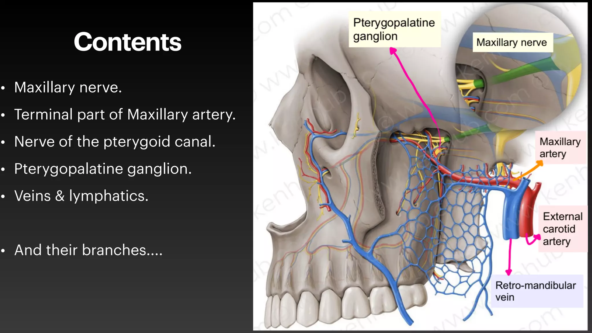
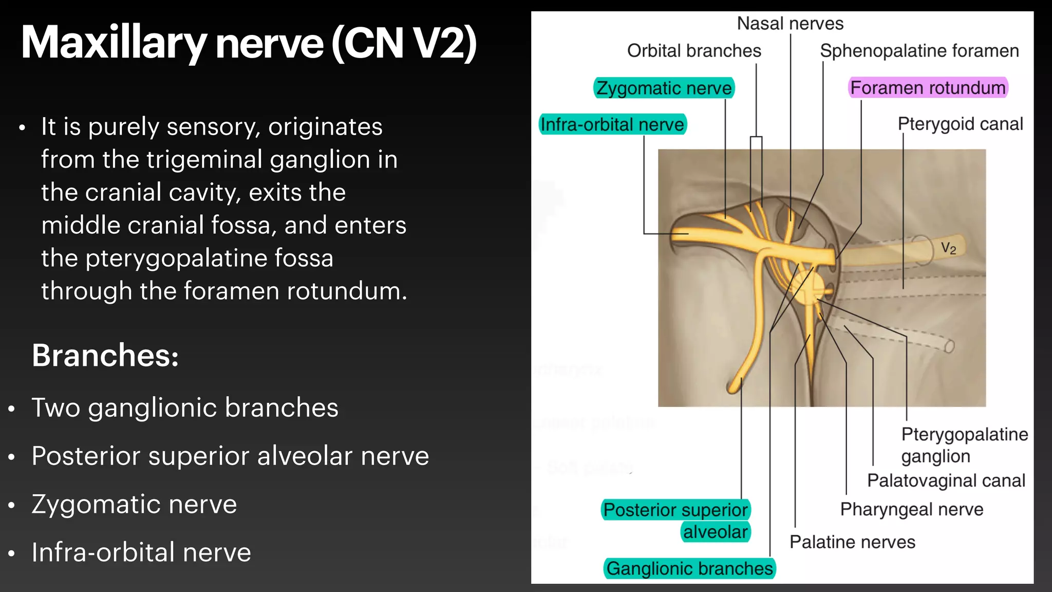
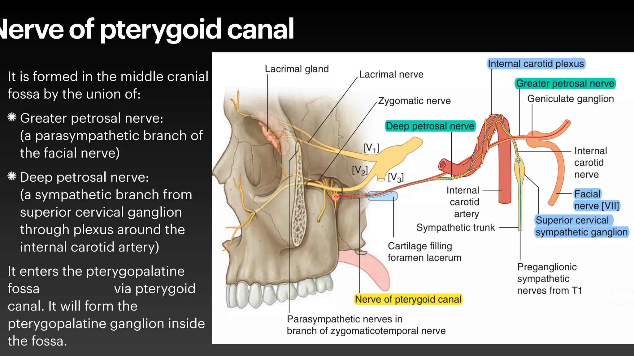
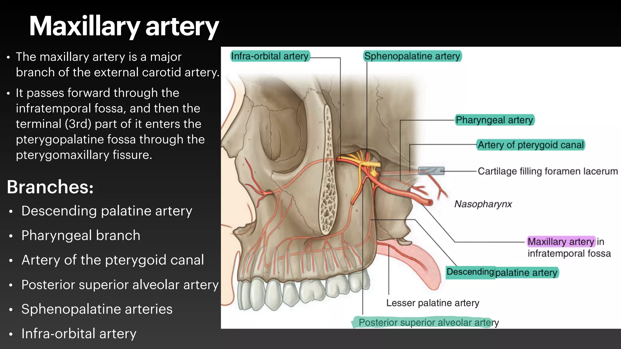
November 22nd, 2020
Halder Jamal
Pterygopalatine fossa
Outline
• What is it?
• Where is it?
• Boundaries?
• Communications?
• Contents?
• Clinical signi
f
icance?
Pterygopalatine fossa
Pterygopalatinefossa
• It is a small pyramidal space
below the apex of the orbit on
the lateral side of the skull.
Placed between the
infratemporal fossa laterally
and the nasopharynx medially.
• It functions as a neurovascular
conduit.
Boundaries
• Anterior wall → the posterior surface
of the maxilla.
• Posterior wall → anterior surface of
the pterygoid process of the
sphenoid bone.
• Medial wall → the lateral surface of
perpendicular plate of palatine bone.
• Laterally → Pterygomaxillary
f
issure.
• Roof → Apex of orbit.
• Floor → pyramidal process of the
palatine bone.
Communications
• Foramen rotundum & Pterygoid canal →
Middle cranial fossa.
• Pterygomaxillary
f
issure → Infratemporal
fossa.
• Inferior orbital
f
issure → Floor of the orbit.
• Sphenopalatine foramen → Lateral wall of
the nasal cavity.
• Palatovaginal canal→ Nasopharynx.
• Pterygopalatine canal → Roof of the oral
cavity.
Contents
• Maxillary nerve.
• Terminal part of Maxillary artery.
• Nerve of the pterygoid canal.
• Pterygopalatine ganglion.
• Veins & lymphatics.
• And their branches....
Maxillarynerve(CNV2)
• Two ganglionic branches
• Posterior superior alveolar nerve
• Zygomatic nerve
• Infra-orbital nerve
Branches:
• It is purely sensory, originates
from the trigeminal ganglion in
the cranial cavity, exits the
middle cranial fossa, and enters
the pterygopalatine fossa
through the foramen rotundum.
Nerve of pterygoid canal
It is formed in the middle cranial
fossa by the union of:
Greater petrosal nerve:
(a parasympathetic branch of
the facial nerve)
Deep petrosal nerve:
(a sympathetic branch from
superior cervical ganglion
through plexus around the
internal carotid artery)
It enters the pterygopalatine
fossa via pterygoid
canal. It will form the
pterygopalatine ganglion inside
the fossa.
Pterygopalatineganglion
• It is the largest of the four parasympathetic
ganglia in the head and is formed by:
• Nerve of pterygoid canal
• Two ganglionic branches of the maxillary
nerve
Branches:
• Nasal nerves
• Orbital branches
• Palatine nerves
• Pharyngeal nerve
Maxillary artery
• Descending palatine artery
• Pharyngeal branch
• Artery of the pterygoid canal
• Posterior superior alveolar artery
• Sphenopalatine arteries
• Infra-orbital artery
• The maxillary artery is a major
branch of the external carotid artery.
• It passes forward through the
infratemporal fossa, and then the
terminal (3rd) part of it enters the
pterygopalatine fossa through the
pterygomaxillary
f
issure.
Branches:
Autonomic efferent pathways
Clinical signi
f
icance
• lesion of parasympathetic
f
ibers of facial nerve → dry eye, this can cause vision
loss in the affected eye if left untreated.
• Because of the location of fossa and associated connections, It is often involved
in the spread of tumors, infections, and in
f
lammations caused by neoplastic
diseases in the head and neck (such as juvenile nasopharyngeal angio
f
ibroma,
nasopharyngeal carcinoma, bacterial sinusitis).
• Maxillary nerve block may be required in cases of extensive dental surgery. The
maxillary nerve in the pterygopalatine fossa is most often approached intra-orally
via the greater palatine canal.
• Ligation of sphenopalatine artery as a treatment of chronic epistaxis. It can be
surgically approached via the maxillary sinus.
Thank you

Pterygopalatine Fossa.pdf

  • 1.
    November 22nd, 2020 HalderJamal Pterygopalatine fossa
  • 2.
    Outline • What isit? • Where is it? • Boundaries? • Communications? • Contents? • Clinical signi f icance? Pterygopalatine fossa
  • 3.
    Pterygopalatinefossa • It isa small pyramidal space below the apex of the orbit on the lateral side of the skull. Placed between the infratemporal fossa laterally and the nasopharynx medially. • It functions as a neurovascular conduit.
  • 4.
    Boundaries • Anterior wall→ the posterior surface of the maxilla. • Posterior wall → anterior surface of the pterygoid process of the sphenoid bone. • Medial wall → the lateral surface of perpendicular plate of palatine bone. • Laterally → Pterygomaxillary f issure. • Roof → Apex of orbit. • Floor → pyramidal process of the palatine bone.
  • 5.
    Communications • Foramen rotundum& Pterygoid canal → Middle cranial fossa. • Pterygomaxillary f issure → Infratemporal fossa. • Inferior orbital f issure → Floor of the orbit. • Sphenopalatine foramen → Lateral wall of the nasal cavity. • Palatovaginal canal→ Nasopharynx. • Pterygopalatine canal → Roof of the oral cavity.
  • 6.
    Contents • Maxillary nerve. •Terminal part of Maxillary artery. • Nerve of the pterygoid canal. • Pterygopalatine ganglion. • Veins & lymphatics. • And their branches....
  • 7.
    Maxillarynerve(CNV2) • Two ganglionicbranches • Posterior superior alveolar nerve • Zygomatic nerve • Infra-orbital nerve Branches: • It is purely sensory, originates from the trigeminal ganglion in the cranial cavity, exits the middle cranial fossa, and enters the pterygopalatine fossa through the foramen rotundum.
  • 8.
    Nerve of pterygoidcanal It is formed in the middle cranial fossa by the union of: Greater petrosal nerve: (a parasympathetic branch of the facial nerve) Deep petrosal nerve: (a sympathetic branch from superior cervical ganglion through plexus around the internal carotid artery) It enters the pterygopalatine fossa via pterygoid canal. It will form the pterygopalatine ganglion inside the fossa.
  • 9.
    Pterygopalatineganglion • It isthe largest of the four parasympathetic ganglia in the head and is formed by: • Nerve of pterygoid canal • Two ganglionic branches of the maxillary nerve Branches: • Nasal nerves • Orbital branches • Palatine nerves • Pharyngeal nerve
  • 10.
    Maxillary artery • Descendingpalatine artery • Pharyngeal branch • Artery of the pterygoid canal • Posterior superior alveolar artery • Sphenopalatine arteries • Infra-orbital artery • The maxillary artery is a major branch of the external carotid artery. • It passes forward through the infratemporal fossa, and then the terminal (3rd) part of it enters the pterygopalatine fossa through the pterygomaxillary f issure. Branches:
  • 11.
  • 12.
    Clinical signi f icance • lesionof parasympathetic f ibers of facial nerve → dry eye, this can cause vision loss in the affected eye if left untreated. • Because of the location of fossa and associated connections, It is often involved in the spread of tumors, infections, and in f lammations caused by neoplastic diseases in the head and neck (such as juvenile nasopharyngeal angio f ibroma, nasopharyngeal carcinoma, bacterial sinusitis). • Maxillary nerve block may be required in cases of extensive dental surgery. The maxillary nerve in the pterygopalatine fossa is most often approached intra-orally via the greater palatine canal. • Ligation of sphenopalatine artery as a treatment of chronic epistaxis. It can be surgically approached via the maxillary sinus.
  • 13.