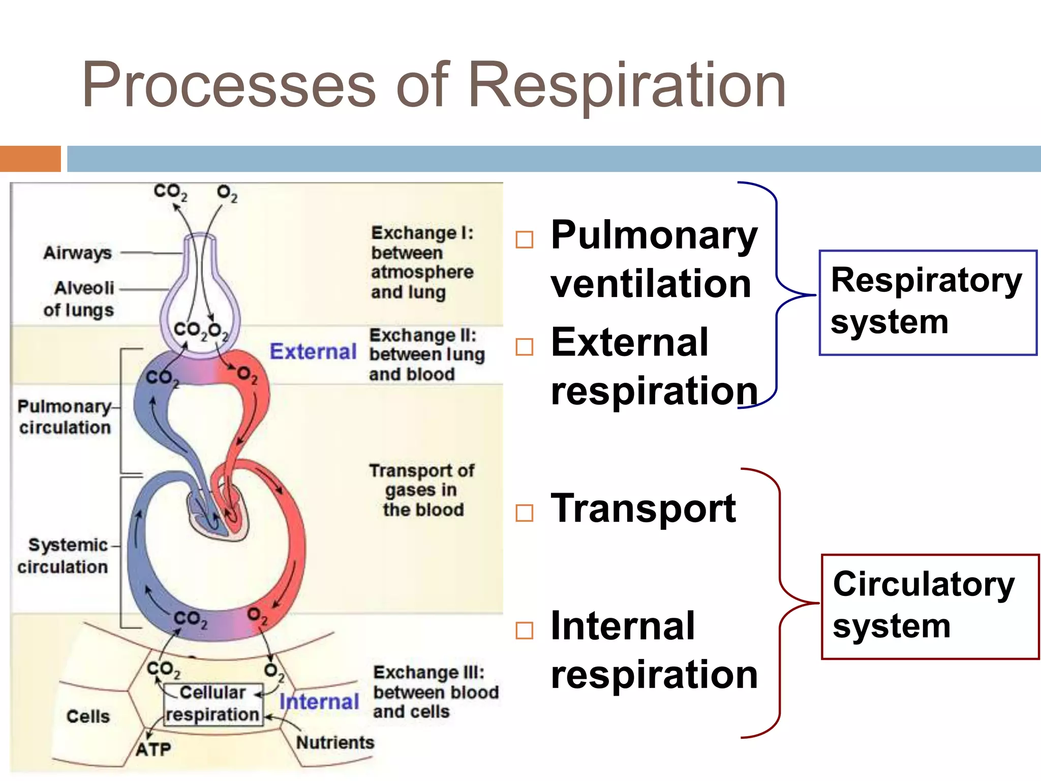
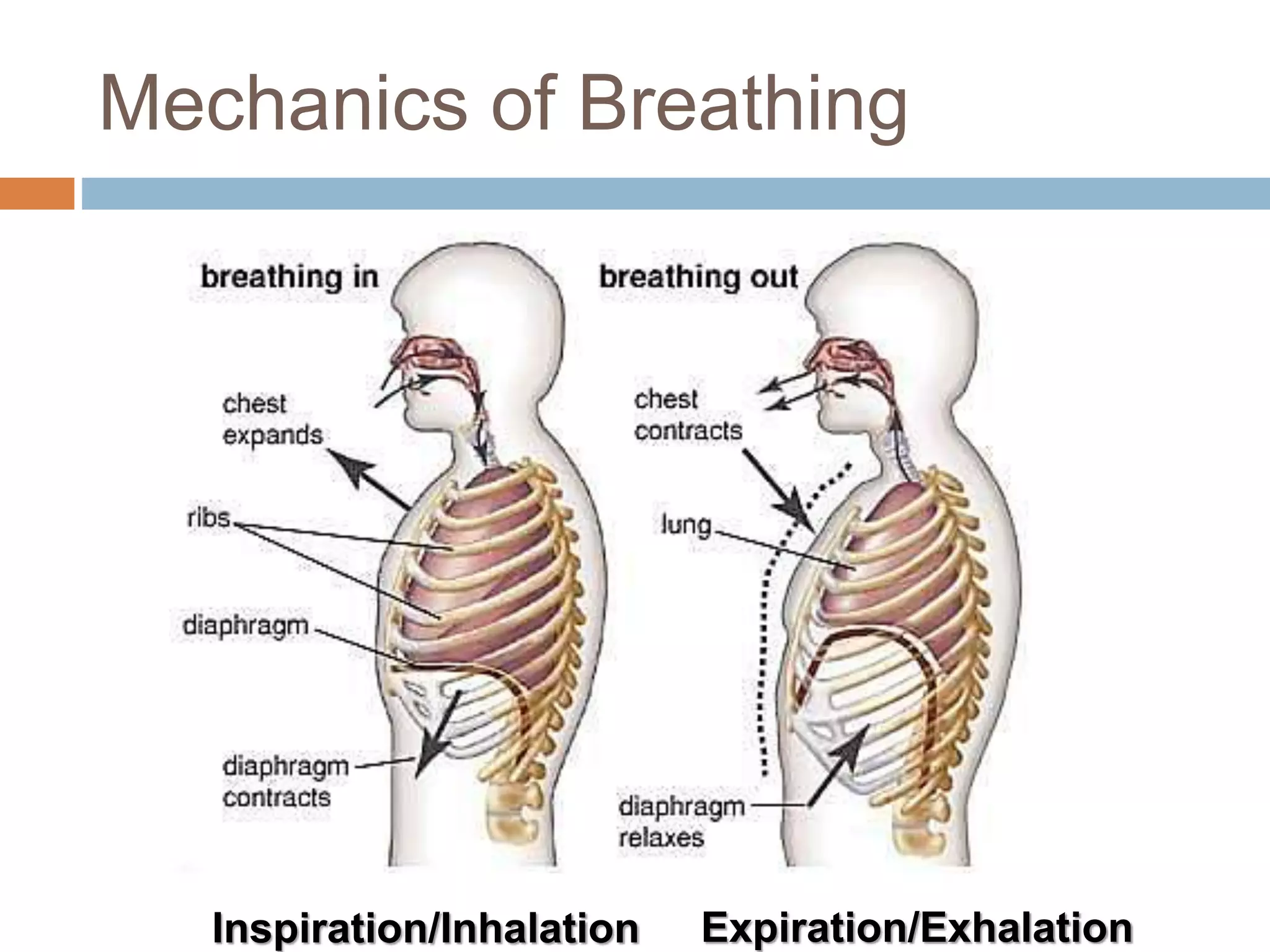
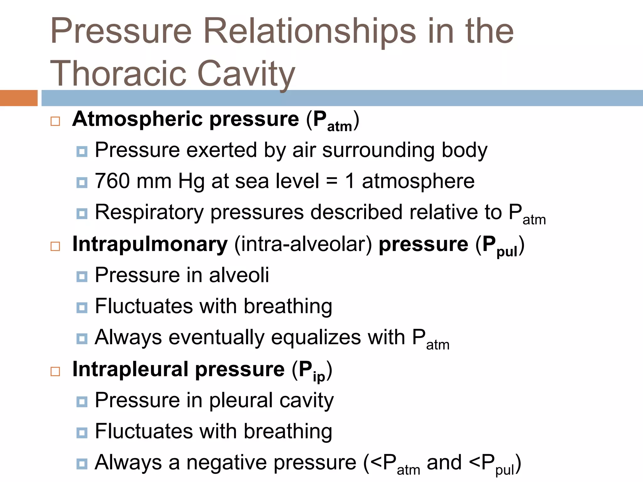
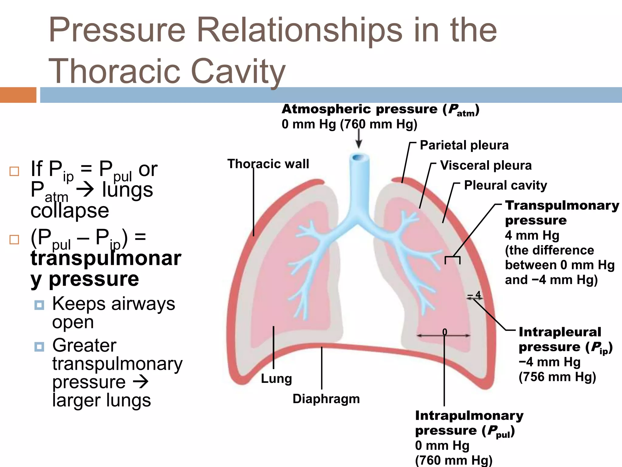
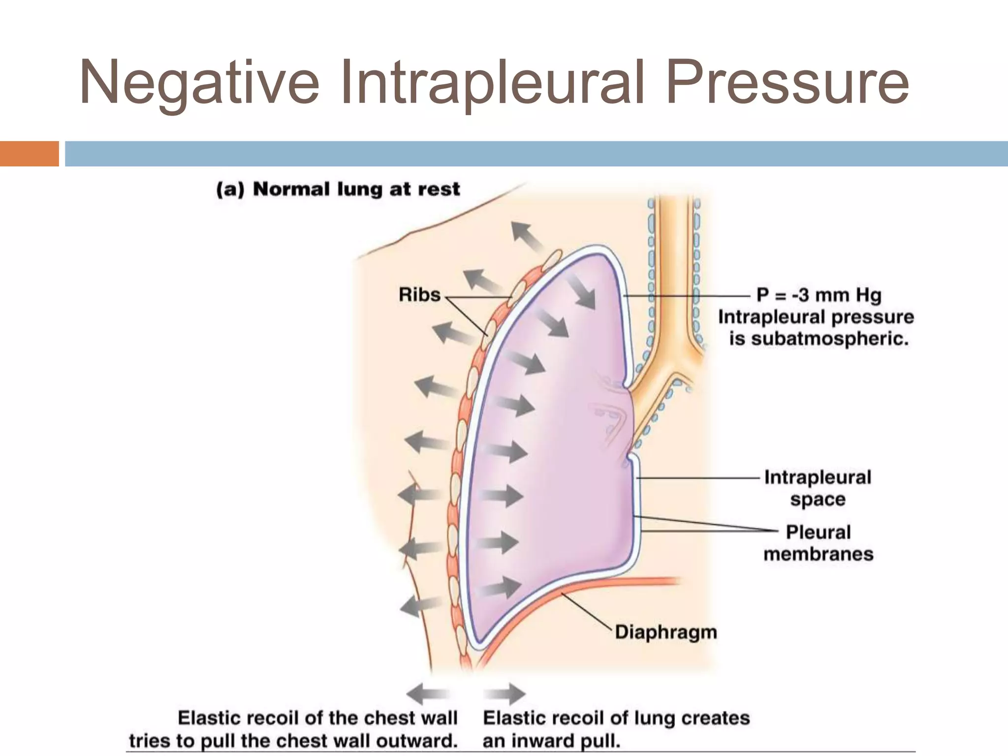
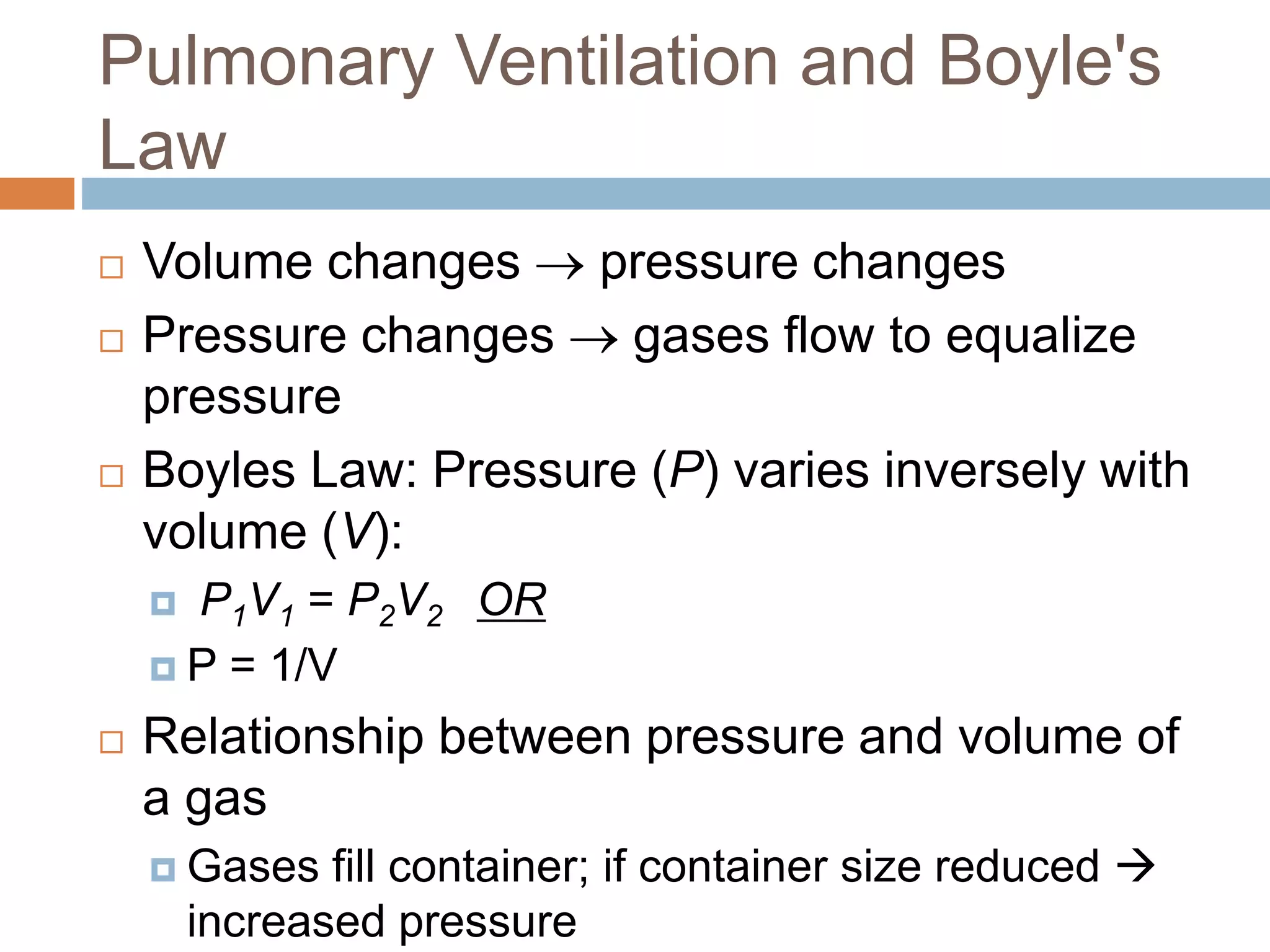
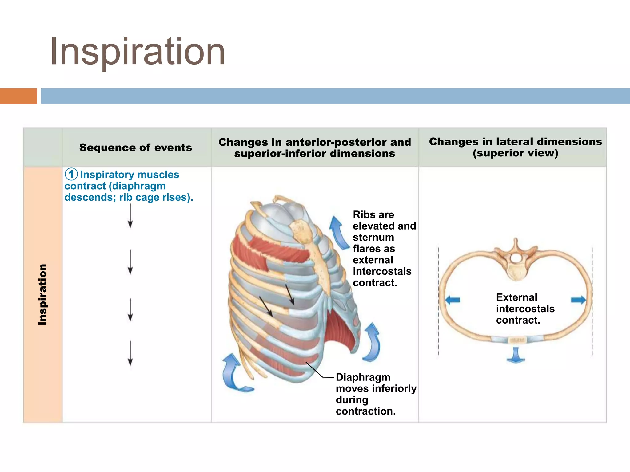
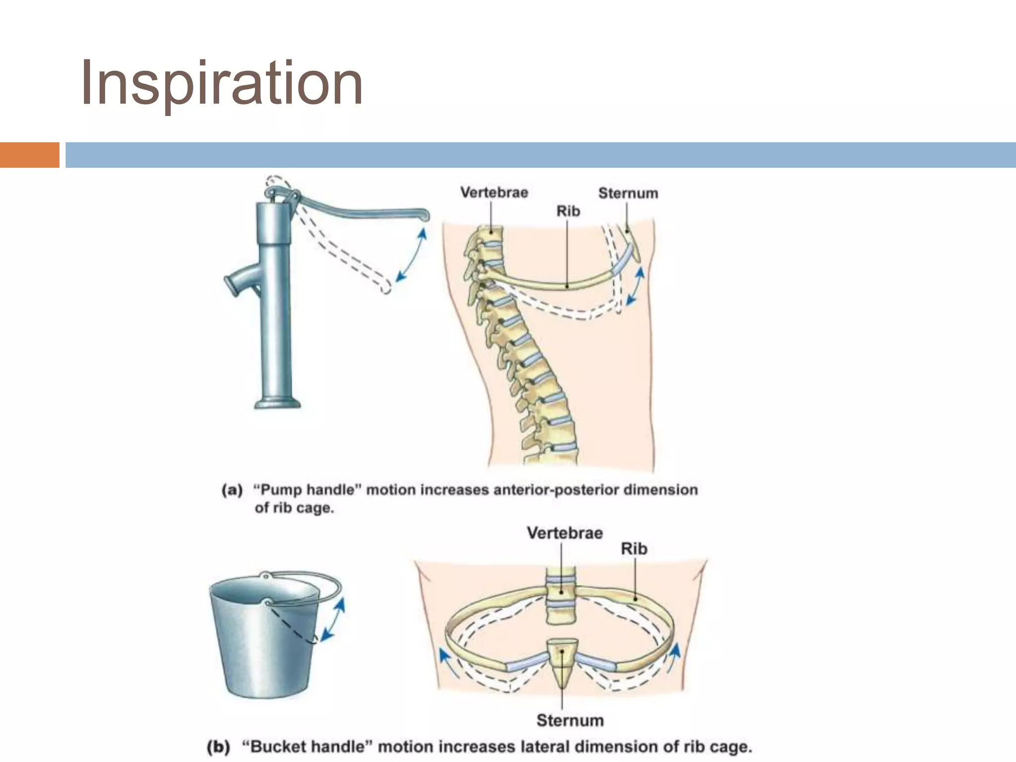
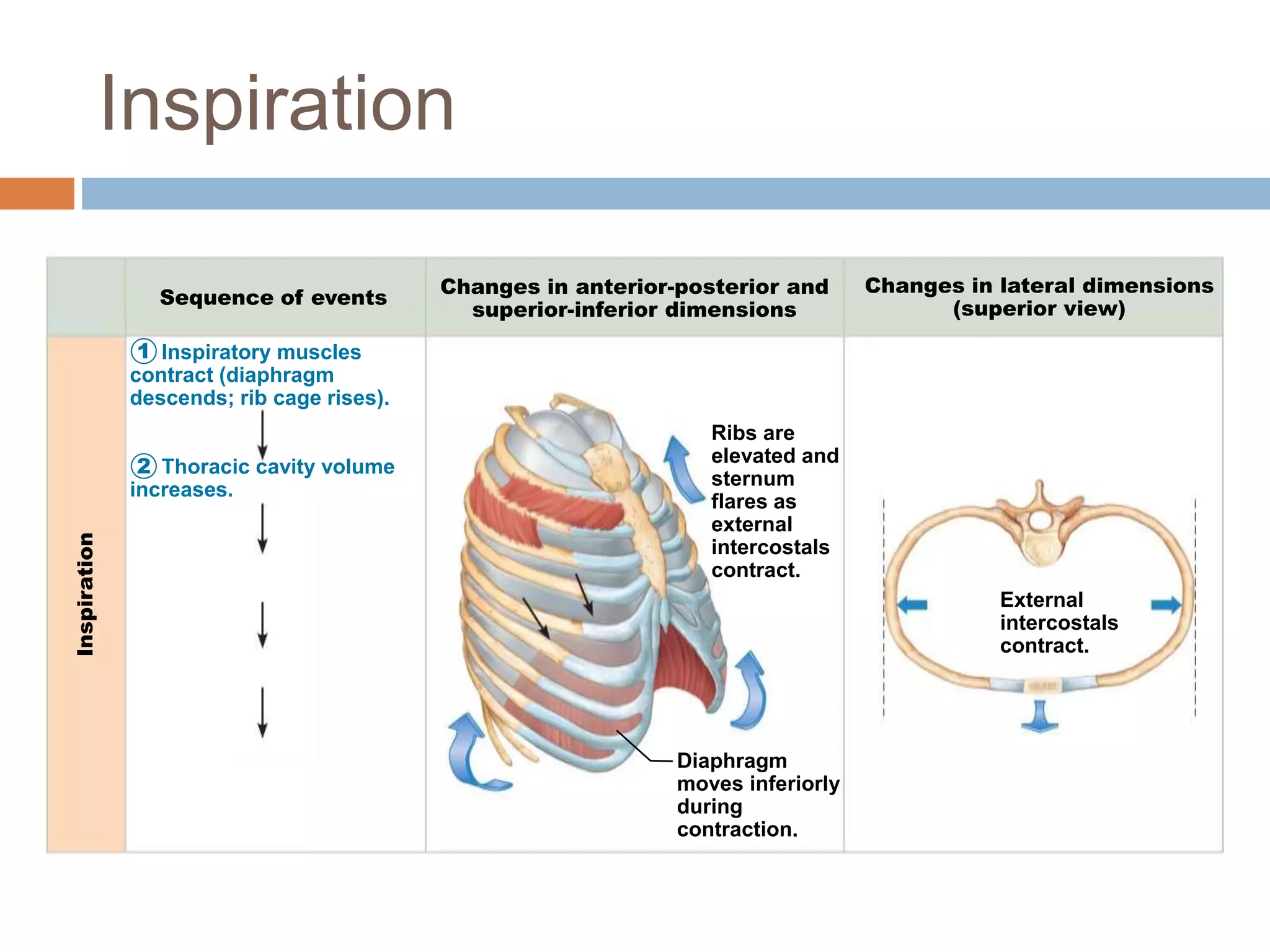
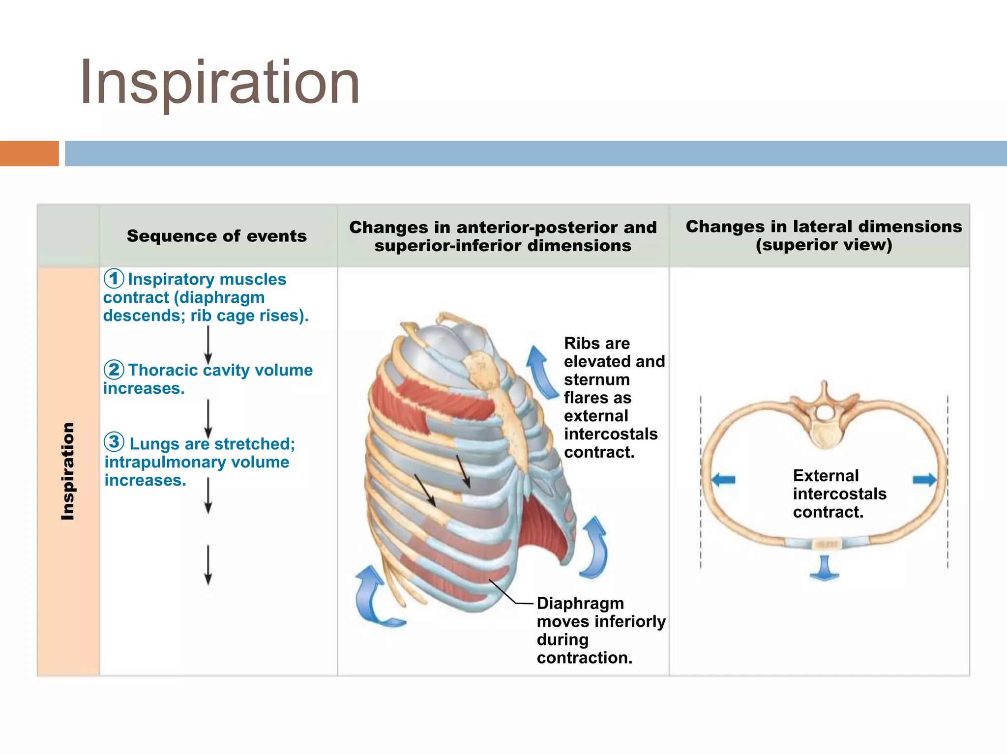
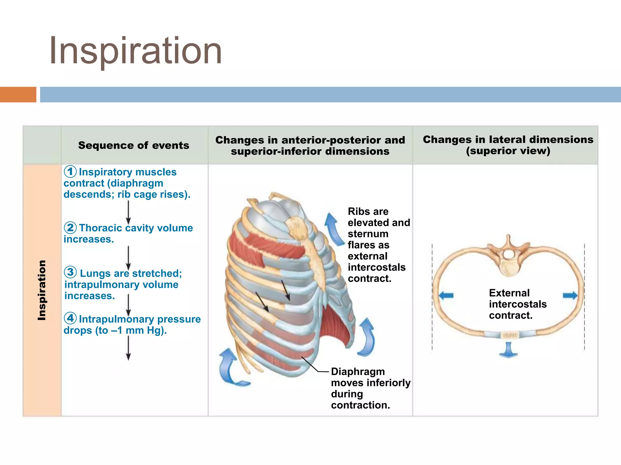
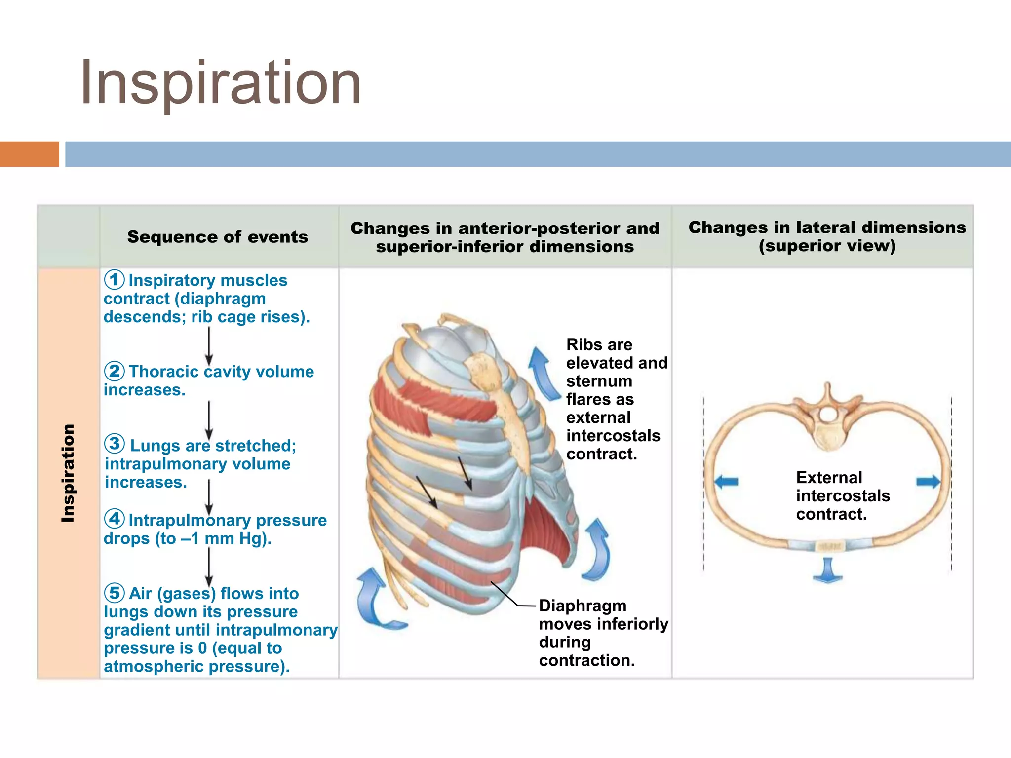
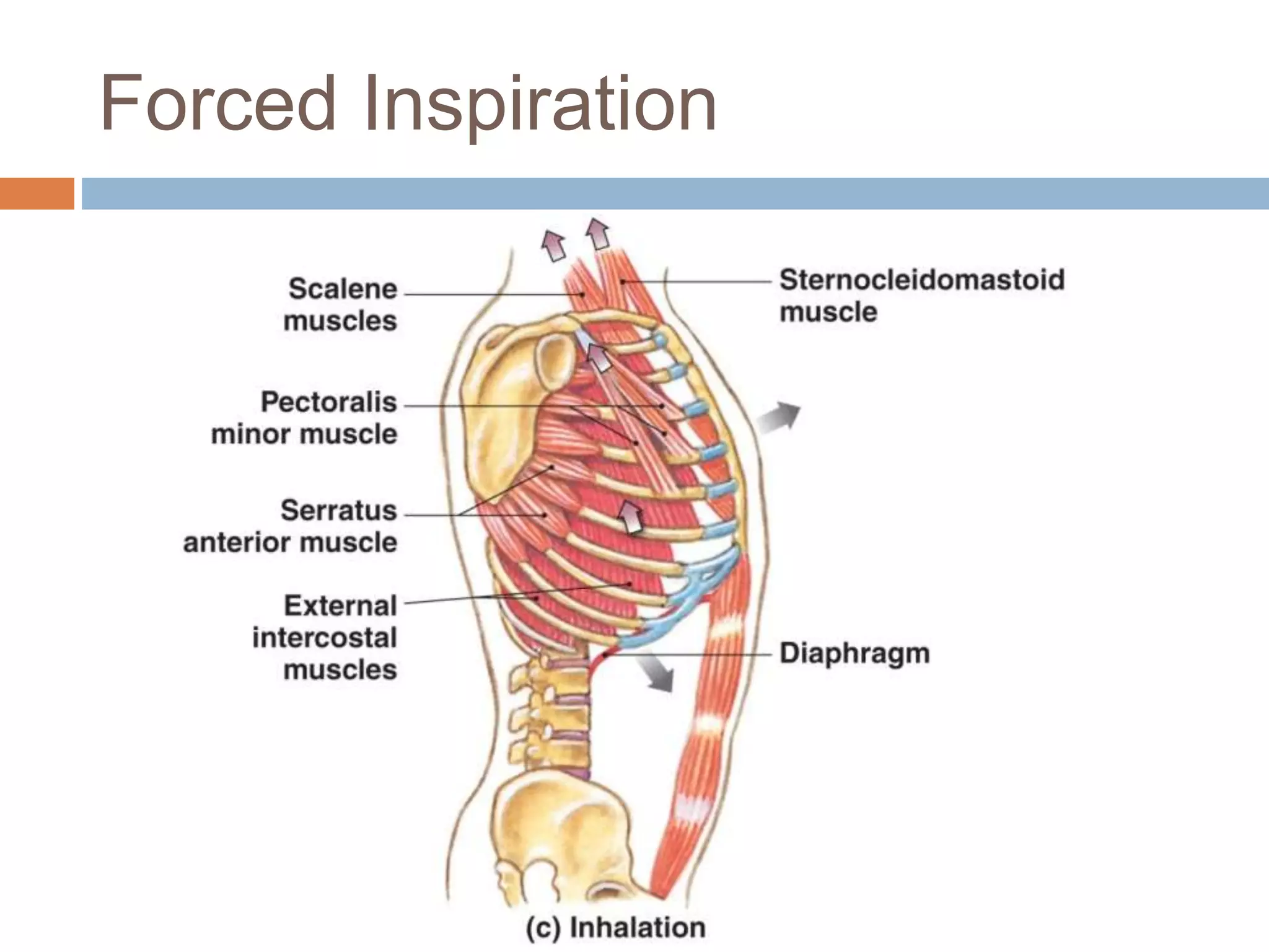
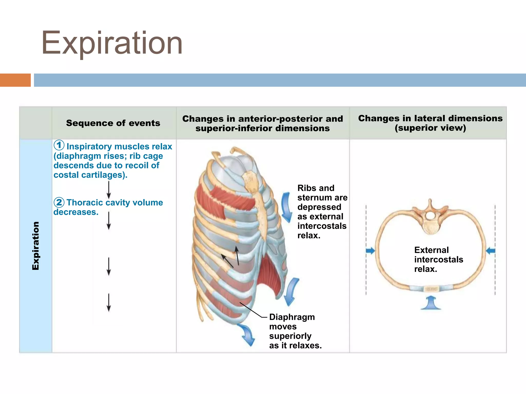
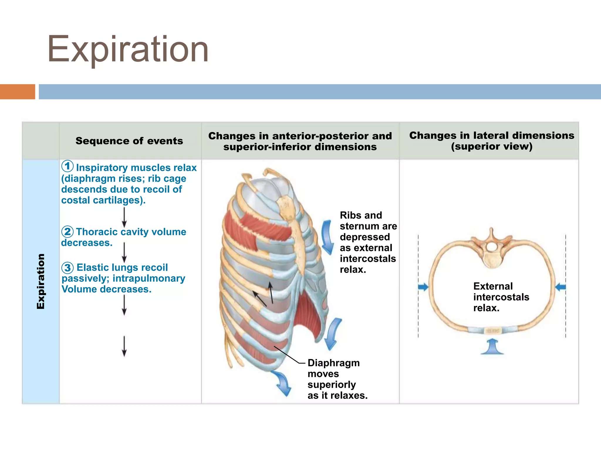
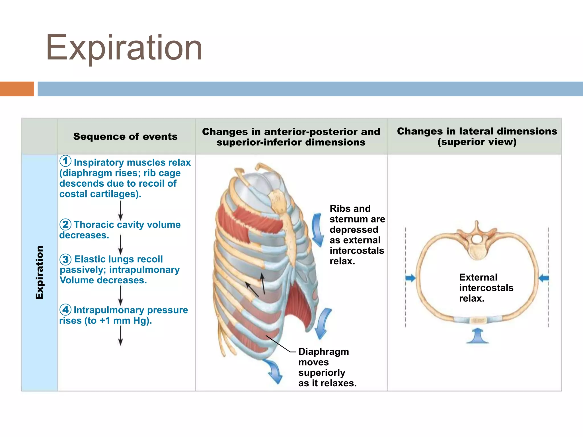
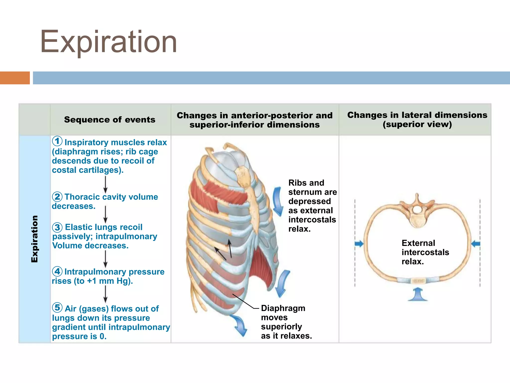
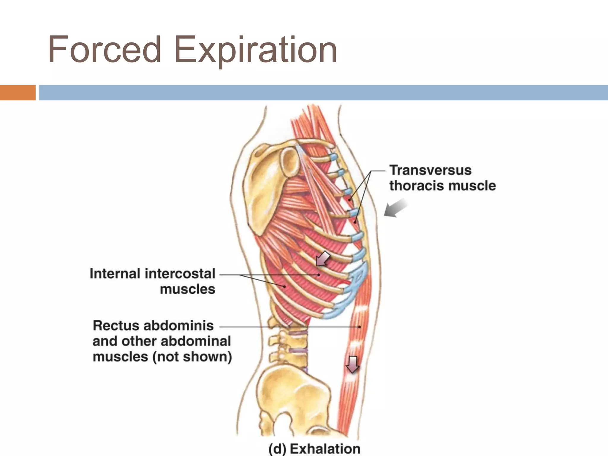
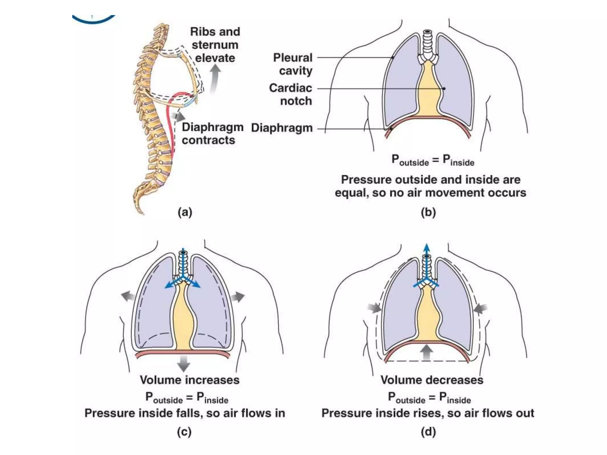
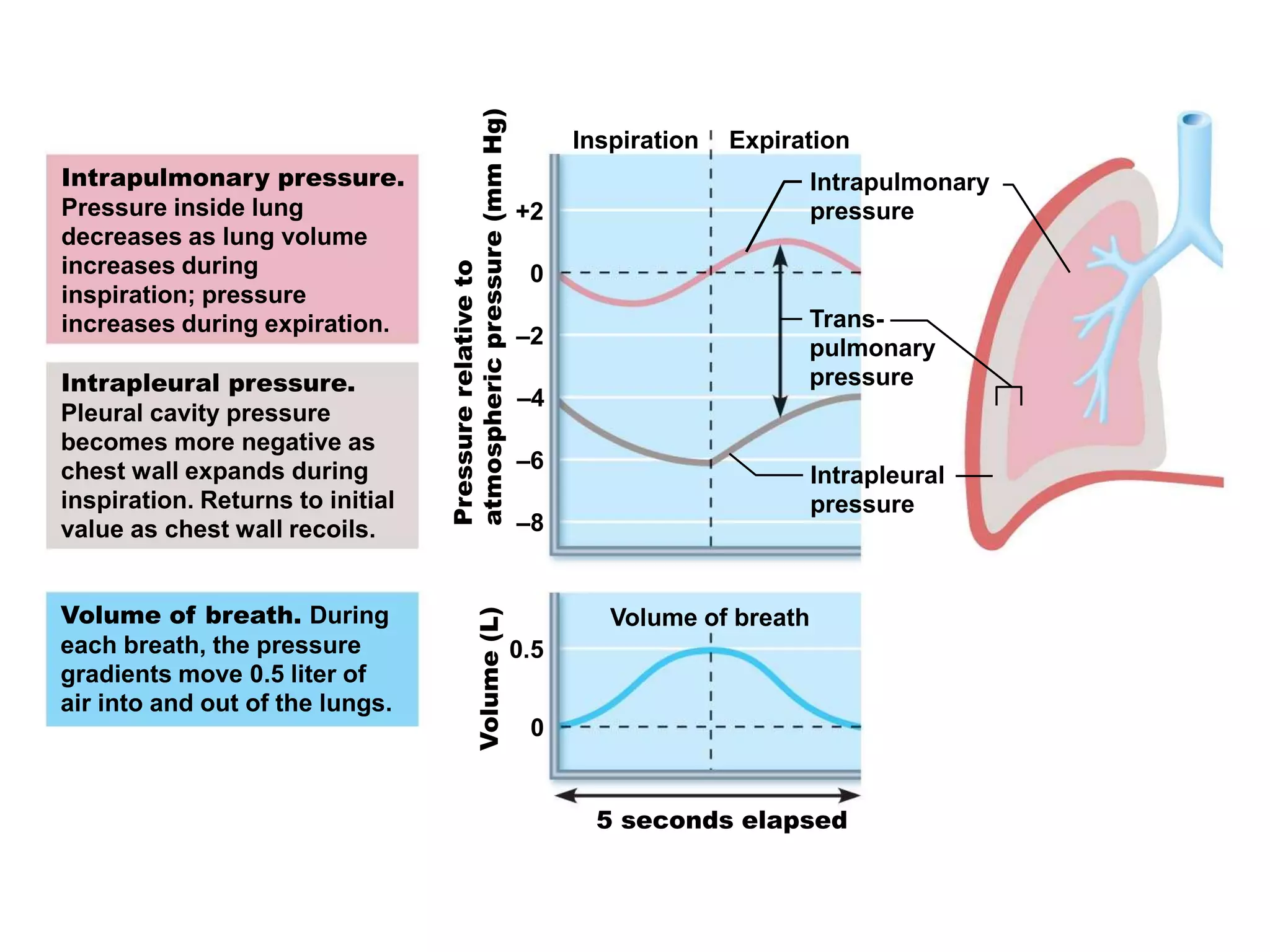
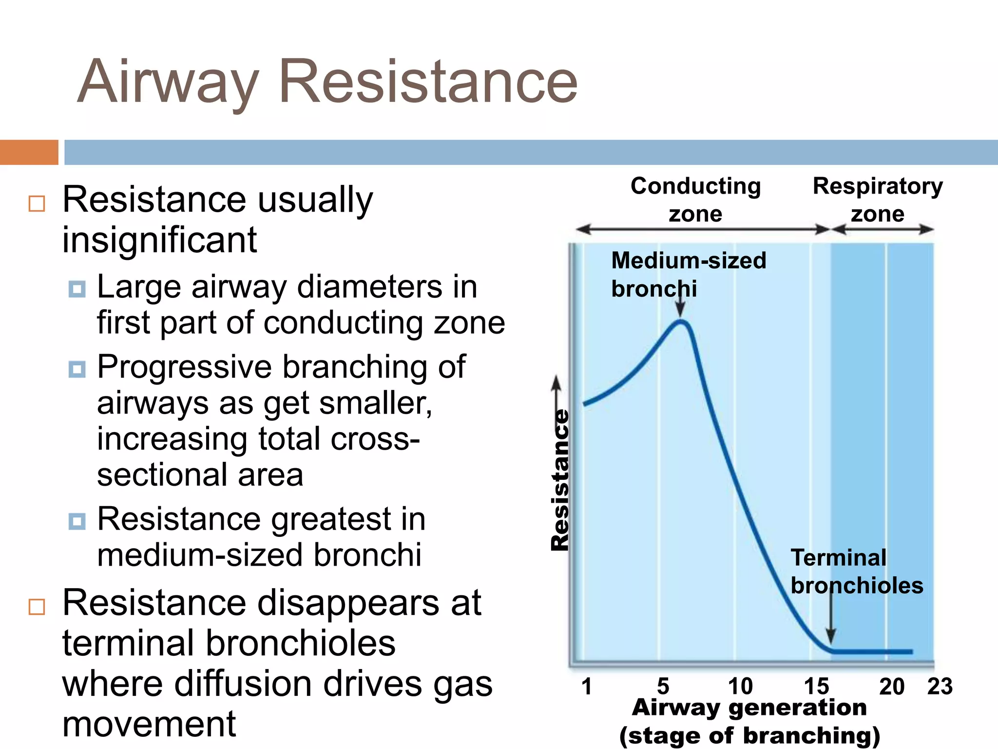
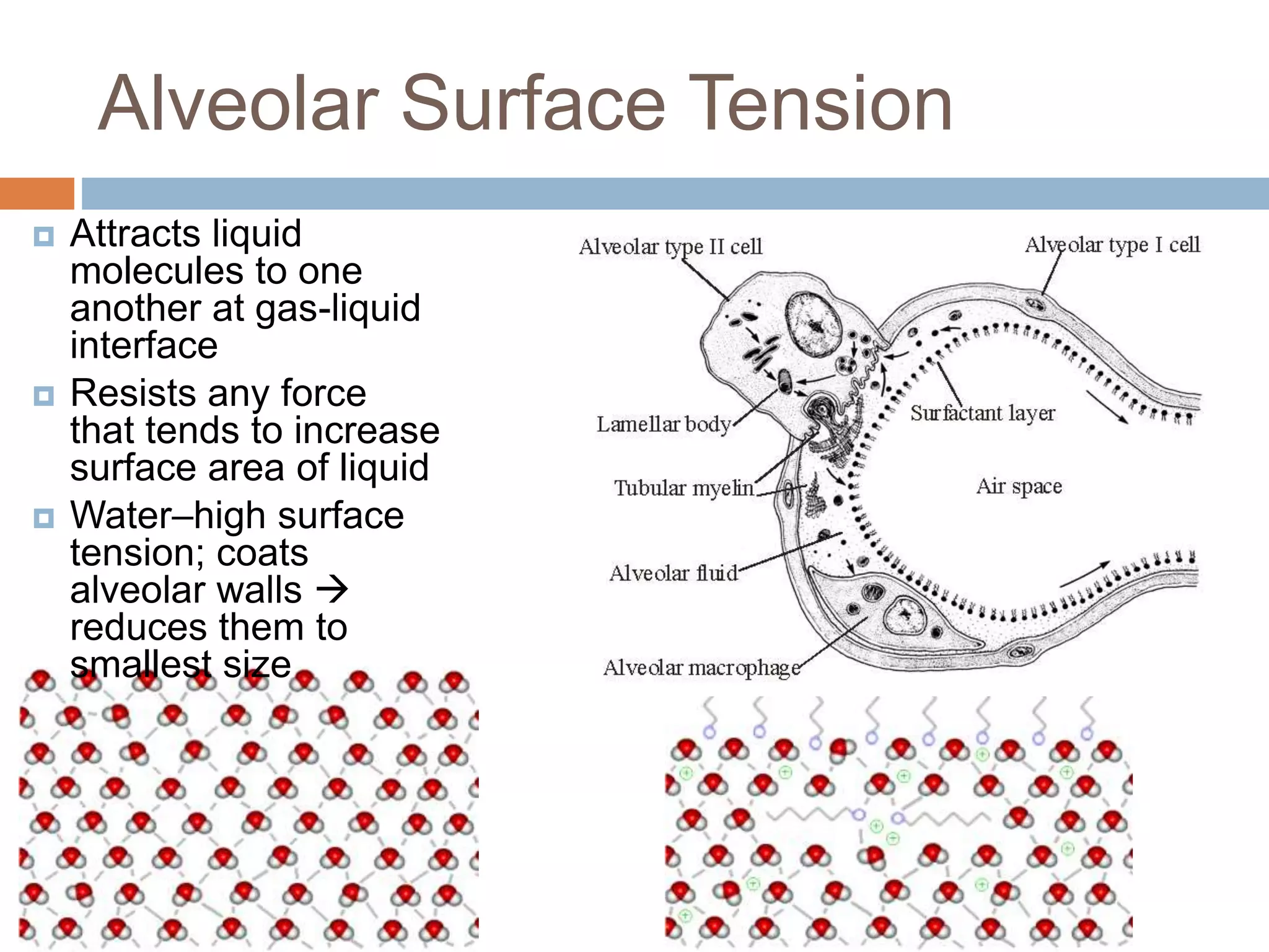
CHAPTER 22:
RESPIRATORY SYSTEM
(2): MECHANICS OF
VENTILATION
Human Anatomy and Physiology II –
BIOL153
Processes of Respiration
 Pulmonary
ventilation
 External
respiration
 Transport
 Internal
respiration
Respiratory
system
Circulatory
system
Goals/Objectives
 Explain the functional importance of the partial vacuum
that exists in the intrapleural space
 Relate Boyle’s law to the events of inspiration and
expiration
 Explain the relative roles of the respiratory muscles
and lung elasticity in producing the volume changes
that cause air to flow into and out of the lungs
 List several physical factors that influence pulmonary
ventilation
 Explain and compare the various lung volumes and
capacities
 Define dead space
Mechanics of Breathing
Inspiration/Inhalation Expiration/Exhalation
Pressure Relationships in the
Thoracic Cavity
 Atmospheric pressure (Patm)
 Pressure exerted by air surrounding body
 760 mm Hg at sea level = 1 atmosphere
 Respiratory pressures described relative to Patm
 Intrapulmonary (intra-alveolar) pressure (Ppul)
 Pressure in alveoli
 Fluctuates with breathing
 Always eventually equalizes with Patm
 Intrapleural pressure (Pip)
 Pressure in pleural cavity
 Fluctuates with breathing
 Always a negative pressure (<Patm and <Ppul)
Pressure Relationships in the
Thoracic Cavity
Atmospheric pressure (Patm)
0 mm Hg (760 mm Hg)
Thoracic wall
Parietal pleura
Visceral pleura
Pleural cavity
Transpulmonary
pressure
4 mm Hg
(the difference
between 0 mm Hg
and −4 mm Hg)
Intrapleural
pressure (Pip)
−4 mm Hg
(756 mm Hg)
Intrapulmonary
pressure (Ppul)
0 mm Hg
(760 mm Hg)
Diaphragm
Lung
0
– 4
 If Pip = Ppul or
Patm  lungs
collapse
 (Ppul – Pip) =
transpulmonar
y pressure
 Keeps airways
open
 Greater
transpulmonary
pressure 
larger lungs
Negative Intrapleural Pressure
Atelectasis (lung collapse)
 Plugged bronchioles  collapse of
alveoli
 Pneumothorax-air in pleural cavity
 From either wound in parietal or
rupture of visceral pleura
 Treated by removing air with chest
tubes; pleurae heal  lung reinflates
Pulmonary Ventilation and Boyle's
Law
 Volume changes  pressure changes
 Pressure changes  gases flow to equalize
pressure
 Boyles Law: Pressure (P) varies inversely with
volume (V):
 P1V1 = P2V2 OR
 P = 1/V
 Relationship between pressure and volume of
a gas
 Gases fill container; if container size reduced 
increased pressure
Inspiration
Sequence of events
Changes in anterior-posterior and
superior-inferior dimensions
Changes in lateral dimensions
(superior view)
1
Diaphragm
moves inferiorly
during
contraction.
Ribs are
elevated and
sternum
flares as
external
intercostals
contract.
External
intercostals
contract.
Inspiratory muscles
contract (diaphragm
descends; rib cage rises).
Inspiration
Inspiration
Inspiratory muscles
contract (diaphragm
descends; rib cage rises).
Thoracic cavity volume
increases.
Inspiration
Sequence of events
Changes in anterior-posterior and
superior-inferior dimensions
Changes in lateral dimensions
(superior view)
1
2
Diaphragm
moves inferiorly
during
contraction.
Ribs are
elevated and
sternum
flares as
external
intercostals
contract.
External
intercostals
contract.
Inspiration
Inspiratory muscles
contract (diaphragm
descends; rib cage rises).
Thoracic cavity volume
increases.
Inspiration
Sequence of events
Changes in anterior-posterior and
superior-inferior dimensions
Changes in lateral dimensions
(superior view)
1
2
3
Diaphragm
moves inferiorly
during
contraction.
Ribs are
elevated and
sternum
flares as
external
intercostals
contract.
External
intercostals
contract.
Lungs are stretched;
intrapulmonary volume
increases.
Inspiration
Inspiratory muscles
contract (diaphragm
descends; rib cage rises).
Thoracic cavity volume
increases.
Lungs are stretched;
intrapulmonary volume
increases.
Inspiration
Sequence of events
Changes in anterior-posterior and
superior-inferior dimensions
Changes in lateral dimensions
(superior view)
1
2
3
4
Diaphragm
moves inferiorly
during
contraction.
Ribs are
elevated and
sternum
flares as
external
intercostals
contract.
External
intercostals
contract.Intrapulmonary pressure
drops (to –1 mm Hg).
Inspiration
Inspiratory muscles
contract (diaphragm
descends; rib cage rises).
Thoracic cavity volume
increases.
Lungs are stretched;
intrapulmonary volume
increases.
Intrapulmonary pressure
drops (to –1 mm Hg).
Air (gases) flows into
lungs down its pressure
gradient until intrapulmonary
pressure is 0 (equal to
atmospheric pressure).
Inspiration
Sequence of events
Changes in anterior-posterior and
superior-inferior dimensions
Changes in lateral dimensions
(superior view)
1
2
3
4
5
Diaphragm
moves inferiorly
during
contraction.
Ribs are
elevated and
sternum
flares as
external
intercostals
contract.
External
intercostals
contract.
Inspiration
Forced Inspiration
1
Expiration
Sequence of events Changes in anterior-posterior and
superior-inferior dimensions
Changes in lateral dimensions
(superior view)
Diaphragm
moves
superiorly
as it relaxes.
Ribs and
sternum are
depressed
as external
intercostals
relax.
External
intercostals
relax.
Inspiratory muscles relax
(diaphragm rises; rib cage
descends due to recoil of
costal cartilages).
Expiration
1
Expiration
Sequence of events Changes in anterior-posterior and
superior-inferior dimensions
Changes in lateral dimensions
(superior view)
2
Diaphragm
moves
superiorly
as it relaxes.
Ribs and
sternum are
depressed
as external
intercostals
relax.
External
intercostals
relax.
Inspiratory muscles relax
(diaphragm rises; rib cage
descends due to recoil of
costal cartilages).
Thoracic cavity volume
decreases.
Expiration
1
Expiration
Sequence of events Changes in anterior-posterior and
superior-inferior dimensions
Changes in lateral dimensions
(superior view)
2
3
Diaphragm
moves
superiorly
as it relaxes.
Ribs and
sternum are
depressed
as external
intercostals
relax.
External
intercostals
relax.
Inspiratory muscles relax
(diaphragm rises; rib cage
descends due to recoil of
costal cartilages).
Thoracic cavity volume
decreases.
Elastic lungs recoil
passively; intrapulmonary
Volume decreases.
Expiration
1
Expiration
Sequence of events Changes in anterior-posterior and
superior-inferior dimensions
Changes in lateral dimensions
(superior view)
2
3
4
Diaphragm
moves
superiorly
as it relaxes.
Ribs and
sternum are
depressed
as external
intercostals
relax.
External
intercostals
relax.
Inspiratory muscles relax
(diaphragm rises; rib cage
descends due to recoil of
costal cartilages).
Thoracic cavity volume
decreases.
Elastic lungs recoil
passively; intrapulmonary
Volume decreases.
Intrapulmonary pressure
rises (to +1 mm Hg).
Expiration
1
Expiration
Sequence of events Changes in anterior-posterior and
superior-inferior dimensions
Changes in lateral dimensions
(superior view)
2
3
4
5 Diaphragm
moves
superiorly
as it relaxes.
Ribs and
sternum are
depressed
as external
intercostals
relax.
External
intercostals
relax.
Inspiratory muscles relax
(diaphragm rises; rib cage
descends due to recoil of
costal cartilages).
Thoracic cavity volume
decreases.
Elastic lungs recoil
passively; intrapulmonary
Volume decreases.
Intrapulmonary pressure
rises (to +1 mm Hg).
Air (gases) flows out of
lungs down its pressure
gradient until intrapulmonary
pressure is 0.
Expiration
Forced Expiration
Intrapulmonary pressure.
Pressure inside lung
decreases as lung volume
increases during
inspiration; pressure
increases during expiration.
Intrapleural pressure.
Pleural cavity pressure
becomes more negative as
chest wall expands during
inspiration. Returns to initial
value as chest wall recoils.
Volume of breath. During
each breath, the pressure
gradients move 0.5 liter of
air into and out of the lungs.
Pressurerelativeto
atmosphericpressure(mmHg)Volume(L)
Inspiration Expiration
Intrapulmonary
pressure
Trans-
pulmonary
pressure
Intrapleural
pressure
Volume of breath
5 seconds elapsed
+2
0
–2
–4
–6
–8
0.5
0
Clicker Question
The pressure in the pleural cavity is known as
__________.
a) intrapleural pressure
b) intrapulmonary pressure
c) transpulmonary pressure
d) atmospheric pressure
Goals/Objectives
 Explain the functional importance of the partial vacuum
that exists in the intrapleural space
 Relate Boyle’s law to the events of inspiration and
expiration
 Explain the relative roles of the respiratory muscles
and lung elasticity in producing the volume changes
that cause air to flow into and out of the lungs
 List several physical factors that influence pulmonary
ventilation
 Explain and compare the various lung volumes and
capacities
 Define dead space
Physical Factors Influencing
Pulmonary Ventilation
 Three physical factors influence the ease of air
passage and the amount of energy required
for ventilation.
 Airway resistance
 Alveolar surface tension
 Lung compliance
Airway Resistance
 Relationship between flow (F), pressure
(P), and resistance (R) is:
 ∆P - pressure gradient between atmosphere
and alveoli (2 mm Hg or less during normal
quiet breathing)
 Gas flow changes inversely with resistance
Conducting
zone
Respiratory
zone
Medium-sized
bronchi
Resistance Terminal
bronchioles
1 5 10 15 20 23
Airway generation
(stage of branching)
Airway Resistance
 Resistance usually
insignificant
 Large airway diameters in
first part of conducting zone
 Progressive branching of
airways as get smaller,
increasing total cross-
sectional area
 Resistance greatest in
medium-sized bronchi
 Resistance disappears at
terminal bronchioles
where diffusion drives gas
movement
Alveolar Surface Tension
 Attracts liquid
molecules to one
another at gas-liquid
interface
 Resists any force
that tends to increase
surface area of liquid
 Water–high surface
tension; coats
alveolar walls 
reduces them to
smallest size
Lung Compliance
 Measure of change in lung volume that occurs
with given change in transpulmonary pressure
 Higher lung compliance  easier to expand
lungs
 Normally high due to
 Distensibility of lung tissue
 Surfactant, which decreases alveolar surface
tension
 Diminished by
 Nonelastic scar tissue replacing lung tissue
(fibrosis)
Respiratory Volumes and
Capacities
 Used to assess respiratory status
 Tidal volume (TV)
 Inspiratory reserve volume (IRV)
 Expiratory reserve volume (ERV)
 Residual volume (RV)
 Combinations of respiratory volumes
 Inspiratory capacity (IC)
 Functional residual capacity (FRC)
 Vital capacity (VC)
 Total lung capacity (TLC)
Measurement
Adult male
average value
Adult female
average value Description
Respiratory
volumes
Respiratory
capacities
Summary of respiratory volumes and capacities for males and females
Tidal volume (TV)
Inspiratory reserve
volume (IRV)
Expiratory reserve
volume (ERV)
Residual volume (RV)
500 ml 500 ml
3100 ml
1200 ml
1200 ml
1900 ml
700 ml
1100 ml
Amount of air inhaled or exhaled with each breath under resting
conditions
Amount of air that can be forcefully inhaled after a normal tidal
volume inspiration
Amount of air that can be forcefully exhaled after a normal tidal
volume expiration
Amount of air remaining in the lungs after a forced expiration
Maximum amount of air contained in lungs after a maximum
inspiratory effort: TLC = TV + IRV + ERV + RV
Maximum amount of air that can be expired after a maximum
inspiratory effort: VC = TV + IRV + ERV
Maximum amount of air that can be inspired after a normal tidal
volume expiration: IC = TV + IRV
Volume of air remaining in the lungs after a normal tidal volume
expiration: FRC = ERV + RV
6000 ml
4800 ml
3600 ml
2400 ml
4200 ml
3100 ml
2400 ml
1800 ml
Total lung capacity (TLC)
Vital capacity (VC)
Inspiratory capacity (IC)
Functional residual
capacity (FRC)
Respiratory Volumes and
Capacities
5000
4000
3000
2000
1000
0
Milliliters(ml)
Spirographic record for a male
6000
Inspiratory
reserve volume
3100 ml
Expiratory
reserve volume
1200 ml
Residual volume
1200 ml
Inspiratory
capacity
3600 ml
Functional
residual
capacity
2400 ml
Vital
capacity
4800 ml
Total lung
capacity
6000 ml
Tidal volume 500 ml
Respiratory Volumes and
Capacities
Dead Space
 Anatomical dead space
 No contribution to gas exchange
 Air remaining in passageways; ~150 ml
 Alveolar dead space–non-functional
alveoli due to collapse or obstruction
 Total dead space-sum of anatomical and
alveolar dead space
Pulmonary Function Tests
 Spirometer-instrument for measuring
respiratory volumes and capacities
Clicker Question
During pulmonary tests, a patient is asked to
breath in normally and then inhale additionally
as much as possible into the spirometer. The
capacity being measured is the:
a) Inspiratory capacity
b) Functional residual capacity
c) Vital capacity
d) Total lung capacity
Pulmonary Function Tests
 To measure rate of gas movement
 Forced vital capacity (FVC)—gas forcibly
expelled after taking deep breath
 Forced expiratory volume (FEV)—amount of
gas expelled during specific time intervals of
FVC

Chapter 22: Respiratory System (#2)

  • 1.
    CHAPTER 22: RESPIRATORY SYSTEM (2):MECHANICS OF VENTILATION Human Anatomy and Physiology II – BIOL153
  • 2.
    Processes of Respiration Pulmonary ventilation  External respiration  Transport  Internal respiration Respiratory system Circulatory system
  • 3.
    Goals/Objectives  Explain thefunctional importance of the partial vacuum that exists in the intrapleural space  Relate Boyle’s law to the events of inspiration and expiration  Explain the relative roles of the respiratory muscles and lung elasticity in producing the volume changes that cause air to flow into and out of the lungs  List several physical factors that influence pulmonary ventilation  Explain and compare the various lung volumes and capacities  Define dead space
  • 4.
  • 5.
    Pressure Relationships inthe Thoracic Cavity  Atmospheric pressure (Patm)  Pressure exerted by air surrounding body  760 mm Hg at sea level = 1 atmosphere  Respiratory pressures described relative to Patm  Intrapulmonary (intra-alveolar) pressure (Ppul)  Pressure in alveoli  Fluctuates with breathing  Always eventually equalizes with Patm  Intrapleural pressure (Pip)  Pressure in pleural cavity  Fluctuates with breathing  Always a negative pressure (<Patm and <Ppul)
  • 6.
    Pressure Relationships inthe Thoracic Cavity Atmospheric pressure (Patm) 0 mm Hg (760 mm Hg) Thoracic wall Parietal pleura Visceral pleura Pleural cavity Transpulmonary pressure 4 mm Hg (the difference between 0 mm Hg and −4 mm Hg) Intrapleural pressure (Pip) −4 mm Hg (756 mm Hg) Intrapulmonary pressure (Ppul) 0 mm Hg (760 mm Hg) Diaphragm Lung 0 – 4  If Pip = Ppul or Patm  lungs collapse  (Ppul – Pip) = transpulmonar y pressure  Keeps airways open  Greater transpulmonary pressure  larger lungs
  • 7.
  • 8.
    Atelectasis (lung collapse) Plugged bronchioles  collapse of alveoli  Pneumothorax-air in pleural cavity  From either wound in parietal or rupture of visceral pleura  Treated by removing air with chest tubes; pleurae heal  lung reinflates
  • 9.
    Pulmonary Ventilation andBoyle's Law  Volume changes  pressure changes  Pressure changes  gases flow to equalize pressure  Boyles Law: Pressure (P) varies inversely with volume (V):  P1V1 = P2V2 OR  P = 1/V  Relationship between pressure and volume of a gas  Gases fill container; if container size reduced  increased pressure
  • 10.
    Inspiration Sequence of events Changesin anterior-posterior and superior-inferior dimensions Changes in lateral dimensions (superior view) 1 Diaphragm moves inferiorly during contraction. Ribs are elevated and sternum flares as external intercostals contract. External intercostals contract. Inspiratory muscles contract (diaphragm descends; rib cage rises). Inspiration
  • 11.
  • 12.
    Inspiratory muscles contract (diaphragm descends;rib cage rises). Thoracic cavity volume increases. Inspiration Sequence of events Changes in anterior-posterior and superior-inferior dimensions Changes in lateral dimensions (superior view) 1 2 Diaphragm moves inferiorly during contraction. Ribs are elevated and sternum flares as external intercostals contract. External intercostals contract. Inspiration
  • 13.
    Inspiratory muscles contract (diaphragm descends;rib cage rises). Thoracic cavity volume increases. Inspiration Sequence of events Changes in anterior-posterior and superior-inferior dimensions Changes in lateral dimensions (superior view) 1 2 3 Diaphragm moves inferiorly during contraction. Ribs are elevated and sternum flares as external intercostals contract. External intercostals contract. Lungs are stretched; intrapulmonary volume increases. Inspiration
  • 14.
    Inspiratory muscles contract (diaphragm descends;rib cage rises). Thoracic cavity volume increases. Lungs are stretched; intrapulmonary volume increases. Inspiration Sequence of events Changes in anterior-posterior and superior-inferior dimensions Changes in lateral dimensions (superior view) 1 2 3 4 Diaphragm moves inferiorly during contraction. Ribs are elevated and sternum flares as external intercostals contract. External intercostals contract.Intrapulmonary pressure drops (to –1 mm Hg). Inspiration
  • 15.
    Inspiratory muscles contract (diaphragm descends;rib cage rises). Thoracic cavity volume increases. Lungs are stretched; intrapulmonary volume increases. Intrapulmonary pressure drops (to –1 mm Hg). Air (gases) flows into lungs down its pressure gradient until intrapulmonary pressure is 0 (equal to atmospheric pressure). Inspiration Sequence of events Changes in anterior-posterior and superior-inferior dimensions Changes in lateral dimensions (superior view) 1 2 3 4 5 Diaphragm moves inferiorly during contraction. Ribs are elevated and sternum flares as external intercostals contract. External intercostals contract. Inspiration
  • 16.
  • 17.
    1 Expiration Sequence of eventsChanges in anterior-posterior and superior-inferior dimensions Changes in lateral dimensions (superior view) Diaphragm moves superiorly as it relaxes. Ribs and sternum are depressed as external intercostals relax. External intercostals relax. Inspiratory muscles relax (diaphragm rises; rib cage descends due to recoil of costal cartilages). Expiration
  • 18.
    1 Expiration Sequence of eventsChanges in anterior-posterior and superior-inferior dimensions Changes in lateral dimensions (superior view) 2 Diaphragm moves superiorly as it relaxes. Ribs and sternum are depressed as external intercostals relax. External intercostals relax. Inspiratory muscles relax (diaphragm rises; rib cage descends due to recoil of costal cartilages). Thoracic cavity volume decreases. Expiration
  • 19.
    1 Expiration Sequence of eventsChanges in anterior-posterior and superior-inferior dimensions Changes in lateral dimensions (superior view) 2 3 Diaphragm moves superiorly as it relaxes. Ribs and sternum are depressed as external intercostals relax. External intercostals relax. Inspiratory muscles relax (diaphragm rises; rib cage descends due to recoil of costal cartilages). Thoracic cavity volume decreases. Elastic lungs recoil passively; intrapulmonary Volume decreases. Expiration
  • 20.
    1 Expiration Sequence of eventsChanges in anterior-posterior and superior-inferior dimensions Changes in lateral dimensions (superior view) 2 3 4 Diaphragm moves superiorly as it relaxes. Ribs and sternum are depressed as external intercostals relax. External intercostals relax. Inspiratory muscles relax (diaphragm rises; rib cage descends due to recoil of costal cartilages). Thoracic cavity volume decreases. Elastic lungs recoil passively; intrapulmonary Volume decreases. Intrapulmonary pressure rises (to +1 mm Hg). Expiration
  • 21.
    1 Expiration Sequence of eventsChanges in anterior-posterior and superior-inferior dimensions Changes in lateral dimensions (superior view) 2 3 4 5 Diaphragm moves superiorly as it relaxes. Ribs and sternum are depressed as external intercostals relax. External intercostals relax. Inspiratory muscles relax (diaphragm rises; rib cage descends due to recoil of costal cartilages). Thoracic cavity volume decreases. Elastic lungs recoil passively; intrapulmonary Volume decreases. Intrapulmonary pressure rises (to +1 mm Hg). Air (gases) flows out of lungs down its pressure gradient until intrapulmonary pressure is 0. Expiration
  • 22.
  • 24.
    Intrapulmonary pressure. Pressure insidelung decreases as lung volume increases during inspiration; pressure increases during expiration. Intrapleural pressure. Pleural cavity pressure becomes more negative as chest wall expands during inspiration. Returns to initial value as chest wall recoils. Volume of breath. During each breath, the pressure gradients move 0.5 liter of air into and out of the lungs. Pressurerelativeto atmosphericpressure(mmHg)Volume(L) Inspiration Expiration Intrapulmonary pressure Trans- pulmonary pressure Intrapleural pressure Volume of breath 5 seconds elapsed +2 0 –2 –4 –6 –8 0.5 0
  • 25.
    Clicker Question The pressurein the pleural cavity is known as __________. a) intrapleural pressure b) intrapulmonary pressure c) transpulmonary pressure d) atmospheric pressure
  • 26.
    Goals/Objectives  Explain thefunctional importance of the partial vacuum that exists in the intrapleural space  Relate Boyle’s law to the events of inspiration and expiration  Explain the relative roles of the respiratory muscles and lung elasticity in producing the volume changes that cause air to flow into and out of the lungs  List several physical factors that influence pulmonary ventilation  Explain and compare the various lung volumes and capacities  Define dead space
  • 27.
    Physical Factors Influencing PulmonaryVentilation  Three physical factors influence the ease of air passage and the amount of energy required for ventilation.  Airway resistance  Alveolar surface tension  Lung compliance
  • 28.
    Airway Resistance  Relationshipbetween flow (F), pressure (P), and resistance (R) is:  ∆P - pressure gradient between atmosphere and alveoli (2 mm Hg or less during normal quiet breathing)  Gas flow changes inversely with resistance
  • 29.
    Conducting zone Respiratory zone Medium-sized bronchi Resistance Terminal bronchioles 1 510 15 20 23 Airway generation (stage of branching) Airway Resistance  Resistance usually insignificant  Large airway diameters in first part of conducting zone  Progressive branching of airways as get smaller, increasing total cross- sectional area  Resistance greatest in medium-sized bronchi  Resistance disappears at terminal bronchioles where diffusion drives gas movement
  • 30.
    Alveolar Surface Tension Attracts liquid molecules to one another at gas-liquid interface  Resists any force that tends to increase surface area of liquid  Water–high surface tension; coats alveolar walls  reduces them to smallest size
  • 31.
    Lung Compliance  Measureof change in lung volume that occurs with given change in transpulmonary pressure  Higher lung compliance  easier to expand lungs  Normally high due to  Distensibility of lung tissue  Surfactant, which decreases alveolar surface tension  Diminished by  Nonelastic scar tissue replacing lung tissue (fibrosis)
  • 32.
    Respiratory Volumes and Capacities Used to assess respiratory status  Tidal volume (TV)  Inspiratory reserve volume (IRV)  Expiratory reserve volume (ERV)  Residual volume (RV)  Combinations of respiratory volumes  Inspiratory capacity (IC)  Functional residual capacity (FRC)  Vital capacity (VC)  Total lung capacity (TLC)
  • 33.
    Measurement Adult male average value Adultfemale average value Description Respiratory volumes Respiratory capacities Summary of respiratory volumes and capacities for males and females Tidal volume (TV) Inspiratory reserve volume (IRV) Expiratory reserve volume (ERV) Residual volume (RV) 500 ml 500 ml 3100 ml 1200 ml 1200 ml 1900 ml 700 ml 1100 ml Amount of air inhaled or exhaled with each breath under resting conditions Amount of air that can be forcefully inhaled after a normal tidal volume inspiration Amount of air that can be forcefully exhaled after a normal tidal volume expiration Amount of air remaining in the lungs after a forced expiration Maximum amount of air contained in lungs after a maximum inspiratory effort: TLC = TV + IRV + ERV + RV Maximum amount of air that can be expired after a maximum inspiratory effort: VC = TV + IRV + ERV Maximum amount of air that can be inspired after a normal tidal volume expiration: IC = TV + IRV Volume of air remaining in the lungs after a normal tidal volume expiration: FRC = ERV + RV 6000 ml 4800 ml 3600 ml 2400 ml 4200 ml 3100 ml 2400 ml 1800 ml Total lung capacity (TLC) Vital capacity (VC) Inspiratory capacity (IC) Functional residual capacity (FRC) Respiratory Volumes and Capacities
  • 34.
    5000 4000 3000 2000 1000 0 Milliliters(ml) Spirographic record fora male 6000 Inspiratory reserve volume 3100 ml Expiratory reserve volume 1200 ml Residual volume 1200 ml Inspiratory capacity 3600 ml Functional residual capacity 2400 ml Vital capacity 4800 ml Total lung capacity 6000 ml Tidal volume 500 ml Respiratory Volumes and Capacities
  • 35.
    Dead Space  Anatomicaldead space  No contribution to gas exchange  Air remaining in passageways; ~150 ml  Alveolar dead space–non-functional alveoli due to collapse or obstruction  Total dead space-sum of anatomical and alveolar dead space
  • 36.
    Pulmonary Function Tests Spirometer-instrument for measuring respiratory volumes and capacities
  • 37.
    Clicker Question During pulmonarytests, a patient is asked to breath in normally and then inhale additionally as much as possible into the spirometer. The capacity being measured is the: a) Inspiratory capacity b) Functional residual capacity c) Vital capacity d) Total lung capacity
  • 38.
    Pulmonary Function Tests To measure rate of gas movement  Forced vital capacity (FVC)—gas forcibly expelled after taking deep breath  Forced expiratory volume (FEV)—amount of gas expelled during specific time intervals of FVC