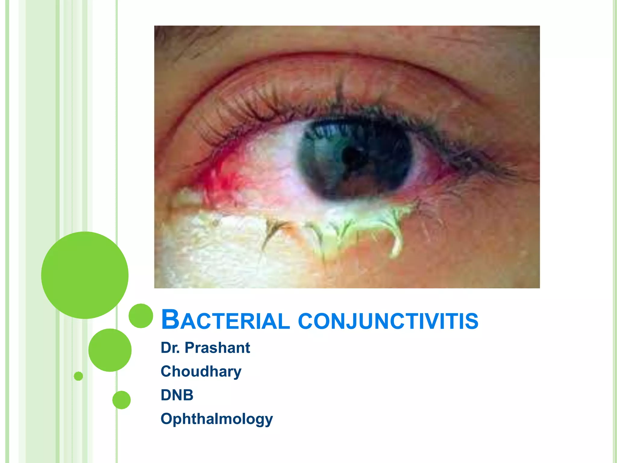
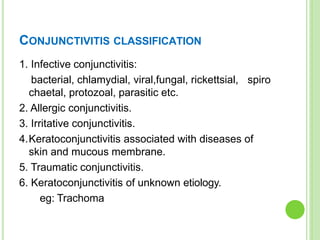
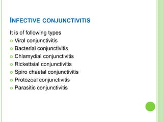
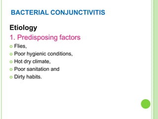
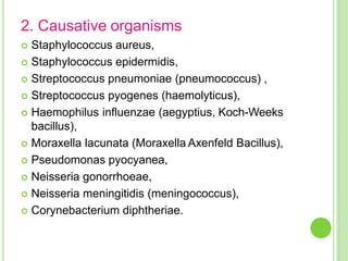
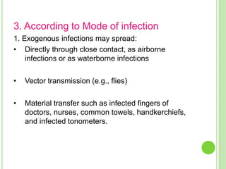
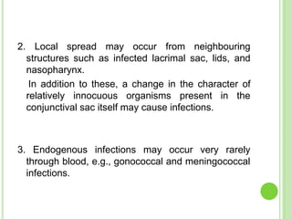
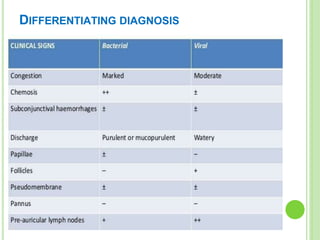
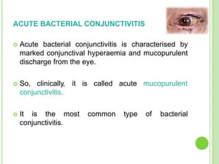
BACTERIAL CONJUNCTIVITIS
Dr. Prashant
Choudhary
DNB
Ophthalmology
CONJUNCTIVITIS CLASSIFICATION
1. Infective conjunctivitis:
bacterial, chlamydial, viral,fungal, rickettsial,
chaetal, protozoal, parasitic etc.
2. Allergic conjunctivitis.
3. Irritative conjunctivitis.
spiro
4.Keratoconjunctivitis associated with diseases of
skin and mucous membrane.
5. Traumatic conjunctivitis.
6. Keratoconjunctivitis of unknown etiology.
eg: Trachoma
INFECTIVE CONJUNCTIVITIS
It is of following types
 Viral conjunctivitis
 Bacterial conjunctivitis
 Chlamydial conjunctivitis
 Rickettsial conjunctivitis
 Spiro chaetal conjunctivitis
 Protozoal conjunctivitis
 Parasitic conjunctivitis
BACTERIAL CONJUNCTIVITIS
 Bacterial conjunctivitis is a common type of pink
eye, caused by bacteria that infect the eye through
various sources of contamination.
 The bacteria can be spread through contact with
an infected individual, exposure to contaminated
surfaces or through other means such as sinus or
ear infections.
BACTERIAL CONJUNCTIVITIS
Etiology
1. Predisposing factors
 Flies,
 Poor hygienic conditions,
 Hot dry climate,
 Poor sanitation and
 Dirty habits.
2. Causative organisms
 Staphylococcus aureus,
 Staphylococcus epidermidis,
 Streptococcus pneumoniae (pneumococcus) ,
 Streptococcus pyogenes (haemolyticus),
 Haemophilus influenzae (aegyptius, Koch-Weeks
bacillus),
 Moraxella lacunata (Moraxella Axenfeld Bacillus),
 Pseudomonas pyocyanea,
 Neisseria gonorrhoeae,
 Neisseria meningitidis (meningococcus),
 Corynebacterium diphtheriae.
3. According to Mode of infection
1. Exogenous infections may spread:
• Directly through close contact, as airborne
infections or as waterborne infections
• Vector transmission (e.g., flies)
• Material transfer such as infected fingers of
doctors, nurses, common towels, handkerchiefs,
and infected tonometers.
2. Local spread may occur from neighbouring
structures such as infected lacrimal sac, lids, and
nasopharynx.
In addition to these, a change in the character of
relatively innocuous organisms present in the
conjunctival sac itself may cause infections.
3. Endogenous infections may occur very rarely
through blood, e.g., gonococcal and meningococcal
infections.
PATHOPHYSIOLOGY
Vascular response
Cellular response
Conjuctival tissue response
Conjuctival discharge
DIFFERENTIATING DIAGNOSIS
CLINICAL TYPES OF BACTERIAL
CONJUNCTIVITIS
 Acute bacterial conjunctivitis,
 Hyperacute bacterial conjunctivitis,
 Chronic bacterial conjunctivitis, and
 Angular bacterial conjunctivitis.
ACUTE BACTERIAL CONJUNCTIVITIS
 Acute bacterial conjunctivitis is characterised by
marked conjunctival hyperaemia and mucopurulent
discharge from the eye.
 So, clinically, it is called acute mucopurulent
conjunctivitis.
 It is the most common type of bacterial
conjunctivitis.
CAUSATIVE AGENTS
 Staphylococcus aureus,
 Koch-Weeks Bacillus,
 Pneumococcus and
 Streptococcus.
 Mucopurulent conjunctivitis generally accompanies
exanthemata such as measles and scarlet fever.
CLINICAL FEATURES
Symptoms
 Discomfort, foreign body,
grittiness, blurring and redness
of sudden onset
 Mild photophobia
 Mucopurulent discharge from
the eyes
 Sticking together of lid margins
with discharge during sleep.
 Slight blurring of vision due to
mucous flakes in front of
cornea.
 Coloured halos
Signs
 Flakes of mucopus
 Congenctival congestion
‘fiery red eye’
 circumcorneal zone
congestion
 Chemosis
 Fine type Papillae
 Petechial haemorrhages
 Cilia with yellow crusts.
 Eyelids may be slightly
oedematous.
COMPLICATIONS
 Marginal corneal ulcer
 Superficial keratitis
 Blepharitis or dacryocystitis
TREATMENT
 Topical antibiotics- broad specturm antibiotics
 Irrigation of conjunctival sac
 Dark goggles
 No steroids should be applied
 No bandage
 Anti-inflammatory and analgesic drugs
ACUTE PURULENT CONJUNCTIVITIS
Other names
-Hyperacute bacterial conjunctivitis
-Acute blenorrhea
Clinical picture:
 Stage of infiltraton
 Stage of blenorrhoea
 Stage of slow healing
HYPERACUTE CONJUNCTIVITIS
OF ADULTS
(GONOCOCCAL CONJUNCTIVITIS)
Etiology
 The disease affects adults, predominantly males.
 Gonococcal infection directly spreads from genitals
to eye.
 Presently, incidence of gonococcal conjunctivitis
has markedly decreased.
CLINICAL FEATURE
Symptoms
 Onset is hyper
acute (12 to 24
hours)
 Pain which is
moderate to severe
 Purulent discharge,
which is usually
copious,
 Swelling of eyelids,
which is usually
marked.
Signs
 Tense and swollen eyelids
 Tenderness
 Discharge is thick purulent,
copius trickling down the cheeks.
 Chemosis, congestion and
papillae, giving bright red velvety
appearance.
 Pseudomembrane appearance
conjunctival surface
 Enlarged and tender preauricular
lymph nodes
 Urethritis and arthritis.
COMPLICATIONS
1. Corneal involvement - Diffuse haze and oedema,
central necrosis, corneal ulceration or even
perforation.
2. Iridocyclitis
3.Systemic complications - Gonorrhoea arthritis,
endocarditis and septicaemia.
TREATMENT
1. Systemic therapy
 Third generation cephalosporin
 Quinolones
 Spectinomycin
2. Topical antibiotic therapy
3. Irrigation
4. Other general measures
5. Topical atropine
CHRO NIC BACTERIAL CONJUNCTIVITIS/
CHRONIC CATARRHAL CONJUNCTIVITIS/ ‘SIMPLE
CHRONIC CONJUNCTIVITIS’
A. Predisposing factors
1. Chronic exposure to dust, smoke, and chemical
irritants.
2.Local cause of irritation such as trichiasis,
concretions, foreign body and seborrhoeic scales.
3.Eye strain due to refractive errors, phorias or
convergence insufficiency.
4.Abuse of alcohol, insomnia and metabolic
disorders.
B. Causative organisms
 Staphylococcus aureus is the commonest cause of
chronic bacterial conjunctivitis.
 Gram negative rods such as Proteus mirabilis,
Klebsiella pneumoniae, Escherichia coli and
Moraxella lacunata are other rare causes.
C. Source and mode of infection.
1. As continuation of acute mucopurulent conjunctivitis
when untreated or partially treated.
dacryocystitis,
2. As chronic infection
chronic
from associated
rhinitis or chronic
chronic
upper
respiratory catarrh.
3. As a mild exogenous infection which results from
direct contact, airborne or material transfer of
infection.
CLINICAL FEATURES
Symptoms
 Burning and grittiness in the
eyes, especially in the
evening.
 Mild chronic redness in the
eyes.
 Feeling of heat and dryness
on the lid margins.
 Difficulty in keeping the eyes
open.
 Mild mucoid discharge
 off and on Watering,
 Feeling of sleepiness and
tiredness in the eyes.
Signs
 Grossly the eyes look
normal but careful
examination may reveal
following signs:
 Congestion of posterior
conjunctival vessels.
 Mild papillary hypertrophy of
the palpebral conjunctiva.
 Sticky look of surface of the
conjunctiva.
 Lid margins may be
congested.
TREATMENT
 1. Eliminate predisposing factors when associated.
 2. Topical antibiotics such as chloramphenicol,
tobramycin or gentamicin should be instilled 3–4
times a day for about 2 weeks to eliminate the mild
chronic infection.
 3. Astringent eye drops such as zinc-boric acid
drops provide symptomatic relief.
ANGULAR BACTERIAL
CONJUNCTIVITIS
1.Predisposing factors are same as for ‘simple
chronic conjunctivitis’.
2. Causative organisms
Moraxella Axenfield (MA) is the commonest causative
organism.
MA bacilli are placed end to end, so the disease is
also called ‘diplobacillary conjunctivitis’.
Rarely, staphylococci may also cause angular
conjunctivitis.
3. Source of infection
- usually nasal cavity.
4. Mode of infection.
- Infection is transmitted from nasal cavity to the eyes
by contaminated fingers or handkerchief.
PATHOLOGY
 The causative organism, i.e., MA bacillus produces
a proteolytic enzyme which acts by macerating the
epithelium.
 This proteolytic enzyme collects at the angles by
the action of tears and thus macerates the
epithelium of the conjunctiva, lid margin and the
skin, the surrounding angles of eye.
 The maceration is followed by vascular and cellular
responses in the form of mild grade chronic
inflammation. Skin may show eczematous changes.
CLINICAL FEATURES
Symptoms
 Irritation
 Burning sensation and
feeling of discomfort in the
eyes
 History of collection of
dirty-white foamy
discharge at the angles
 Redness in the angles of
eyes.
Signs
 Hyperaemia of bulbar
conjunctiva near the
canthi.
 Hyperaemia of lid
margins near the angles
 Excoriation of the skin
around the angles.
 Foamy mucopurulent
discharge at the angles is
usually present.
COMPLICATIONS
 Blepharitis and
 Shallow marginal catarrhal corneal ulceration.
TREATMENT
A.Prophylaxis includes treatment of associated nasal
infection and good personal hygiene.
B. Curative treatment consists of:
 1. Oxytetracycline (1%) eye ointment, 2–3 times a
day for 9–14 days will eradicate the infection.
 2. Zinc lotion instilled in day time and zinc oxide
ointment at bed time inhibits the proteolytic ferment
and thus helps in reducing the maceration.
THANK YOU

Bacterialvmconj.pptx

  • 1.
  • 2.
    CONJUNCTIVITIS CLASSIFICATION 1. Infectiveconjunctivitis: bacterial, chlamydial, viral,fungal, rickettsial, chaetal, protozoal, parasitic etc. 2. Allergic conjunctivitis. 3. Irritative conjunctivitis. spiro 4.Keratoconjunctivitis associated with diseases of skin and mucous membrane. 5. Traumatic conjunctivitis. 6. Keratoconjunctivitis of unknown etiology. eg: Trachoma
  • 3.
    INFECTIVE CONJUNCTIVITIS It isof following types  Viral conjunctivitis  Bacterial conjunctivitis  Chlamydial conjunctivitis  Rickettsial conjunctivitis  Spiro chaetal conjunctivitis  Protozoal conjunctivitis  Parasitic conjunctivitis
  • 4.
    BACTERIAL CONJUNCTIVITIS  Bacterialconjunctivitis is a common type of pink eye, caused by bacteria that infect the eye through various sources of contamination.  The bacteria can be spread through contact with an infected individual, exposure to contaminated surfaces or through other means such as sinus or ear infections.
  • 5.
    BACTERIAL CONJUNCTIVITIS Etiology 1. Predisposingfactors  Flies,  Poor hygienic conditions,  Hot dry climate,  Poor sanitation and  Dirty habits.
  • 6.
    2. Causative organisms Staphylococcus aureus,  Staphylococcus epidermidis,  Streptococcus pneumoniae (pneumococcus) ,  Streptococcus pyogenes (haemolyticus),  Haemophilus influenzae (aegyptius, Koch-Weeks bacillus),  Moraxella lacunata (Moraxella Axenfeld Bacillus),  Pseudomonas pyocyanea,  Neisseria gonorrhoeae,  Neisseria meningitidis (meningococcus),  Corynebacterium diphtheriae.
  • 7.
    3. According toMode of infection 1. Exogenous infections may spread: • Directly through close contact, as airborne infections or as waterborne infections • Vector transmission (e.g., flies) • Material transfer such as infected fingers of doctors, nurses, common towels, handkerchiefs, and infected tonometers.
  • 8.
    2. Local spreadmay occur from neighbouring structures such as infected lacrimal sac, lids, and nasopharynx. In addition to these, a change in the character of relatively innocuous organisms present in the conjunctival sac itself may cause infections. 3. Endogenous infections may occur very rarely through blood, e.g., gonococcal and meningococcal infections.
  • 9.
  • 10.
  • 11.
    CLINICAL TYPES OFBACTERIAL CONJUNCTIVITIS  Acute bacterial conjunctivitis,  Hyperacute bacterial conjunctivitis,  Chronic bacterial conjunctivitis, and  Angular bacterial conjunctivitis.
  • 12.
    ACUTE BACTERIAL CONJUNCTIVITIS Acute bacterial conjunctivitis is characterised by marked conjunctival hyperaemia and mucopurulent discharge from the eye.  So, clinically, it is called acute mucopurulent conjunctivitis.  It is the most common type of bacterial conjunctivitis.
  • 13.
    CAUSATIVE AGENTS  Staphylococcusaureus,  Koch-Weeks Bacillus,  Pneumococcus and  Streptococcus.  Mucopurulent conjunctivitis generally accompanies exanthemata such as measles and scarlet fever.
  • 14.
    CLINICAL FEATURES Symptoms  Discomfort,foreign body, grittiness, blurring and redness of sudden onset  Mild photophobia  Mucopurulent discharge from the eyes  Sticking together of lid margins with discharge during sleep.  Slight blurring of vision due to mucous flakes in front of cornea.  Coloured halos Signs  Flakes of mucopus  Congenctival congestion ‘fiery red eye’  circumcorneal zone congestion  Chemosis  Fine type Papillae  Petechial haemorrhages  Cilia with yellow crusts.  Eyelids may be slightly oedematous.
  • 15.
    COMPLICATIONS  Marginal cornealulcer  Superficial keratitis  Blepharitis or dacryocystitis
  • 16.
    TREATMENT  Topical antibiotics-broad specturm antibiotics  Irrigation of conjunctival sac  Dark goggles  No steroids should be applied  No bandage  Anti-inflammatory and analgesic drugs
  • 17.
    ACUTE PURULENT CONJUNCTIVITIS Othernames -Hyperacute bacterial conjunctivitis -Acute blenorrhea Clinical picture:  Stage of infiltraton  Stage of blenorrhoea  Stage of slow healing
  • 18.
    HYPERACUTE CONJUNCTIVITIS OF ADULTS (GONOCOCCALCONJUNCTIVITIS) Etiology  The disease affects adults, predominantly males.  Gonococcal infection directly spreads from genitals to eye.  Presently, incidence of gonococcal conjunctivitis has markedly decreased.
  • 19.
    CLINICAL FEATURE Symptoms  Onsetis hyper acute (12 to 24 hours)  Pain which is moderate to severe  Purulent discharge, which is usually copious,  Swelling of eyelids, which is usually marked. Signs  Tense and swollen eyelids  Tenderness  Discharge is thick purulent, copius trickling down the cheeks.  Chemosis, congestion and papillae, giving bright red velvety appearance.  Pseudomembrane appearance conjunctival surface  Enlarged and tender preauricular lymph nodes  Urethritis and arthritis.
  • 20.
    COMPLICATIONS 1. Corneal involvement- Diffuse haze and oedema, central necrosis, corneal ulceration or even perforation. 2. Iridocyclitis 3.Systemic complications - Gonorrhoea arthritis, endocarditis and septicaemia.
  • 21.
    TREATMENT 1. Systemic therapy Third generation cephalosporin  Quinolones  Spectinomycin 2. Topical antibiotic therapy 3. Irrigation 4. Other general measures 5. Topical atropine
  • 22.
    CHRO NIC BACTERIALCONJUNCTIVITIS/ CHRONIC CATARRHAL CONJUNCTIVITIS/ ‘SIMPLE CHRONIC CONJUNCTIVITIS’ A. Predisposing factors 1. Chronic exposure to dust, smoke, and chemical irritants. 2.Local cause of irritation such as trichiasis, concretions, foreign body and seborrhoeic scales. 3.Eye strain due to refractive errors, phorias or convergence insufficiency. 4.Abuse of alcohol, insomnia and metabolic disorders.
  • 23.
    B. Causative organisms Staphylococcus aureus is the commonest cause of chronic bacterial conjunctivitis.  Gram negative rods such as Proteus mirabilis, Klebsiella pneumoniae, Escherichia coli and Moraxella lacunata are other rare causes.
  • 24.
    C. Source andmode of infection. 1. As continuation of acute mucopurulent conjunctivitis when untreated or partially treated. dacryocystitis, 2. As chronic infection chronic from associated rhinitis or chronic chronic upper respiratory catarrh. 3. As a mild exogenous infection which results from direct contact, airborne or material transfer of infection.
  • 25.
    CLINICAL FEATURES Symptoms  Burningand grittiness in the eyes, especially in the evening.  Mild chronic redness in the eyes.  Feeling of heat and dryness on the lid margins.  Difficulty in keeping the eyes open.  Mild mucoid discharge  off and on Watering,  Feeling of sleepiness and tiredness in the eyes. Signs  Grossly the eyes look normal but careful examination may reveal following signs:  Congestion of posterior conjunctival vessels.  Mild papillary hypertrophy of the palpebral conjunctiva.  Sticky look of surface of the conjunctiva.  Lid margins may be congested.
  • 26.
    TREATMENT  1. Eliminatepredisposing factors when associated.  2. Topical antibiotics such as chloramphenicol, tobramycin or gentamicin should be instilled 3–4 times a day for about 2 weeks to eliminate the mild chronic infection.  3. Astringent eye drops such as zinc-boric acid drops provide symptomatic relief.
  • 27.
    ANGULAR BACTERIAL CONJUNCTIVITIS 1.Predisposing factorsare same as for ‘simple chronic conjunctivitis’. 2. Causative organisms Moraxella Axenfield (MA) is the commonest causative organism. MA bacilli are placed end to end, so the disease is also called ‘diplobacillary conjunctivitis’. Rarely, staphylococci may also cause angular conjunctivitis.
  • 28.
    3. Source ofinfection - usually nasal cavity. 4. Mode of infection. - Infection is transmitted from nasal cavity to the eyes by contaminated fingers or handkerchief.
  • 29.
    PATHOLOGY  The causativeorganism, i.e., MA bacillus produces a proteolytic enzyme which acts by macerating the epithelium.  This proteolytic enzyme collects at the angles by the action of tears and thus macerates the epithelium of the conjunctiva, lid margin and the skin, the surrounding angles of eye.  The maceration is followed by vascular and cellular responses in the form of mild grade chronic inflammation. Skin may show eczematous changes.
  • 30.
    CLINICAL FEATURES Symptoms  Irritation Burning sensation and feeling of discomfort in the eyes  History of collection of dirty-white foamy discharge at the angles  Redness in the angles of eyes. Signs  Hyperaemia of bulbar conjunctiva near the canthi.  Hyperaemia of lid margins near the angles  Excoriation of the skin around the angles.  Foamy mucopurulent discharge at the angles is usually present.
  • 31.
    COMPLICATIONS  Blepharitis and Shallow marginal catarrhal corneal ulceration.
  • 32.
    TREATMENT A.Prophylaxis includes treatmentof associated nasal infection and good personal hygiene. B. Curative treatment consists of:  1. Oxytetracycline (1%) eye ointment, 2–3 times a day for 9–14 days will eradicate the infection.  2. Zinc lotion instilled in day time and zinc oxide ointment at bed time inhibits the proteolytic ferment and thus helps in reducing the maceration.
  • 33.