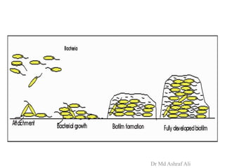
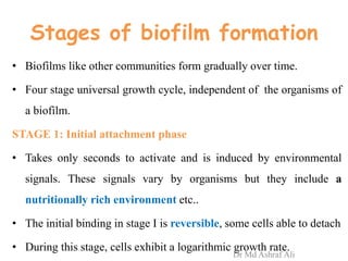
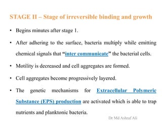
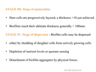
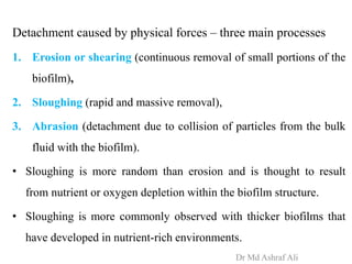
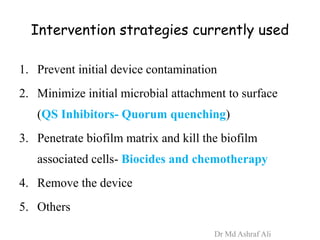
BIOFILMS
Microbial life on surfaces
• INTRODUCTION
• DEFINITION
• CHARACTERISTICS
• BIOFILM STRUCTURE
• FORMATION
• QUORUM SENSING
• BIOFILMS ON MEDICAL DEVICES
• RESISTANCE TO ANTIMICROBIAL AGENTS
• METHODS OF DETECTION
• INTERVENTION STRATEGIES
• FUTURE PROSPECTS Dr Md Ashraf Ali
Introduction
• Higher organisms, including humans are known to
flock together and form colonies for various reasons.
• Even microbes also could form colonies and function
as groups and that individuals within the group could
respond to the group, as a whole.
Dr Md Ashraf Ali
Signals:
• This phenomenon is mediated through various
chemical signals facilitating interaction and
coordination of group bacterial activities.
• This phenomenon is known as “quorum sensing” and
often results in the formation of physical structures
with unique characteristics known as “biofilms”.
Dr Md Ashraf Ali
History
• In 1684, Antony van leeuwenhook utilizing one of his own
microscopes remarked in a report to the Royal Society of London on
the vast no. of microorganisms he was able to see in dental plaque :
"The number of these animalcules in the scurf of a man's teeth are
so many that I believe they exceed the number of men in a
kingdom."
• In some ways it's too bad that his early work did not receive more
recognition in our day.
• The word scurf used by van Leewenhoek,much more colorful
than biofilm and animalcules much cuter than microbes.
Dr Md Ashraf Ali
History
• 1978- The term ‘Biofilm’ was coined by Bill Costerton.
• Heukelekian and Heller observed the “bottle effect” for marine
microorganisms, i.e., bacterial growth and activity were
enhanced by the incorporation of a surface to which these
organisms could attach.
• In 2002, Donlan and Costerton offered the most salient
description of a biofilm.
Dr Md Ashraf Ali
DEFINITION
• Biofilm is a sessile community of microbes, that are
irreversibly attached to a surface or to each other, and
are embedded in a matrix of Extracellular Polymeric
Substances (EPS) produced by the organism.
• Exhibit an altered phenotype with respect to growth
rate and gene transcription.
Dr Md Ashraf Ali
Differences from their free
living counterparts
1. Generation of EPS
2. Reduced growth rates
3. Regulation of certain genes.
Dr Md Ashraf Ali
Characteristics
Several aspects of biofilms make their formation a clinically
relevant process:
1. Protection: Resistant to antimicrobial agents;
2. Plasmid- They may allow exchange of resistance plasmids.
3. Persistent source of infection;
4. Pathogenic organisms- may be harbored
Dr Md Ashraf Ali
Pathogenic Mechanisms
• Evade host defenses such as phagocytosis;
• Obtain a high density of microorganisms;
• “Division of labor” increases metabolic efficiency of the
community;
• Exchange genes that can result in more virulent strains of
microorganisms;
• Produce a large concentration of toxins;
• Detachment of microbial aggregates transmits microbes to other
sites. Dr Md Ashraf Ali
STRUCTURE
Consists of
• Extracellular Polymeric Substance (EPS)
• Microbial cells
– monomicrobial aggregation- single organism
– polymicrobial aggergation- numerous species
Dr Md Ashraf Ali
Extracellular polymeric substances (EPSs)
• Extracellular polymeric substances (EPSs) consist primarily of
polysaccharides. EPSs provide the matrix or structure for the
biofilm.
• They are highly hydrated (98% water) and tenaciously bound to
the underlying surface.
• Has “water channels” that allow transport of essential nutrients
and oxygen to the cells growing within the biofilm
• Biofilms act as filters to entrap particles of various kinds,
including minerals and host components such as fibrin, RBCs, and
platelets. Dr Md Ashraf Ali
• EPS may associate with metal ions, divalent cations, other
macromolecules (such as proteins, DNA, lipids, and even
humic substances).
• EPS production is known to be affected by nutrient status of
the growth medium; excess available carbon promote EPS
synthesis and excess nitrogen, potassium, or phosphate
decrease its production.
• Slow bacterial growth will also enhance EPS production
Dr Md Ashraf Ali
Microorganism Isolated from
Candida albicans Artificial voice prosthesis
Central venous catheter
Intrauterine devices
Coagulase-negative
staphylococci
Artificial hip prosthesis
Artificial voice prosthesis
Central venous catheter
Intrauterine devices
Prosthetic heart valve
Urinary catheter
Enterococcus spp. Artificial hip prosthesis
Central venous catheter
Intrauterine devices
Prosthetic heart valve
Urinary catheter
Dr Md Ashraf Ali
Klebsiella pneumonia Central venous catheter
Urinary catheter
Pseudomonas aeruginosa Artificial hip prosthesis
Central venous catheter
Urinary catheter
Staphylococcus aureus Artificial hip prosthesis
Central venous catheter
Intrauterine devices
Prosthetic heart valve
Dr Md Ashraf Ali
BIOFILM FORMATION
Dr Md Ashraf Ali
Dr Md Ashraf Ali
Stages of biofilm formation
• Biofilms like other communities form gradually over time.
• Four stage universal growth cycle, independent of the organisms of
a biofilm.
STAGE 1: Initial attachment phase
• Takes only seconds to activate and is induced by environmental
signals. These signals vary by organisms but they include a
nutritionally rich environment etc..
• The initial binding in stage I is reversible, some cells able to detach
• During this stage, cells exhibit a logarithmic growth rate.
Dr Md Ashraf Ali
STAGE II – Stage of irreversible binding and growth
• Begins minutes after stage 1.
• After adhering to the surface, bacteria multiply while emitting
chemical signals that “inter communicate” the bacterial cells.
• Motility is decreased and cell aggregates are formed.
• Cell aggregates become progressively layered.
• The genetic mechanisms for Extracellular Polymeric
Substance (EPS) production are activated which is able to trap
nutrients and planktonic bacteria.
Dr Md Ashraf Ali
STAGE III- Stage of maturation
• Here cells are progressively layered, a thickness >10 μm achieved.
• Biofilms reach their ultimate thickness generally > 100mm.
STAGE IV- Stage of dispersion - Biofilm cells may be dispersed
• either by shedding of daughter cells from actively growing cells
• Depletion of nutrient levels or quorum sensing
• Detachment of biofilm aggregates by physical forces.
Dr Md Ashraf Ali
Detachment caused by physical forces – three main processes
1. Erosion or shearing (continuous removal of small portions of the
biofilm),
2. Sloughing (rapid and massive removal),
3. Abrasion (detachment due to collision of particles from the bulk
fluid with the biofilm).
• Sloughing is more random than erosion and is thought to result
from nutrient or oxygen depletion within the biofilm structure.
• Sloughing is more commonly observed with thicker biofilms that
have developed in nutrient-rich environments.
Dr Md Ashraf Ali
Dr Md Ashraf Ali
Attachment
• Depends on the following properties.
1. Properties of the substratum / surface
2. Conditioning films forming on the substratum
3. Hydrodynamics of the aqueous medium
4. Characteristics of the medium
5. Properties of the cell surface.
Dr Md Ashraf Ali
1. The substratum / surface
• Significant effect on rate and extent of attachment of microbes.
• In general the rougher and more hydrophobic materials will
develop biofilms more rapidly. (although there are exceptions).
hydrophobic materials hydrophilic materials
1. Teflon
2. various plastics
3. latex
4. silicone
1. Glass
2. various metals
Dr Md Ashraf Ali
2. Conditioning Films
• A material surface exposed in an aqueous medium will
immediately become conditioned or coated by polymers from
that medium, and the resulting in formation of a FILM.
Examples
• A proteinaceous conditioning film which develops on tooth
enamel and comprises albumin, lysozyme, glycoproteins, etc.
Bacteria from oral cavity colonize these surfaces within hours.
• Other host-produced conditioning films are blood, urine, saliva,
and respiratory secretions.
Dr Md Ashraf Ali
4. Characteristics of the Aqueous Medium
• Concentration of cations - an increase in (sodium, calcium,
ferric iron) affects the attachment of Pseudomonas fluorescens
to glass surfaces. Cations cross-link the anionic groups of EPS.
• Nutrient concentration - an increase is correlated with an
increase in the number of attached bacterial cells.
• pH- Increased pH lead to a higher biofilm production
Dr Md Ashraf Ali
5. Properties of the Cell
Adhesion was greater on hydrophobic materials.
1. Presence of flagella facilitated attachment of gram-negative
bacteria to surfaces.
2. Fimbriae, i.e., nonflagellar appendages other than those
involved in transfer of viral or bacterial nucleic acids (pili),
contribute to cell surface hydrophobicity.
3. Cell surface hydrophobicity
Dr Md Ashraf Ali
Other factors contributing to biofilm
formation
• Quorum sensing
Dr Md Ashraf Ali
Quorum Sensing: Cell-cell Communication
• The process by which bacteria coordinate their behaviors in a
density-dependent manner, using signaling molecules.
• “Quorate”--- ‘Having the necessary number of people present
for decisions to be allowed to be made’
• 3 well described systems
– Autoinducer- 1(AI-1) system (Acyl-HSL)
– Peptide –based
– Autoinducer- 2(AI-2) system
Dr Md Ashraf Ali
AI-1 System
• Gram - negative bacteria
• Intraspecies communication
• Acylated Homoserine Lactone
AI-2 System (Autoinducer 2)
• Gram negative and gram positive bacteria
• Interspecies communication
• Furanosyl borate diester
Dr Md Ashraf Ali
• most of the above interactions probably evolved to allow co-
existence of a bacterium and its host
• pathogens seem to have ‘hijacked’ these communication
systems to control their virulence traits.
• Quorum sensing inhibition or quorum quenching has been
pursued as one of such novel strategies. While antibiotics kill
or slow down the growth of bacteria, quorum sensing
inhibitors (QSIs) or quorum quenchers (QQs) attenuate
bacterial virulence.
Dr Md Ashraf Ali
BIOFILMS ON MEDICAL DEVICES
• Prosthetic heart valves
• Central venous Catheters
• Urinary Cathters
• Contact lense
• Intrauterine devices
Dr Md Ashraf Ali
Prosthetic heart valves
Prosthetic valve endocarditis
• Microbial infection of the valve and surrounding tissues of heart.
• Early colonizers- preominantly CoNS
• Late PVE (>12m of valve replacement)
– Streptococci- viridans group
– CoNS
– S. aureus
– GNB
– Fungi- Candida
• Detection: Blood culture, transesophageal echo(biofilm)
Dr Md Ashraf Ali
Central venous catheters
• Risk greater than any other indwelling medical device (Blood)
• Biofilms present
– External surface (organisms originate from skin insertion site)
– Inner lumen (Manipulation by health care worker)
• As device is in direct contact with bloodstream (which acts as a
conditioning film).
• Colonization and biofilm formation may occur within 3 days
• Organisms associated
– S. epidermidis
– S. aureus
– P.aeruginosa
– K.pneumonia
– E.faecalis
– Candida albicans
Dr Md Ashraf Ali
Methods of Detection of biofilm in CVC
Method Procedure Advantage Limitation
Roll-plate -Distal tip of
catheter removed
-Roll the catheter
tip over Blood agar
plate
East to perform Only outer
surface
biofilm
Vortexing of
catheter tips
Catheter section in
PBS, vortexed and
cultured
Measures
intraluminal and
extraluminal
biofilm
Acridine
orange direct
staininng
Following culture,
catheter tips can be
stained with
acridine orange
Rapid results No
quantification
Dr Md Ashraf Ali
Urinary catheters
• Initially cathters are colonized by single species, such as
S.epidermidis, Enterococcus faecalis, E.coli or Proteus mirabilis.
• As the catheter remains in place, the no. & diversity of organisms
increase.
• Mixed communities develop, containing organisms such as
Providencis stuartii, P.aeruginosa, Proteus mirabilis, Klebsiella
pneumoniae and Enterobacter aerogenes.
• Rare organisms include Acinetobacter calcoaceticus, M.morganii.
Dr Md Ashraf Ali
• Urease producing organisms like Proteus spp, K. Pneumoniae
urea ammonia Increase pH precipitation of
minerals in catheter biofilms
(k/a mineral encrustation)
Catheter
blockage
Dr Md Ashraf Ali
Contact lenses
Organisms
• P.aeruginosa, S.aureus, S.epidermidis, Serratia spp, E.coli, Proteus
spp., and Candida spp
• Soft and hard contact lenses, in both biofilms can be formed
• Biofilms develop on contact lens storage cases.
• Lens case has been implicated as primary source of organisms
– Biofilm related keratitis
– Contamination of lens disinfectant solutions
• Acanthamoeba may be a component of these biofilms, these
organisms feed on the biofilm bacteria and cause microbial keratitis
Dr Md Ashraf Ali
Intrauterine devices (IUD) - Pelvic inflammatory Diseases
IUD have a tail that fcilitates locating the device for removal
• IUD removed form asymptomatic woman have been contaminated
with S.epidermidis, Enterococci and anaerobic lactobacilli
• IUD removed from women wiith PID have shown to contain β-
hemolytic streptococci, S.aureus, E.coli and some anaerobes.
• Tail portion of IUD may be the primary source of contamination
• Tail portion can act as a wick to allow bacterianto travel by
capillary action into the endometrial cavity
Dr Md Ashraf Ali
ANTIBIOTIC RESISTANCE
Mechanisms responsible for resistance may be one or more of
the following:
1. delayed penetration of the antimicrobial agent through the
biofilm matrix.
2. altered growth rate of biofilm organisms.
3. other physiological changes due to the biofilm mode of growth.
Dr Md Ashraf Ali
Pseudomonas aeruginosa Biofilms
• principal pathogen in lungs of patients wid cystic fibrosis (CF).
• Biofilm matrix of Pseudomonas strains produce alginate
• Two new polymers have been found in Pseudomonas polymers
– pelA-G gene produces a glucose rich polymer
– pslA-O genes produce a mannose rich polymer
• In P.aeruginosa; a complex quorum sensing hierarchy plays a
central role in the regulation of virulence and contributes to the
late stages of biofilm maturation.
• P. aeruginosa possess 2 AHL – dependent quorum sensing systems
• Antibiotic therapy in patients colonized with P. aeruginosa often
gives a measure of relief from symptoms but fails to cure
infection Dr Md Ashraf Ali
Staphylococcal Biofilms
• A hallmark is the production of the slime substance PIA
(polysaccharide intercellular adhesion)
• a polysaccharide, in which the cells are embedded and protected
against the host’s immune defence and antibiotic treatment
• Proteins have been identified that are also involved in biofilm
formation
– accumulation-associated protein (AAP),
– the clumping factor A (Clf A),
– the staphylococcal surface protein (SSP1)
– biofilm associated protein (Bap).
• Intercellular adhesions with in biofilms of Staphylococcus
epidermidis, a major cause of medical device related infections,
is mediated by the PIA
Dr Md Ashraf Ali
Candida Biofilms
• Most manifestations of candidiasis are associated with the
formation of Candida biofilms on surfaces and it is also
associated with infections at both mucosal and systemic sites.
• Candida biofilms share several properties with bacterial biofilms.
• C. albicans biofilm formation has 3 distinct developmental
phases: early (0-11 h), intermediate (12-30 h) and mature (38-
72h).
• The detailed structure of mature C. albicans biofilms consists of a
dense network of yeast, hyphae and pseudohyphae.
Dr Md Ashraf Ali
• C. dubliniensis has the ability to adhere to and form biofilms with
typical microcolony and water channel architecture similar to
bacterial biofilms and C. albicans biofilms.
• Indwelling intravascular catheters represent a risk factor that is
associated with nosocomial Candida infections.
• The devices become colonised by the microorganisms that form a
biofilm of cells, the detachment of which results in septicaemia.
• Antifungal drug resistance- Major genes that contribute to drug
resistance in C.albicans and C. dubliniensis are CDR genes (CDR 1
and CDR 2) and MDR genes. These genes have been demonstrated
to be upregulated during biofilm formation and development
Dr Md Ashraf Ali
METHODS OF DETECTION
Dr Md Ashraf Ali
MICROSCOPY
1. Electron microscope
• Scanning electron microscope
• Transmission electron microscope
• Confocal electron microscope
2. Epi-fluorescence microscope
RECOVERYAND MEASUREMENT
1. Tissue culture plate method
2. Tube method
3. Congo Red Agar method
Dr Md Ashraf Ali
Scanning electron microscope
Dehydration of samples required
• Alcohol, acetone and xylene
Importance
• Early investigative work on biofilms
was done.
Limitation
• Sample distortion and artifacts
• EPS will appear as Fibers, rather than
as gelatinous matrix
• Nature of these extracellular fibres
couldn’t be appreciated
• No details about internal composition
Dr Md Ashraf Ali
• Scanning electron micrograph of a staphylococcal biofilm on the
inner surface of an indwelling medical device.
Courtesy: Lippincott Williams & Wilkins.Dr Md Ashraf Ali
Transmission electon microscope along with
polysaccharide (EPS) stains like ruthenium red
• Identifies nature of extracellular fibres in
biofilms
Dr Md Ashraf Ali
Confocal laser scanning
microscope
• Organisms stained with a
fluorescennt dye
• Biofilm matrix unaltered and
intact
• Images are acquired point by
point and reconstructed with a
computer
• 3D reconstructions can be
obtained
Dr Md Ashraf Ali
Panels depict 3D
reconstructions of confocal
stacks of images of 24h (A),
48h (B) and 72h (C)
biofilms of C. albicans
grown on cover slips
Dr Md Ashraf Ali
Fluorescence microscopy
Stains used are
• Nucleic acid stains
– DAPI (di-amidino-phenyl indole)
– Acridine orange
– Syto 9
• These will stain DNA and RNA regardless of their viabilty.
• Propidium Iodide is taken up by cells with damaged cell
membrane.
• Cyano-tetrazolium chloride is taken up and metabolized only
by viable cells – helps to quantify the viable cells.
Dr Md Ashraf Ali
Polymicrobic biofilm stained with 4,6-diamidino-2-phenylindole
(DAPI) and examined by epifluorescence microscopyDr Md Ashraf Ali
Recovery of organisms from biofilm
Tissue culture plate method
• A quantitative test
• Considered the gold standard method for biofilm
detection.
Procedure
• Organisms isolated from fresh agar plates are inoculated
in 10 mL of trypticase soy broth with 1% glucose.
• Broths are incubated at 37°C for 24 h.
Dr Md Ashraf Ali
• The cultures are diluted 1:100 with fresh broth.
• Microtitre plate wells filled with 200 μL of diluted cultures.
• The plates incubated at 37°C for 24 h.
• After incubation, contents of each well removed by gentle
tapping.
• The wells washed with 0.2 mL of phosphate buffer saline to
remove free floating bacteria.
Dr Md Ashraf Ali
• Biofilm formed by bacteria adherent to the wells fixed by
2% sodium acetate
• stained by crystal violet (0.1%).
• Excess stain removed and Optical density (OD) of
stained adherent biofilm obtained.
• Interpretation: the higher the stain intensity, the more
biofilm present
Dr Md Ashraf Ali
Tube method
• Entire procedure same
as TCP ,
• Insteead of microtitre
plate the test carried out
in a test tube
Dr Md Ashraf Ali
Congo Red Agar method
• simple qualitative method to detect biofilm
production by using Congo Red Agar (CRA)
medium.
• CRA medium is prepared with brain heart
infusion broth, sucrose, agar and Congo Red
indicator.
• CRA plates were inoculated with test organisms
and incubated at 37°C for 24 h aerobically.
Dr Md Ashraf Ali
• Black colonies with a dry crystalline consistency indicated
biofilm production.
Disdvantage
• Low sensitivity
Dr Md Ashraf Ali
Other methods for deetection
Bioluminescent assay
• Monitor the conditioning films that facilitate early harbing of
biofilm formation.
Piezoelectric Sensors
• Such as quart with crystal microbalances monitor
• Detects mass accumulation on sensors
• Frequencyn shifts are observed as mass accumulates on the
sensor surface.
Dr Md Ashraf Ali
INTERVENTIONAL
STRATEGIES
Dr Md Ashraf Ali
Intervention strategies currently used
1. Prevent initial device contamination
2. Minimize initial microbial attachment to surface
(QS Inhibitors- Quorum quenching)
3. Penetrate biofilm matrix and kill the biofilm
associated cells- Biocides and chemotherapy
4. Remove the device
5. Others
Dr Md Ashraf Ali
1. Prevent device contamination
• CVC coateed with silver sulfa and chlorhexidine
• Prosthetic valves- Silizone coating
• Urinary catheters- silver oxide coating
Dr Md Ashraf Ali
2. Quorum quenching
QS system is a target for anti-virulence therapy
• In contrast to classic antibiotics, quorum-quenching compounds
are inhibitors of bacterial virulence, rather than of bacterial
growth.
• first studies on QS inhibitors, the halogenated furanones
• Two compounds, named yayurea A and B, excreted from
Staphylococcus delphini quench the QS regulation in a wide
spectrum of Gram-negative bacteria
• Apolipoprotein B & AIP Sequesters against Staphylococcal
biofilms Dr Md Ashraf Ali
Against pseudomonal biofilms
• QS signal antagonists- against lasR gene
• Ionic silver
– High concentrations of ionic silver disperses biofilms
• Gallium- compete out Fe, an important factor for initiation of
biofilm formation
• NO-releasing silica nanoparticles
– Silica nanoparticles provide a means for rapid diffusion of
toxic NO for better biofilm dispersion
Dr Md Ashraf Ali
3. Agents for the destruction of
biofilms( Industrial biocides)
• Alexidine, chlorhexidine, polyhexamethylene biguanides,
monophenylethers (phenoxyethanol)
• Quaternary amonium compounds (cetrimide, benzalkoniums)
and have demonstrated biochemical bases for the activities and
associated mammalian cell toxicities of thiol interactive agents
(bronopol, isothiazolones).
Dr Md Ashraf Ali
Other Possible Strategies for Eradication of Biofilms
For eradication, combination of strategies have been used:
1. Mechanical disruption / removal (sonication);
2. Immune modulation (Azithromycin and low dose doxycycline);
3. Antimicrobial agents (silver and tobramycin);
4.Amphotericin B lipid formulations and the Echinocandins
against the Candida biofilms.
• The effective control will require a concerted effort to develop
therapeutic agents that target both the biofilm phenotype and the
community signalling
Dr Md Ashraf Ali
FUTURE PROSPECTS
Dr Md Ashraf Ali
Bacteriophages (possible treatment?)
—◦ Are ubiquitous
◦ Are viruses that infect bacteria
◦ highly specific as to which bacteria they infect
◦ “enemies” of biofilm-associated bacteria
◦ Pose no risk to mammals
◦ Have potential to infect bacteria in a biofilm
themselves die in mammalian tissue
◦ Are possible therapy against multi-drug resistant
strains of many bacteria
Dr Md Ashraf Ali
Dr Md Ashraf Ali

Biofilms ashraf ..

  • 1.
  • 2.
    • INTRODUCTION • DEFINITION •CHARACTERISTICS • BIOFILM STRUCTURE • FORMATION • QUORUM SENSING • BIOFILMS ON MEDICAL DEVICES • RESISTANCE TO ANTIMICROBIAL AGENTS • METHODS OF DETECTION • INTERVENTION STRATEGIES • FUTURE PROSPECTS Dr Md Ashraf Ali
  • 3.
    Introduction • Higher organisms,including humans are known to flock together and form colonies for various reasons. • Even microbes also could form colonies and function as groups and that individuals within the group could respond to the group, as a whole. Dr Md Ashraf Ali
  • 4.
    Signals: • This phenomenonis mediated through various chemical signals facilitating interaction and coordination of group bacterial activities. • This phenomenon is known as “quorum sensing” and often results in the formation of physical structures with unique characteristics known as “biofilms”. Dr Md Ashraf Ali
  • 5.
    History • In 1684,Antony van leeuwenhook utilizing one of his own microscopes remarked in a report to the Royal Society of London on the vast no. of microorganisms he was able to see in dental plaque : "The number of these animalcules in the scurf of a man's teeth are so many that I believe they exceed the number of men in a kingdom." • In some ways it's too bad that his early work did not receive more recognition in our day. • The word scurf used by van Leewenhoek,much more colorful than biofilm and animalcules much cuter than microbes. Dr Md Ashraf Ali
  • 6.
    History • 1978- Theterm ‘Biofilm’ was coined by Bill Costerton. • Heukelekian and Heller observed the “bottle effect” for marine microorganisms, i.e., bacterial growth and activity were enhanced by the incorporation of a surface to which these organisms could attach. • In 2002, Donlan and Costerton offered the most salient description of a biofilm. Dr Md Ashraf Ali
  • 7.
    DEFINITION • Biofilm isa sessile community of microbes, that are irreversibly attached to a surface or to each other, and are embedded in a matrix of Extracellular Polymeric Substances (EPS) produced by the organism. • Exhibit an altered phenotype with respect to growth rate and gene transcription. Dr Md Ashraf Ali
  • 8.
    Differences from theirfree living counterparts 1. Generation of EPS 2. Reduced growth rates 3. Regulation of certain genes. Dr Md Ashraf Ali
  • 9.
    Characteristics Several aspects ofbiofilms make their formation a clinically relevant process: 1. Protection: Resistant to antimicrobial agents; 2. Plasmid- They may allow exchange of resistance plasmids. 3. Persistent source of infection; 4. Pathogenic organisms- may be harbored Dr Md Ashraf Ali
  • 10.
    Pathogenic Mechanisms • Evadehost defenses such as phagocytosis; • Obtain a high density of microorganisms; • “Division of labor” increases metabolic efficiency of the community; • Exchange genes that can result in more virulent strains of microorganisms; • Produce a large concentration of toxins; • Detachment of microbial aggregates transmits microbes to other sites. Dr Md Ashraf Ali
  • 11.
    STRUCTURE Consists of • ExtracellularPolymeric Substance (EPS) • Microbial cells – monomicrobial aggregation- single organism – polymicrobial aggergation- numerous species Dr Md Ashraf Ali
  • 12.
    Extracellular polymeric substances(EPSs) • Extracellular polymeric substances (EPSs) consist primarily of polysaccharides. EPSs provide the matrix or structure for the biofilm. • They are highly hydrated (98% water) and tenaciously bound to the underlying surface. • Has “water channels” that allow transport of essential nutrients and oxygen to the cells growing within the biofilm • Biofilms act as filters to entrap particles of various kinds, including minerals and host components such as fibrin, RBCs, and platelets. Dr Md Ashraf Ali
  • 13.
    • EPS mayassociate with metal ions, divalent cations, other macromolecules (such as proteins, DNA, lipids, and even humic substances). • EPS production is known to be affected by nutrient status of the growth medium; excess available carbon promote EPS synthesis and excess nitrogen, potassium, or phosphate decrease its production. • Slow bacterial growth will also enhance EPS production Dr Md Ashraf Ali
  • 14.
    Microorganism Isolated from Candidaalbicans Artificial voice prosthesis Central venous catheter Intrauterine devices Coagulase-negative staphylococci Artificial hip prosthesis Artificial voice prosthesis Central venous catheter Intrauterine devices Prosthetic heart valve Urinary catheter Enterococcus spp. Artificial hip prosthesis Central venous catheter Intrauterine devices Prosthetic heart valve Urinary catheter Dr Md Ashraf Ali
  • 15.
    Klebsiella pneumonia Centralvenous catheter Urinary catheter Pseudomonas aeruginosa Artificial hip prosthesis Central venous catheter Urinary catheter Staphylococcus aureus Artificial hip prosthesis Central venous catheter Intrauterine devices Prosthetic heart valve Dr Md Ashraf Ali
  • 16.
  • 17.
  • 18.
    Stages of biofilmformation • Biofilms like other communities form gradually over time. • Four stage universal growth cycle, independent of the organisms of a biofilm. STAGE 1: Initial attachment phase • Takes only seconds to activate and is induced by environmental signals. These signals vary by organisms but they include a nutritionally rich environment etc.. • The initial binding in stage I is reversible, some cells able to detach • During this stage, cells exhibit a logarithmic growth rate. Dr Md Ashraf Ali
  • 19.
    STAGE II –Stage of irreversible binding and growth • Begins minutes after stage 1. • After adhering to the surface, bacteria multiply while emitting chemical signals that “inter communicate” the bacterial cells. • Motility is decreased and cell aggregates are formed. • Cell aggregates become progressively layered. • The genetic mechanisms for Extracellular Polymeric Substance (EPS) production are activated which is able to trap nutrients and planktonic bacteria. Dr Md Ashraf Ali
  • 20.
    STAGE III- Stageof maturation • Here cells are progressively layered, a thickness >10 μm achieved. • Biofilms reach their ultimate thickness generally > 100mm. STAGE IV- Stage of dispersion - Biofilm cells may be dispersed • either by shedding of daughter cells from actively growing cells • Depletion of nutrient levels or quorum sensing • Detachment of biofilm aggregates by physical forces. Dr Md Ashraf Ali
  • 21.
    Detachment caused byphysical forces – three main processes 1. Erosion or shearing (continuous removal of small portions of the biofilm), 2. Sloughing (rapid and massive removal), 3. Abrasion (detachment due to collision of particles from the bulk fluid with the biofilm). • Sloughing is more random than erosion and is thought to result from nutrient or oxygen depletion within the biofilm structure. • Sloughing is more commonly observed with thicker biofilms that have developed in nutrient-rich environments. Dr Md Ashraf Ali
  • 22.
  • 23.
    Attachment • Depends onthe following properties. 1. Properties of the substratum / surface 2. Conditioning films forming on the substratum 3. Hydrodynamics of the aqueous medium 4. Characteristics of the medium 5. Properties of the cell surface. Dr Md Ashraf Ali
  • 24.
    1. The substratum/ surface • Significant effect on rate and extent of attachment of microbes. • In general the rougher and more hydrophobic materials will develop biofilms more rapidly. (although there are exceptions). hydrophobic materials hydrophilic materials 1. Teflon 2. various plastics 3. latex 4. silicone 1. Glass 2. various metals Dr Md Ashraf Ali
  • 25.
    2. Conditioning Films •A material surface exposed in an aqueous medium will immediately become conditioned or coated by polymers from that medium, and the resulting in formation of a FILM. Examples • A proteinaceous conditioning film which develops on tooth enamel and comprises albumin, lysozyme, glycoproteins, etc. Bacteria from oral cavity colonize these surfaces within hours. • Other host-produced conditioning films are blood, urine, saliva, and respiratory secretions. Dr Md Ashraf Ali
  • 26.
    4. Characteristics ofthe Aqueous Medium • Concentration of cations - an increase in (sodium, calcium, ferric iron) affects the attachment of Pseudomonas fluorescens to glass surfaces. Cations cross-link the anionic groups of EPS. • Nutrient concentration - an increase is correlated with an increase in the number of attached bacterial cells. • pH- Increased pH lead to a higher biofilm production Dr Md Ashraf Ali
  • 27.
    5. Properties ofthe Cell Adhesion was greater on hydrophobic materials. 1. Presence of flagella facilitated attachment of gram-negative bacteria to surfaces. 2. Fimbriae, i.e., nonflagellar appendages other than those involved in transfer of viral or bacterial nucleic acids (pili), contribute to cell surface hydrophobicity. 3. Cell surface hydrophobicity Dr Md Ashraf Ali
  • 28.
    Other factors contributingto biofilm formation • Quorum sensing Dr Md Ashraf Ali
  • 29.
    Quorum Sensing: Cell-cellCommunication • The process by which bacteria coordinate their behaviors in a density-dependent manner, using signaling molecules. • “Quorate”--- ‘Having the necessary number of people present for decisions to be allowed to be made’ • 3 well described systems – Autoinducer- 1(AI-1) system (Acyl-HSL) – Peptide –based – Autoinducer- 2(AI-2) system Dr Md Ashraf Ali
  • 30.
    AI-1 System • Gram- negative bacteria • Intraspecies communication • Acylated Homoserine Lactone AI-2 System (Autoinducer 2) • Gram negative and gram positive bacteria • Interspecies communication • Furanosyl borate diester Dr Md Ashraf Ali
  • 31.
    • most ofthe above interactions probably evolved to allow co- existence of a bacterium and its host • pathogens seem to have ‘hijacked’ these communication systems to control their virulence traits. • Quorum sensing inhibition or quorum quenching has been pursued as one of such novel strategies. While antibiotics kill or slow down the growth of bacteria, quorum sensing inhibitors (QSIs) or quorum quenchers (QQs) attenuate bacterial virulence. Dr Md Ashraf Ali
  • 32.
    BIOFILMS ON MEDICALDEVICES • Prosthetic heart valves • Central venous Catheters • Urinary Cathters • Contact lense • Intrauterine devices Dr Md Ashraf Ali
  • 33.
    Prosthetic heart valves Prostheticvalve endocarditis • Microbial infection of the valve and surrounding tissues of heart. • Early colonizers- preominantly CoNS • Late PVE (>12m of valve replacement) – Streptococci- viridans group – CoNS – S. aureus – GNB – Fungi- Candida • Detection: Blood culture, transesophageal echo(biofilm) Dr Md Ashraf Ali
  • 34.
    Central venous catheters •Risk greater than any other indwelling medical device (Blood) • Biofilms present – External surface (organisms originate from skin insertion site) – Inner lumen (Manipulation by health care worker) • As device is in direct contact with bloodstream (which acts as a conditioning film). • Colonization and biofilm formation may occur within 3 days • Organisms associated – S. epidermidis – S. aureus – P.aeruginosa – K.pneumonia – E.faecalis – Candida albicans Dr Md Ashraf Ali
  • 35.
    Methods of Detectionof biofilm in CVC Method Procedure Advantage Limitation Roll-plate -Distal tip of catheter removed -Roll the catheter tip over Blood agar plate East to perform Only outer surface biofilm Vortexing of catheter tips Catheter section in PBS, vortexed and cultured Measures intraluminal and extraluminal biofilm Acridine orange direct staininng Following culture, catheter tips can be stained with acridine orange Rapid results No quantification Dr Md Ashraf Ali
  • 36.
    Urinary catheters • Initiallycathters are colonized by single species, such as S.epidermidis, Enterococcus faecalis, E.coli or Proteus mirabilis. • As the catheter remains in place, the no. & diversity of organisms increase. • Mixed communities develop, containing organisms such as Providencis stuartii, P.aeruginosa, Proteus mirabilis, Klebsiella pneumoniae and Enterobacter aerogenes. • Rare organisms include Acinetobacter calcoaceticus, M.morganii. Dr Md Ashraf Ali
  • 37.
    • Urease producingorganisms like Proteus spp, K. Pneumoniae urea ammonia Increase pH precipitation of minerals in catheter biofilms (k/a mineral encrustation) Catheter blockage Dr Md Ashraf Ali
  • 38.
    Contact lenses Organisms • P.aeruginosa,S.aureus, S.epidermidis, Serratia spp, E.coli, Proteus spp., and Candida spp • Soft and hard contact lenses, in both biofilms can be formed • Biofilms develop on contact lens storage cases. • Lens case has been implicated as primary source of organisms – Biofilm related keratitis – Contamination of lens disinfectant solutions • Acanthamoeba may be a component of these biofilms, these organisms feed on the biofilm bacteria and cause microbial keratitis Dr Md Ashraf Ali
  • 39.
    Intrauterine devices (IUD)- Pelvic inflammatory Diseases IUD have a tail that fcilitates locating the device for removal • IUD removed form asymptomatic woman have been contaminated with S.epidermidis, Enterococci and anaerobic lactobacilli • IUD removed from women wiith PID have shown to contain β- hemolytic streptococci, S.aureus, E.coli and some anaerobes. • Tail portion of IUD may be the primary source of contamination • Tail portion can act as a wick to allow bacterianto travel by capillary action into the endometrial cavity Dr Md Ashraf Ali
  • 40.
    ANTIBIOTIC RESISTANCE Mechanisms responsiblefor resistance may be one or more of the following: 1. delayed penetration of the antimicrobial agent through the biofilm matrix. 2. altered growth rate of biofilm organisms. 3. other physiological changes due to the biofilm mode of growth. Dr Md Ashraf Ali
  • 41.
    Pseudomonas aeruginosa Biofilms •principal pathogen in lungs of patients wid cystic fibrosis (CF). • Biofilm matrix of Pseudomonas strains produce alginate • Two new polymers have been found in Pseudomonas polymers – pelA-G gene produces a glucose rich polymer – pslA-O genes produce a mannose rich polymer • In P.aeruginosa; a complex quorum sensing hierarchy plays a central role in the regulation of virulence and contributes to the late stages of biofilm maturation. • P. aeruginosa possess 2 AHL – dependent quorum sensing systems • Antibiotic therapy in patients colonized with P. aeruginosa often gives a measure of relief from symptoms but fails to cure infection Dr Md Ashraf Ali
  • 42.
    Staphylococcal Biofilms • Ahallmark is the production of the slime substance PIA (polysaccharide intercellular adhesion) • a polysaccharide, in which the cells are embedded and protected against the host’s immune defence and antibiotic treatment • Proteins have been identified that are also involved in biofilm formation – accumulation-associated protein (AAP), – the clumping factor A (Clf A), – the staphylococcal surface protein (SSP1) – biofilm associated protein (Bap). • Intercellular adhesions with in biofilms of Staphylococcus epidermidis, a major cause of medical device related infections, is mediated by the PIA Dr Md Ashraf Ali
  • 43.
    Candida Biofilms • Mostmanifestations of candidiasis are associated with the formation of Candida biofilms on surfaces and it is also associated with infections at both mucosal and systemic sites. • Candida biofilms share several properties with bacterial biofilms. • C. albicans biofilm formation has 3 distinct developmental phases: early (0-11 h), intermediate (12-30 h) and mature (38- 72h). • The detailed structure of mature C. albicans biofilms consists of a dense network of yeast, hyphae and pseudohyphae. Dr Md Ashraf Ali
  • 44.
    • C. dubliniensishas the ability to adhere to and form biofilms with typical microcolony and water channel architecture similar to bacterial biofilms and C. albicans biofilms. • Indwelling intravascular catheters represent a risk factor that is associated with nosocomial Candida infections. • The devices become colonised by the microorganisms that form a biofilm of cells, the detachment of which results in septicaemia. • Antifungal drug resistance- Major genes that contribute to drug resistance in C.albicans and C. dubliniensis are CDR genes (CDR 1 and CDR 2) and MDR genes. These genes have been demonstrated to be upregulated during biofilm formation and development Dr Md Ashraf Ali
  • 45.
  • 46.
    MICROSCOPY 1. Electron microscope •Scanning electron microscope • Transmission electron microscope • Confocal electron microscope 2. Epi-fluorescence microscope RECOVERYAND MEASUREMENT 1. Tissue culture plate method 2. Tube method 3. Congo Red Agar method Dr Md Ashraf Ali
  • 47.
    Scanning electron microscope Dehydrationof samples required • Alcohol, acetone and xylene Importance • Early investigative work on biofilms was done. Limitation • Sample distortion and artifacts • EPS will appear as Fibers, rather than as gelatinous matrix • Nature of these extracellular fibres couldn’t be appreciated • No details about internal composition Dr Md Ashraf Ali
  • 48.
    • Scanning electronmicrograph of a staphylococcal biofilm on the inner surface of an indwelling medical device. Courtesy: Lippincott Williams & Wilkins.Dr Md Ashraf Ali
  • 49.
    Transmission electon microscopealong with polysaccharide (EPS) stains like ruthenium red • Identifies nature of extracellular fibres in biofilms Dr Md Ashraf Ali
  • 50.
    Confocal laser scanning microscope •Organisms stained with a fluorescennt dye • Biofilm matrix unaltered and intact • Images are acquired point by point and reconstructed with a computer • 3D reconstructions can be obtained Dr Md Ashraf Ali
  • 51.
    Panels depict 3D reconstructionsof confocal stacks of images of 24h (A), 48h (B) and 72h (C) biofilms of C. albicans grown on cover slips Dr Md Ashraf Ali
  • 52.
    Fluorescence microscopy Stains usedare • Nucleic acid stains – DAPI (di-amidino-phenyl indole) – Acridine orange – Syto 9 • These will stain DNA and RNA regardless of their viabilty. • Propidium Iodide is taken up by cells with damaged cell membrane. • Cyano-tetrazolium chloride is taken up and metabolized only by viable cells – helps to quantify the viable cells. Dr Md Ashraf Ali
  • 53.
    Polymicrobic biofilm stainedwith 4,6-diamidino-2-phenylindole (DAPI) and examined by epifluorescence microscopyDr Md Ashraf Ali
  • 54.
    Recovery of organismsfrom biofilm Tissue culture plate method • A quantitative test • Considered the gold standard method for biofilm detection. Procedure • Organisms isolated from fresh agar plates are inoculated in 10 mL of trypticase soy broth with 1% glucose. • Broths are incubated at 37°C for 24 h. Dr Md Ashraf Ali
  • 55.
    • The culturesare diluted 1:100 with fresh broth. • Microtitre plate wells filled with 200 μL of diluted cultures. • The plates incubated at 37°C for 24 h. • After incubation, contents of each well removed by gentle tapping. • The wells washed with 0.2 mL of phosphate buffer saline to remove free floating bacteria. Dr Md Ashraf Ali
  • 56.
    • Biofilm formedby bacteria adherent to the wells fixed by 2% sodium acetate • stained by crystal violet (0.1%). • Excess stain removed and Optical density (OD) of stained adherent biofilm obtained. • Interpretation: the higher the stain intensity, the more biofilm present Dr Md Ashraf Ali
  • 57.
    Tube method • Entireprocedure same as TCP , • Insteead of microtitre plate the test carried out in a test tube Dr Md Ashraf Ali
  • 58.
    Congo Red Agarmethod • simple qualitative method to detect biofilm production by using Congo Red Agar (CRA) medium. • CRA medium is prepared with brain heart infusion broth, sucrose, agar and Congo Red indicator. • CRA plates were inoculated with test organisms and incubated at 37°C for 24 h aerobically. Dr Md Ashraf Ali
  • 59.
    • Black colonieswith a dry crystalline consistency indicated biofilm production. Disdvantage • Low sensitivity Dr Md Ashraf Ali
  • 60.
    Other methods fordeetection Bioluminescent assay • Monitor the conditioning films that facilitate early harbing of biofilm formation. Piezoelectric Sensors • Such as quart with crystal microbalances monitor • Detects mass accumulation on sensors • Frequencyn shifts are observed as mass accumulates on the sensor surface. Dr Md Ashraf Ali
  • 61.
  • 62.
    Intervention strategies currentlyused 1. Prevent initial device contamination 2. Minimize initial microbial attachment to surface (QS Inhibitors- Quorum quenching) 3. Penetrate biofilm matrix and kill the biofilm associated cells- Biocides and chemotherapy 4. Remove the device 5. Others Dr Md Ashraf Ali
  • 63.
    1. Prevent devicecontamination • CVC coateed with silver sulfa and chlorhexidine • Prosthetic valves- Silizone coating • Urinary catheters- silver oxide coating Dr Md Ashraf Ali
  • 64.
    2. Quorum quenching QSsystem is a target for anti-virulence therapy • In contrast to classic antibiotics, quorum-quenching compounds are inhibitors of bacterial virulence, rather than of bacterial growth. • first studies on QS inhibitors, the halogenated furanones • Two compounds, named yayurea A and B, excreted from Staphylococcus delphini quench the QS regulation in a wide spectrum of Gram-negative bacteria • Apolipoprotein B & AIP Sequesters against Staphylococcal biofilms Dr Md Ashraf Ali
  • 65.
    Against pseudomonal biofilms •QS signal antagonists- against lasR gene • Ionic silver – High concentrations of ionic silver disperses biofilms • Gallium- compete out Fe, an important factor for initiation of biofilm formation • NO-releasing silica nanoparticles – Silica nanoparticles provide a means for rapid diffusion of toxic NO for better biofilm dispersion Dr Md Ashraf Ali
  • 66.
    3. Agents forthe destruction of biofilms( Industrial biocides) • Alexidine, chlorhexidine, polyhexamethylene biguanides, monophenylethers (phenoxyethanol) • Quaternary amonium compounds (cetrimide, benzalkoniums) and have demonstrated biochemical bases for the activities and associated mammalian cell toxicities of thiol interactive agents (bronopol, isothiazolones). Dr Md Ashraf Ali
  • 67.
    Other Possible Strategiesfor Eradication of Biofilms For eradication, combination of strategies have been used: 1. Mechanical disruption / removal (sonication); 2. Immune modulation (Azithromycin and low dose doxycycline); 3. Antimicrobial agents (silver and tobramycin); 4.Amphotericin B lipid formulations and the Echinocandins against the Candida biofilms. • The effective control will require a concerted effort to develop therapeutic agents that target both the biofilm phenotype and the community signalling Dr Md Ashraf Ali
  • 68.
  • 69.
    Bacteriophages (possible treatment?) —◦Are ubiquitous ◦ Are viruses that infect bacteria ◦ highly specific as to which bacteria they infect ◦ “enemies” of biofilm-associated bacteria ◦ Pose no risk to mammals ◦ Have potential to infect bacteria in a biofilm themselves die in mammalian tissue ◦ Are possible therapy against multi-drug resistant strains of many bacteria Dr Md Ashraf Ali
  • 70.