This document provides information on the structure and function of prokaryotic cells, with a focus on bacterial cell walls and flagella. It discusses the key components of gram positive and gram negative cell walls, including peptidoglycan, teichoic acids, lipopolysaccharides, and porins. It also describes the structure of bacterial flagella, including the filament, hook, basal body, and motor mechanisms involved in rotation and direction switching. Additional appendages like pili are discussed in terms of their role in adhesion and genetic transfer.
Detailed description about viroid, virusoid and prions are described in a simple and detailed manner, will be very to understand about different plant pathogens
Bergey's Manual and it's classification. A brief concised presentation prepared for taking seminar and classes.
Volume II (Edition 2) described more in detail.
A fimbria (Latin for 'fringe', plural fimbriae), also referred to as an "attachment pilus" by some scientists, is an appendage that can be found on many Gram-negative and some Gram-positive bacteria, that is thinner and shorter than a flagellum. This appendage ranges from 3–10 nanometers in diameter and can be up to several micrometers long. Fimbriae are used by bacteria to adhere to one another and to adhere to animal cells and some inanimate objects. A bacterium can have as many as 1,000 fimbriae. Fimbriae are only visible with the use of an electron microscope. They may be straight or flexible.
A pilus (Latin for 'hair'; plural: pili) is a hair-like appendage found on the surface of many bacteria and archaea.[1] The terms pilus and fimbria (Latin for 'fringe'; plural: fimbriae) can be used interchangeably, although some researchers reserve the term pilus for the appendage required for bacterial conjugation. All pili in the latter sense are primarily composed of pilin proteins, which are oligomeric.
FOLLOW US ON YOUTUBE # BIOTECH SIMPLIFIED #
General bacteriology / /certified fixed orthodontic courses by Indian dental...Indian dental academy
The Indian Dental Academy is the Leader in continuing dental education , training dentists in all aspects of dentistry and offering a wide range of dental certified courses in different formats.
Indian dental academy provides dental crown & Bridge,rotary endodontics,fixed orthodontics,
Dental implants courses.for details pls visit www.indiandentalacademy.com ,or call
00919248678078
prof . dr. ihsan edan alsaimary
department of microbiology - college of medicine - university of basrah - basrah -IRAQ
ihsanalsaimary@gmail.com
00964 7801410838
WRI’s brand new “Food Service Playbook for Promoting Sustainable Food Choices” gives food service operators the very latest strategies for creating dining environments that empower consumers to choose sustainable, plant-rich dishes. This research builds off our first guide for food service, now with industry experience and insights from nearly 350 academic trials.
Natural farming @ Dr. Siddhartha S. Jena.pptxsidjena70
A brief about organic farming/ Natural farming/ Zero budget natural farming/ Subash Palekar Natural farming which keeps us and environment safe and healthy. Next gen Agricultural practices of chemical free farming.
Willie Nelson Net Worth: A Journey Through Music, Movies, and Business Venturesgreendigital
Willie Nelson is a name that resonates within the world of music and entertainment. Known for his unique voice, and masterful guitar skills. and an extraordinary career spanning several decades. Nelson has become a legend in the country music scene. But, his influence extends far beyond the realm of music. with ventures in acting, writing, activism, and business. This comprehensive article delves into Willie Nelson net worth. exploring the various facets of his career that have contributed to his large fortune.
Follow us on: Pinterest
Introduction
Willie Nelson net worth is a testament to his enduring influence and success in many fields. Born on April 29, 1933, in Abbott, Texas. Nelson's journey from a humble beginning to becoming one of the most iconic figures in American music is nothing short of inspirational. His net worth, which estimated to be around $25 million as of 2024. reflects a career that is as diverse as it is prolific.
Early Life and Musical Beginnings
Humble Origins
Willie Hugh Nelson was born during the Great Depression. a time of significant economic hardship in the United States. Raised by his grandparents. Nelson found solace and inspiration in music from an early age. His grandmother taught him to play the guitar. setting the stage for what would become an illustrious career.
First Steps in Music
Nelson's initial foray into the music industry was fraught with challenges. He moved to Nashville, Tennessee, to pursue his dreams, but success did not come . Working as a songwriter, Nelson penned hits for other artists. which helped him gain a foothold in the competitive music scene. His songwriting skills contributed to his early earnings. laying the foundation for his net worth.
Rise to Stardom
Breakthrough Albums
The 1970s marked a turning point in Willie Nelson's career. His albums "Shotgun Willie" (1973), "Red Headed Stranger" (1975). and "Stardust" (1978) received critical acclaim and commercial success. These albums not only solidified his position in the country music genre. but also introduced his music to a broader audience. The success of these albums played a crucial role in boosting Willie Nelson net worth.
Iconic Songs
Willie Nelson net worth is also attributed to his extensive catalog of hit songs. Tracks like "Blue Eyes Crying in the Rain," "On the Road Again," and "Always on My Mind" have become timeless classics. These songs have not only earned Nelson large royalties but have also ensured his continued relevance in the music industry.
Acting and Film Career
Hollywood Ventures
In addition to his music career, Willie Nelson has also made a mark in Hollywood. His distinctive personality and on-screen presence have landed him roles in several films and television shows. Notable appearances include roles in "The Electric Horseman" (1979), "Honeysuckle Rose" (1980), and "Barbarosa" (1982). These acting gigs have added a significant amount to Willie Nelson net worth.
Television Appearances
Nelson's char
"Understanding the Carbon Cycle: Processes, Human Impacts, and Strategies for...MMariSelvam4
The carbon cycle is a critical component of Earth's environmental system, governing the movement and transformation of carbon through various reservoirs, including the atmosphere, oceans, soil, and living organisms. This complex cycle involves several key processes such as photosynthesis, respiration, decomposition, and carbon sequestration, each contributing to the regulation of carbon levels on the planet.
Human activities, particularly fossil fuel combustion and deforestation, have significantly altered the natural carbon cycle, leading to increased atmospheric carbon dioxide concentrations and driving climate change. Understanding the intricacies of the carbon cycle is essential for assessing the impacts of these changes and developing effective mitigation strategies.
By studying the carbon cycle, scientists can identify carbon sources and sinks, measure carbon fluxes, and predict future trends. This knowledge is crucial for crafting policies aimed at reducing carbon emissions, enhancing carbon storage, and promoting sustainable practices. The carbon cycle's interplay with climate systems, ecosystems, and human activities underscores its importance in maintaining a stable and healthy planet.
In-depth exploration of the carbon cycle reveals the delicate balance required to sustain life and the urgent need to address anthropogenic influences. Through research, education, and policy, we can work towards restoring equilibrium in the carbon cycle and ensuring a sustainable future for generations to come.
UNDERSTANDING WHAT GREEN WASHING IS!.pdfJulietMogola
Many companies today use green washing to lure the public into thinking they are conserving the environment but in real sense they are doing more harm. There have been such several cases from very big companies here in Kenya and also globally. This ranges from various sectors from manufacturing and goes to consumer products. Educating people on greenwashing will enable people to make better choices based on their analysis and not on what they see on marketing sites.
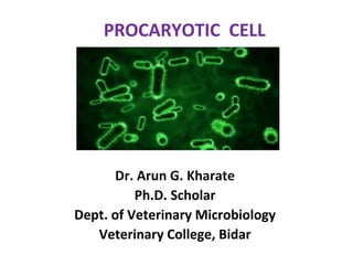
3. CELL WALL
The cell wall is the outer most layer of the cell.
In many cases the cell wall comes in direct contact
with the environment.
Function
• Protection of the cell.
• Maintains the shapes of the cell.
• Maintains the osmotic integrity of the cell.
• Prevents expulsion of ions, molecules and water.
4. • Assist some cells in attaching to other cells or in eluding
antimicrobial drugs.
• Not present in animal cells, so can target cell wall of
bacteria with antibiotics.
• Providing attachment sites for bacteriophages.
• Play an essential role in cell division.
• Providing a rigid platform for surface appendages-
flagella, fimbriae and pili.
6. Peptidoglycan
• Peptidoglycan, also known as murein, is
a polymer consisting of sugars and amino acids that
forms a mesh-like layer outside the cell membrane of
most bacteria forming the cell wall.
• The sugar component consists of alternating residues
of β-(1,4) linked N-acetylglucosamine and N-
acetylmuramic acid.
• These subunits which are related to glucose in their
structure are covalently joined to one another to
form glycan chains.
7. • Attached to the N-acetylmuramic acid is a peptide
chain of four amino acids. The peptide chain can be
cross-linked to the peptide chain of another strand
forming the peptidoglycan.
• Tetra peptide
• L-Alenin
• D-Alenin
• Meso-diaminopimilic acid
• D-Glutamic acid
10. Gram Positive Cell wall
• Usually thick, homogenous, composed mainly
of peptidoglycan.
• It accounts for 50-90% of the dry weight of the
cell wall.
• Contain large amount of teichoic acids
(polymers of glycerol or ribitol joined by
phosphate group).
13. Teichoic acid
• Teichoic acids are connected to either peptidoglycan or to
plasma membrane lipids.
• Absent in gram negative bacteria.
Function of Teichoic Acid:
. Antigenic determinant
-Receptor molecule for bacteriophages.
. Participate in the supply of Mg to the cell by binding Mg++
. Regulate normal cell division.
For most part, protein is not found as a constituent of the G+ cell
wall except M protein on group streptococci.
14. Gram Negative Cell Wall
• Multi layered and more complex than Gram
positive cell walls.
• Peptidoglycan of gram negative bacteria is
thin and comprises only 10% or less of cell
wall.
• Outer membrane lies outside the thin
peptidoglycan layer.
• Most abundant protein is Braun’s lipoprotein.
16. Periplasm:
• The region between the cytoplasmic membrane and
the outer membrane is filled with a gel-like fluid
called periplasm.
• In gram negative bacteria, all secreted proteins are
contained within the periplasm, unless they are
specifically translocated across the outer membrane.
• Periplasm is filled with the proteins that are involved
in various cellular activities, including nutrient
degradation and transport.
17. Outer membrane
• Peptidoglycan layer is surrounded by outer
membrane in the gram negative bacteria.
• Its outside leaflet is made up of lipopolysaccharides,
rather than phospholipids.
• For this reason, the outer membrane is also called
the lipopolysaccharide layer or LPS.
• The outer membrane functions as a protective
barrier and excludes many toxic compounds.
18. • Lipopolysaccharide molecule is extremely important
from a medical stand point.
• It consists of three parts, two of them are medically
significant.
1. Lipid A…..embedded in membrane.
2. Core polysaccharide…..located on the surface of
membrane.
3. O antigens….which are short polysaccharides
extended out from core.
19. • Lipid A: The chemical makeup of lipid A molecule
plays significant role in our body’s ability to recognize
the presence of invading bacteria.
• Contains two glucosamine sugar derevetives.
• It is toxic in nature, as a result the LPS can act as an
endotoxin, causing symptoms like fever, diarrhea
and shock.
• O-antigen: It is composed of carbohydrates,
including glucose, galactose, mannose and some
other sugars in varying combinations.
• The O-antigens can resist react with their specific
antibodies by changing nature of their O side chains
to avoid detection
20. • Porin proteins: Three
porin molecules cluster
together and span the
outer membrane to
form a narrow channel
through which
molecules smaller than
about 600 to 700 Da
can pass.
23. Filament
A. Filament
1. Number of flagella
- Monotrichous,
amphitrichous,
Multitrichous
Three possible locations
Polar, Lateral and
peritrichous
Lophotrichous
• Sheathed flagella
(Vibrio cholerae)
• Periplasmic flagella
(Spirochetes)
26. -Flagella can switch
Physical factors- Toque, temperature,
pH, salt concentration.
Genetic factors- Point mutation
• Polymorphism of flagella-
-some mutant flagella, such as straight
flagella, are too stiff to transform into
another helix.
• Helical transformation is necessary for
untangling a jammed bundle of tangled
flagella
27. 3. Flagellin
Component protein filament is c/a Flagellin
Many bacteria have one kind few have two kind of Flagllin
Mol. Wt. 20-60 kDa.
• Amino Acid Sequences
Terminal Regions -Conserved,
Central Region - Highly Variable
Eg. Salmonella serotype variation
• In the filament, the terminal regions are located at
the innermost radius of a cylindrical structures,
whereas the central region is exposed to the outside.
28. 4. Cap protein
• flagellin can polymerize into flagella-invitro
• flagellin assembly requires Cap protein- invivo
• without Cap protein or Flid the flagellin is
secreted into the medium as monomers.
• located at the tip- pentamer, forming a
star-shaped structure.
29. B.Hook
1.Shape
shorter., more sharply curved
(almost in a right angle)
Length -55 nm(+ 6nm)
A polyhook -indefinite length,
seen in mutants.
2. Hook protein
• Hook- polymer, hook protein or
FlgE.
• Mol. size m 29 kDa (Bacillus
subtilis) to 76 kDa (Helicobacter
pylori),
• 42 kDa for most species.
30. 3. Scaffolding protein
Helper protein-FlgD,
FlgD-polymerize the hook
protein (Sits at tip)
FlgD is c/a Scaffolding protein
bcz of temporary existence
4. Hook-associated protein
• HAPs- two minor proteins
between the hook and
filament.
.
31. C. Basal structure
• The basal body typically consists of four rings and one rod
1. Basal body
• The basal body contains rings and a rod penetrating them.
• four rings –gram negatives,
• two rings - gram-positives,
• The structure of the basal body of S. typhimurium has been
extensively analyzed
32. 2.LP-ring complex
• L ring, - LPS layer of the outer
membrane
• P ring - peptidoglycan layer.
• The component proteins,
• FlgH for the L ring
• FlgI for the P ring, have signal
peptides
• LP-ring complex, resistant to
extremes of pH or temperature.
• Role- Ambiguous, bcz mutants
lacking the complex still can swim,
and LP complex is not found in
gram-positive bacteria
33. 1. MS-ring complex
• single type of protein, FliF, self-assembles into a
complex consisting of the M and S rings and part of the
rod.
• FliF is 65 kDa, the largest of the flagellar proteins
MS-ring complex
• Is the structural center of the basal structure and plays
an important role in flagellar assembly
34. 4. Rod
• The rod is not as simple as its name
suggests; it consists of at least four
distinct proteins.
• No intermediate rod structure
-- a whole rod or no rod at all.
5. C-Ring
The C ring is a fragile component of the basal
structure
• The C ring consists of the switch proteins
(FliG, FliM, and FliN) and so is
sometimes called the switch
complex.
• 20–40 copies of FliG, 20–40 copies of FliM,
and several 100 copies of FliN.
• Role in flagellar formation, torque
generation, and the switching of
35. FUNCTIONS
There is no correlation between bacterial flagella and eukaryotic
flagella,
A. Torque
The rotational force (torque) of the flagellar motor is
difficult to measure directly, but can be estimated
from the rotational speed of flagella
1. Rotational direction
• 70% - by CCW rotation, (Enterobacteriaceae)
• majority CW-Rhodobacter sphaeroides,
2. Rotational speed
• torque of the flagellar motor cannot be directly measured
1. highest speed-200 Hz for S. typhimurium
2. High viscocity slows down the speed. ion
36. B. Energy source
• The energy source of torque generation in the
flagellar motor is not ATP but proton-motive force
(PMF).
• PMF is the electrochemical potential of the
proton,and results in the flow of protons from
outside toinside the cell.
C. Switching of rotational direct
• Switching the rotational direction of flagella is the
primary basis of chemotaxis
• an effector binds to the switch complex in the
flagellar motor. The effector is the phosphorylated
form of CheY, a signalling protein in the sensory
transduction system
37. Genetics
A. Flagellar genes
• There are more than 50 flagellar genes, which are
divided into three types
1. The fla genes: flg, flh, fli, and flj;
one for each of the clusters of genes scattered in several regions
around the chromosome.
2. The mot genes.
• Mutants that produce paralyzed flagella are called
motility deficient (Mot) mutants.
• There are only two mot genes (motA and motB) in S. typhimurium,
3. The che genes.
• Mutants that can produce functional flagella but that cannot show a
normal chemotactic behavior are called chemotaxis deficient (Che)
mutants.
• two types, general chemotaxis mutants and specific chemotaxis
mutants
38. B. Gene clusters in four regions
• Flagellar genes are found in gene clusters on
the chromosome, They are in four regions
• Region I -the flg genes
• Region II- the flh genes and mot and che genes
• Regions IIIa and IIIb- fli genes
39. The Kinetics of Morphogenesis
• In order to achieve coherent cell activities,
flagellar construction has to be synchronized with cell
division
1. Filament growth
• A defined number of flagella have to be supplied at
each cell division.
• The number of flagella must be genetically controlled.
• On the other hand, filament growth seems free from
genetic control, because it continues over generations.
• The elongation rate of filaments is estimated to vary
inversely to the length
41. • Chemotaxis in microbiology
refers to the migration of cells
toward attractant chemicals or
away from repellents.
• Motility involves one or several
flagella, or whether it occurs by a
mechanism such as gliding
motility that does not involve
flagella.
• Attractants: amino acids,
peptides, and sugars
• Repellents: phenol and acid
42. RESPONSE STRATEGY
A. Biased random walk
• In a constant environment,
motile bacteria generally move
in a random walk of straight
runs punctuated by brief
periods of reversal that serve to
randomize
the direction of the next run.
• Individual cells never have to
determine in which direction
they want to move. Instead, they
simply determine whether they
want to continue on course or
change direction.
43.
44. B. Temporal sensing and
memory
• 1970s, through the work of
Macnab, Koshland, Berg, and
others
• chemotaxis depends on a
temporal rather than a spatial
sensing mechanism
• As the cell moves if the
comparison is favorable, the
cell tends to keep going; if not,
it tends to change direction.
45. • C. Excitation and adaptation
• Bacteria must have a way of comparing the past with
the present—they must have memory.
• Bacteria do not respond to absolute concentrations
of attractant and repellent chemicals. They respond
only to changes.
• There is a close relationship between memory and
adaptation.
• The sense and degree of excitation and adaptation
in response to a new place in time are only
determined in relation to the memory of the old one
46. Pili
• Pili, also known as fimbriae, are
proteinaceous, filamentous
polymeric organelles expressed on
the surface of bacteria.
• 5-20ϻm × 2 to 11 nm
• Pili are composed of single or
multiple types of protein subunits,
called pilins or fimbrins, which are
typically arranged in a helical
fashion.
• Pilus architecture varies from thin,
twisting threadlike fibers to thick,
rigid rods with small axial holes.
47. • Pili with diameters of 2–3 nm,
are often referred to as
“fibrillae”. Eg. K88 and K99 pili,
• Pili which tend to coil up into a
fuzzy adhesive mass on the
bacterial surface, are referred
to
as thin aggregative pili or curli.
• Pili are expressed peritrichously
(most)
• pili, can be localized to one
pole- eg. type 4
48. • Pili expressed by gram-negative bacteria have
been extensively characterized, and the
expression of pili by gram-positive bacteria has
also been reported.
• Functions
• Primary function – adhesion , adhesins
- adaptation,
-survival,
-spread of both pathogenic and commensal
bacteria.
• - act as receptors for bacteriophage, facilitate
DNA uptake and transfer (conjugation),
49. History
• Pili were first noted in early electron microscopic
investigations as nonflagellar, filamentous appendages
of bacteria
• In 1955, Duguid -“fimbriae” (plural, from Latin for
thread or fiber) and correlated their presence with the
ability of E. coli to gglutinate red blood cells.
• In 1965 Brinton introduced the term “pilus” (singular,
from Latin for hair) to describe the fibrous structures
(the F pilus) associated with the conjugative transfer of
genetic material between bacteria
50. CLASSIFICATION
• Duguid and co-workers, pili expressed by
different E. coli strains were distinguished on the
basis of their ability to bind to and agglutinate
red blood cells (hemagglutination) in a mannose
sensitive (MS) (Type 1 pili)as opposed to a
mannose resistant (MR) fashion.
• Other bases for classification
• Adhesive and antigenic traits
• Distribution among bacterial strains
• Microscopic charecterization
• Assembly mechanism-Gram negateive 6 types
51. • These pili are very diverse and possess a
myriad of architectures and different receptor
binding specificities and functions
• Pili are now known to be encoded by virtually
all gram-negative organisms and are some of
the best-characterized colonization and
virulence factors in bacteria.
52. Molecular structure
Type 1 pili
• The P pilus tip is a 2-
nmwide structure
composed of a distally
located adhesin PapG,
a tip pilin PapE, and
adaptor pilins PapF and
PapK.
P pili
• type 1 pilus has a short,
3-nm-wide fibrillar tip
made up of the
mannose-binding
adhesin, FimH, and two
additional pilins, FimG
and FimF.
55. • 50% glycerol can cause
the pilus rod to
reversibly unwind into a
2-nm-thick linear fiber
similar in appearance to
the tip fibrillum.
• Bullitt and Makowski
(1995) have proposed
that unwinding will help
to withstand better the
stress, such as shearing
forces from the bulk flow
of fluid through the
urinary tract, without
breaking.
56. Charecters of Pili
• P pili are major virulence factors associated
with pyelonephritis caused by uropathogenic
E. coli. UPEC
• type 1 pili appear to be more rigid and prone
to breaking than P pili.
• K88 and K99 pili are significant virulence
factors expressed by enterotoxigenic E. coli
(ETEC) strains. They are relatively rigid and rod
like.
57. REGULATION OF PILUS
BIOGENESIS
• Pilus biogenesis, in general, is a tightly
regulated process.
• Ideally, the costs in energy and other resources
required for pilus ssembly must be balanced
with any potential benefits.
• Pathogenic and other bacteria must also
control pilus expression, in some cases,
to avoid attachment to unfavorable sites
tissues)
58. Biogenesis depends on
• Temperature,
• Osmolarity,
• Ph,
• Oxygen tension,
• Carbon source, and
• Nutrient availability
59. ROLE OF PILI IN DISEASE PROCESSES
• Adherance- colonization-ETEC,UPEC
• Virulance K88, K99
• Uptake of DNA-Conjugation(Resistance,
Virulance factors)
• Biofilm fomation- Antibiotic Resistance
• To be continued…