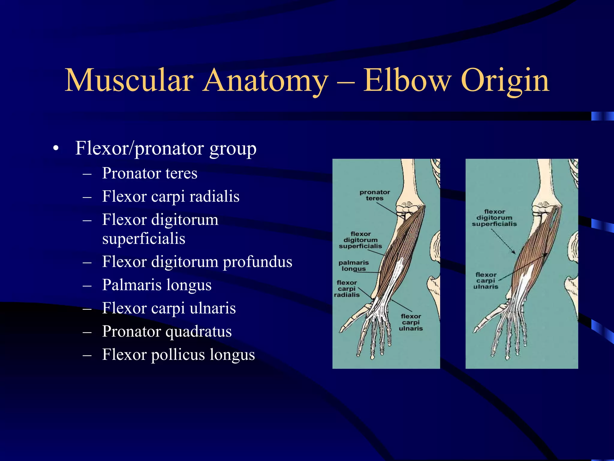
KIN 191B – Advanced Assessment of Upper Extremity Injuries Elbow and Forearm Anatomy  and Evaluation
Anatomy
Anatomy Bony anatomy Articulations and ligamentous anatomy Muscular anatomy Neurological and vascular anatomy Bursa of the elbow
Bony Anatomy
Bony Anatomy: Humerus Distally – 2 condyles forming articular surfaces of trochlea and capitellum Proximally – neck and head articulate with glenoid fossa of scapula
Distal Humerus Anatomy Medial epicondyle proximal to trochlea  – attachment site for UCL and flexor/pronator ms. Lateral epicondyle proximal to capitellum  – attachment site for RCL, extensor/supinator ms. Radial fossa  – accommodates margin of radial head during flexion Coronoid fossa  – accepts coronoid process of ulna during flexion
Distal Humerus – Posterior Olecranon fossa accepts olecranon process of ulna during extension
Ulnar Anatomy Sigmoid/semilunar/ trochlear notch Anteriorly composed of coronoid process Posteriorly composed of olecranon process Articulates with trochlea of humerus
Radial Anatomy Radial head articulates with capitellum Radial neck tapers to radial tuberosity which is insertion for biceps brachii tendon
Articulations and Ligamentous Anatomy
Elbow Joint Articulation -  Elbow consists of 3 articulations: Ulnohumeral (elbow flexion/extension) Radiohumeral (forearm pronation/supination) Radioulnar (forearm pronation/supination)
Elbow Ligamentous Structures
Medial Ligamentous Structures Medial/Ulnar Collateral Ligament Anterior bundle – most discrete segment Posterior bundle – thickening of posterior capsule Transverse bundle – spans medial border of semilunar notch, little/no contribution to elbow stability
Medial Ligamentous Structures Origin of anterior bundle is inferior to axis of motion (flexion/extension), so some fibers tight during flexion and some during extension (major stabilizing component) Origin of posterior bundle is inferior and posterior to the axis, so fibers are tight during flexion and not during extension
Lateral Ligamentous Structures Annular ligament inserts on anterior/posterior margins of lesser (radial) semilunar notch  maintains radial head in contact with ulna (forms 4/5 of fibro-osseous ring)
Muscular Anatomy
Muscular Anatomy Muscles acting on elbow Anterior arm Posterior arm Muscles originating at elbow, acting on forearm, wrist and hand Flexor/pronator group (hand reference) Extensor/supinator group (3 medial, 3 lateral, 3 “outcropping”, 3 “accessory”)
Muscular Anatomy – Anterior Arm Biceps   brachii Origin: long head from supraglenoid rim, short head from coracoid process Insertion: radial tuberosity Innervation: musculocutaneous nerve Action: elbow flexion, forearm supination, shoulder flexion
Muscular Anatomy – Anterior Arm Brachialis Origin: distal anterior humerus Insertion: ulnar tuberosity and coronoid process Innervation: musculocutaneous nerve Action: elbow flexion, forearm supination
Muscular Anatomy – Anterior Arm Coracobrachialis Origin: coracoid process Insertion: medial humerus opposite deltoid tuberosity Innervation: musculocutaneous nerve Action: shoulder flexion, IR
Muscular Anatomy – Posterior Arm Triceps brachii Origin: long head from inferior glenoid rim, lateral head from posterior humeral ridge, medial head from distal 2/3 of posteromedial humerus Insertion: olecranon process Innervation: radial nerve Action: elbow extension
Muscular Anatomy – Elbow Origin Flexor/pronator group Pronator teres Flexor carpi radialis Flexor digitorum superficialis Flexor digitorum profundus Palmaris longus Flexor carpi ulnaris Pronator quadratus Flexor pollicus longus
Hand Reference “ Heel” of hand at medial epicondyle Superficial layer Thumb = pronator teres, index finger = flexor carpi radialis, ring finger = palmaris longus, little finger = flexor carpi ulnaris Intermediate layer Middle finger = flexor digitorum superficialis Deep layer Flexor digitorum profundus, pronator quadratus, flexor pollicus longus
Pronator Teres Origin: common flexor tendon at medial epicondyle and medial coronoid process Insertion: lateral surface of radial shaft Innervation: median nerve Action: forearm pronation
Flexor Carpi Radialis Origin: common flexor tendon at medial epicondyle Insertion: base of 2 nd  and 3 rd  metacarpals Innervation: median nerve Action: flexes and abducst/radial deviate the wrist
Palmaris Longus Present in approximately 70% of population Origin: common flexor tendon at medial epicondyle Insertion: palmar aponeurosis Action: flexes wrist and tenses palmar aponeurosis
Flexor Carpi Ulnaris Origin: common flexor tendon at medial epicondyle and proximal 2/3 of posterior ulnar border Insertion: pisiform, hamate and 5 th  metacarpal Innervation: ulnar Action: flexes and adducts/ulnar deviate the wrist
Flexor Digitorum Superficialis Origin: common flexor tendon at medial epicondyle, medial aspect of coronoid process and oblique line of radius Insertion: sides of middle phalanges of 2 nd  – 5 th  digits Innervation: median nerve Action: flexes PIP joints, assists flexion of MCP and wrist joints
Flexor Digitorum Profundus Origin: anteriomedial proximal ulna Insertion: bases of distal phalanges (anteriorly) of 2 nd -5 th  digits Innervation: 1 st  and 2 nd  tendons by anterior interosseous nerve (median nerve), 3 rd  and 4 th  tendons by ulnar nerve Action: flexes DIP joints, assists in flexion of PIP and MCP joints
Flexor Pollicus Longus Origin: anterior radius Insertion: palmar surface of base of distal phalanx of thumb Innervation: palmar interosseous nerve Action: flexion of 1 st  interphalangeal and metacarpophalangeal joints
Pronator Quadratus Origin: anterior, distal ulna Insertion: lateral, distal radius Innervation: anterior interosseous nerve (median nerve) Action: forearm pronation
Muscular Anatomy – Elbow Origin Extensor/supinator group Brachioradialis Extensor carpi radialis longus Extensor carpi radialis brevis Extensor carpi ulnaris Extensor digitorum Extensor digiti minimi Supinator Anconeus Extensor indicis Extensor pollicus longus Extensor pollicus brevis Abductor pollicus longus
Lateral Muscles Brachioradialis Extensor carpi radialis longus Extensor carpi radialis brevis
Brachioradialis Brachioradialis Origin: lateral supracondylar ridge of humerus Insertion: lateral aspect of radial styloid process Innervation: radial nerve Action: elbow flexion, especially w/ forearm in neutral position
Extensor Carpi Radialis Longus Origin: lateral supracondylar ridge of humerus Insertion: dorsal base of 2 nd  metacarpal Innervation: radial nerve Action: extend and abduct/radial deviate the wrist
Extensor Carpi Radialis Brevis Origin: common extensor tendon at lateral epicondyle Insertion: dorsal base of 3 rd  metacarpal Innervation: radial nerve Action: extend and abduct/radial deviate the wrist
Medial Muscles Extensor digitorum Extensor carpi ulnaris Extensor digiti minimi
Extensor Digitorum Origin: common tendon from lateral epicondyle Insertion: bases of middle and distal phalanges via bands of 4 tendons Innervation: radial nerve Action: MCP/IP joint extension
Extensor Carpi Ulnaris Origin: common extensor tendon at lateral epicondyle Insertion: ulnar side of base of 5 th  metacarpal Innervation: radial nerve Action: extend and adduct/ulnar deviate the wrist
Extensor Digiti Minimi Origin: common extensor tendon at lateral epicondyle Insertion: base of the 5 th  proximal phalanx Innervation: posterior interosseous (radial) nerve Action: extension of 5 th  MP joint
“ Outcropping” Muscles Abductor pollicis longus Extensor pollicis longus Extensor pollicis brevis
Abductor Pollicis Longus Origin: posterior, distal radius and ulna Insertion: base of 1 st  metacarpal Innervation: median nerve Action: extension, abduction of 1 st  carpometacarpal joint
Extensor Pollicis Longus/Brevis Origin: longus – posterior, middle ulna, brevis – posterior, distal radius Insertion: dorsal aspect of base of distal phalanx of thumb Innervation: deep radial nerve Action: extension of 1 st  carpometacarpal and metacarpophalangeal joints
“ Accessory” Muscles Supinator Anconeus Extensor indicis
Supinator Origin: lateral epicondyle, annular ligament/RCL and supinator crest of ulna Insertion: lateral proximal 1/3 of radius Innervation: posterior interosseous nerve (deep branch of radial nerve) Action: forearm supination
Anconeus Anconeus Origin: lateral epicondyle of humerus Insertion: lateral aspect of olecranon and posterior ulna Innervation: radial nerve Action: assists elbow extension
Extensor Indicis Origin: distal, lateral ulna Insertion: dorsal aspect of 2 nd  proximal phalanx Innervation: posterior interosseous (radial) nerve Action: index finger extension and assists with wrist extension
Vascular Anatomy
Vascular Structures Brachial artery Radial artery Ulnar artery Elbow vascular anastamosis
Vascular Structures Brachial artery Descends along arm along medial aspect of brachialis muscle Enters antecubital fossa medial to biceps brachii tendon and lateral to median nerve Terminates at radial head as radial/ulnar arteries
Vascular Structures Radial artery Originates at radial head, emerges from antecubital fossa between brachioradialis and pronator teres muscles Continues laterally along forearm deep to brachioradialis muscle
Vascular Structures Ulnar artery Originates at radial head, continues medially down forearm
Elbow Vascular Anastamosis Laterally  – profunda brachii artery meets radial recurrent artery Medially  – inferior ulnar collateral artery meets anterior ulnar recurrent artery and superior ulnar collateral artery meets posterior ulnar recurrent artery
Neurological Anatomy
Neurological Structures Terminal branches of brachial plexus Axillary Musculocutaneous Median Radial Ulnar Anterior interosseous nerve Dermatomes and myotomes
Musculocutaneous/Axillary Nerves Musculocutaneous nerve Innervates biceps brachii, coracobrachialis and brachialis muscles Sensory distribution is anterior arm and lateral forearm Axillary nerve Innervates deltoid and teres minor muscles Sensory distribution is lateral arm
Median Nerve Median nerve Enters antecubital fossa medial to biceps brachii tendon and brachial artery Courses down medial forearm to hand/wrist distribution Sensory distribution is pad of index finger
Radial Nerve Radial   nerve Enteres antecubital fossa posterior to brachialis muscle Divides into superficial and deep (posterior interosseous) branches Courses down lateral forearm to hand/wrist distribution Sensory distribution is 1 st  dorsal webspace
Ulnar Nerve Ulnar nerve Courses in cubital tunnel posterior to medial epicondyle Superficial and susceptible to compression or entrapment Courses down medial forearm to hand/wrist distribution Sensory distribution is pad of little finger
Anterior Interosseous Nerve Anterior interosseous nerve (branch of median nerve) Passes between 2 heads of pronator teres muscle, may be impinged upon Anterior interosseous nerve syndrome characterized by abnormal pinch deformity (inability to extend DIP of thumb and index finger)
Dermatomes C5 –  lateral arm C6 –  lateral forearm, thumb and index finger C7 –  posterior forearm and middle finger C8 –  medial forearm, ring and little fingers T1 –  medial arm
Myotomes C5  – shoulder abduction C6  – elbow flexion, wrist extension C7  – elbow extension, wrist flexion C8  – finger flexion/grip strength T1  – finger abduction/adduction
Olecranon Bursa Most frequently injured bursa in the elbow Lays between skin and olecranon process Allows unrestricted/fluid movement of skin over olecranon process
Evaluation of Elbow and Forearm Injuries
History
History Location of symptoms Onset of symptoms Mechanism of injury (etiology) Unusual sounds/sensations Prior history of injury/general health Technique (associated with overuse injuries)
Location of Symptoms Generally associated with tissue damaged Must be conscious of potential for referred pain secondary to cervical and/or shoulder pathologies
Onset of Symptoms Acute injury typically associated with traumatic onset – specific mechanism Chronic injury typically associated with gradual onset of symptoms – insiduous mechanism
Mechanism of Injury (Etiology) Fall on outstretched arm Direct trauma or force application Repetitive stresses (throwing, hand/wrist movements)
Unusual Sounds/Sensations Feeling of “giving way” of elbow with throwing motion “ Pop” or “snap” often associated with ligament or tendon rupture Clicking/grating often associated with osteochondritis dissecans (“joint mice”)
Prior History/General Health Must consider residual effects from previous injury on current symptoms Must consider neurovascular, inflammatory and systemic conditions/illnesses (ischemic contracture, neuropathy, arthritis, etc.)
Technique Especially important for overhead activities and athletes Must consider changes in training Duration Intensity Frequency Biomechanics Equipment
Inspection
Inspection Anterior structures Medial structures Lateral structures Posterior structures
Anterior Structures Carrying angle Cubitus valgus (carrying angle) Cubitus varus (gunstock deformity) Cubital fossa
Carrying Angle/Cubitus Valgus Formed by long axis of humerus and midline of forearm Male norms – 11-14 degrees Female norms – 13-16 degrees Larger angles are considered abnormal
Gunstock Deformity/Cubitus Varus Usually develops secondary to condylar humerus fracture
Cubital Fossa Superior border –  imaginary line between medial/lateral epicondyles Medial border –  pronator teres muscle Lateral border –  brachioradialis muscle Contents –  brachial artery and median nerve
Medial Structures Medial epicondyle Flexor/pronator group
Lateral Structures Wrist/forearm alignment Cubital recurvatum (hyperextension) Extensor/supinator group
Posterior Structures Bony alignment Olecranon process/bursa
Bony Alignment With elbow extended, straight line between medial/lateral epicondyles and tip of olecranon process’ With elbow flexed, isosceles triangle connects these points
Palpation
Palpation – Anterior Structures Biceps brachii – tendon Cubital fossa Borders, brachial artery Brachioradialis Elbow flexion with thumb up Flexor-pronator group
Palpation – Medial Structures Medial epicondyle Ulna (length of shaft) Medial (ulnar) collateral ligament Ulnar nerve (cubital tunnel posterior to medial epicondyle)
Palpation – Lateral Structures Lateral epicondyle Radial head (forearm movement) Lateral (radial) collateral ligament Between radial head and lateral epicondyle, difficult to isolate Capitellum (area at articulation with radius) Annular ligament (area) Lateral ulnar collateral ligament (difficult)
Palpation – Posterior Structures Olecranon process Olecranon fossa Elbow flexed and triceps relaxed Triceps brachii Anconeus Between lateral epicondyle and olecranon process Extensor/supinator group
Special Tests
Special Tests Ranges of motion Neurological evaluation Vascular evaluation Ligamentous/capsular stress tests
Range of Motion Flexion/extension  – ginglymus joint (ulnohumeral articulation) Flexion typically 0-150 degrees, stops due to soft tissue approximation Extension typically 0-10 degrees (hyperextension, especially in females), stops due to bony opposition
Range of Motion Forearm pronation and supination  – trochoid joint (radiohumeral and proximal radioulnar articulations) Pronation/supination typical 0-85/90 degrees each from neutral point (thumb up), stops due to tissue tensions/stretch from opposing tissue Elbow ROM has 2 degrees of freedom
Range of Motion Elbow motion/s serve to adjust height and length of limb to reach any point within sphere of shoulder motion and also to rotate forearm to place hand in the most effective operating position
Neurovascular Evaluation Brachial plexus dermatomes/myotomes Peripheral nerve sensory and motor function Pulse points Brachial artery Radial artery Capillary refill Skin color/temperature
Ligamentous Stress Tests Valgus stress test Assesses medial (ulnar) collateral ligament (anterior bundle) Varus stress test Assesses lateral (radial) collateral ligament and annular ligament
Valgus Stress Test Performed in 25-30 degrees of elbow flexion
Varus Stress Test Performed in 25-30 degrees of elbow flexion
Elbow Kinematics One of the most congruous and stable joints In extension, anterior capsule provides most restraint, while MCL becomes primary stabilizer at 90 degrees flexion Valgus stress in extension equally resisted by bone structure (proximal ½ of semilunar notch), MCL and medial joint capsule – in flexion, resisted primarily by MCL (anterior bundle) Varus stress in extension resisted by bone structure (distal ½ of coronoid process), LCL and lateral joint capsule – in flexion, resisted primarily by bone structure
Elbow Biomechanics – Injury Contribution and Rehab Considerations Significant valgus torques present during arm cocking phase of throwing (medial distraction and lateral compression) UCL is static stabilizer and flexor/pronators, triceps and anconeus are dynamic stabilizers to valgus loads Rapid extension velocity during acceleration phase of throwing (low biceps activity and high centrifugal forces from trunk) High distraction forces at elbow during extension countered by muscular activity Large deceleration forces through elbow flexors during deceleration phase of throwing High distraction forces countered by ligamentous stabilization and muscular activity

Kin 191 B Elbow And Forearm Anatomy And Evaluation

  • 1.
    KIN 191B –Advanced Assessment of Upper Extremity Injuries Elbow and Forearm Anatomy and Evaluation
  • 2.
  • 3.
    Anatomy Bony anatomyArticulations and ligamentous anatomy Muscular anatomy Neurological and vascular anatomy Bursa of the elbow
  • 4.
  • 5.
    Bony Anatomy: HumerusDistally – 2 condyles forming articular surfaces of trochlea and capitellum Proximally – neck and head articulate with glenoid fossa of scapula
  • 6.
    Distal Humerus AnatomyMedial epicondyle proximal to trochlea – attachment site for UCL and flexor/pronator ms. Lateral epicondyle proximal to capitellum – attachment site for RCL, extensor/supinator ms. Radial fossa – accommodates margin of radial head during flexion Coronoid fossa – accepts coronoid process of ulna during flexion
  • 7.
    Distal Humerus –Posterior Olecranon fossa accepts olecranon process of ulna during extension
  • 8.
    Ulnar Anatomy Sigmoid/semilunar/trochlear notch Anteriorly composed of coronoid process Posteriorly composed of olecranon process Articulates with trochlea of humerus
  • 9.
    Radial Anatomy Radialhead articulates with capitellum Radial neck tapers to radial tuberosity which is insertion for biceps brachii tendon
  • 10.
  • 11.
    Elbow Joint Articulation- Elbow consists of 3 articulations: Ulnohumeral (elbow flexion/extension) Radiohumeral (forearm pronation/supination) Radioulnar (forearm pronation/supination)
  • 12.
  • 13.
    Medial Ligamentous StructuresMedial/Ulnar Collateral Ligament Anterior bundle – most discrete segment Posterior bundle – thickening of posterior capsule Transverse bundle – spans medial border of semilunar notch, little/no contribution to elbow stability
  • 14.
    Medial Ligamentous StructuresOrigin of anterior bundle is inferior to axis of motion (flexion/extension), so some fibers tight during flexion and some during extension (major stabilizing component) Origin of posterior bundle is inferior and posterior to the axis, so fibers are tight during flexion and not during extension
  • 15.
    Lateral Ligamentous StructuresAnnular ligament inserts on anterior/posterior margins of lesser (radial) semilunar notch maintains radial head in contact with ulna (forms 4/5 of fibro-osseous ring)
  • 16.
  • 17.
    Muscular Anatomy Musclesacting on elbow Anterior arm Posterior arm Muscles originating at elbow, acting on forearm, wrist and hand Flexor/pronator group (hand reference) Extensor/supinator group (3 medial, 3 lateral, 3 “outcropping”, 3 “accessory”)
  • 18.
    Muscular Anatomy –Anterior Arm Biceps brachii Origin: long head from supraglenoid rim, short head from coracoid process Insertion: radial tuberosity Innervation: musculocutaneous nerve Action: elbow flexion, forearm supination, shoulder flexion
  • 19.
    Muscular Anatomy –Anterior Arm Brachialis Origin: distal anterior humerus Insertion: ulnar tuberosity and coronoid process Innervation: musculocutaneous nerve Action: elbow flexion, forearm supination
  • 20.
    Muscular Anatomy –Anterior Arm Coracobrachialis Origin: coracoid process Insertion: medial humerus opposite deltoid tuberosity Innervation: musculocutaneous nerve Action: shoulder flexion, IR
  • 21.
    Muscular Anatomy –Posterior Arm Triceps brachii Origin: long head from inferior glenoid rim, lateral head from posterior humeral ridge, medial head from distal 2/3 of posteromedial humerus Insertion: olecranon process Innervation: radial nerve Action: elbow extension
  • 22.
    Muscular Anatomy –Elbow Origin Flexor/pronator group Pronator teres Flexor carpi radialis Flexor digitorum superficialis Flexor digitorum profundus Palmaris longus Flexor carpi ulnaris Pronator quadratus Flexor pollicus longus
  • 23.
    Hand Reference “Heel” of hand at medial epicondyle Superficial layer Thumb = pronator teres, index finger = flexor carpi radialis, ring finger = palmaris longus, little finger = flexor carpi ulnaris Intermediate layer Middle finger = flexor digitorum superficialis Deep layer Flexor digitorum profundus, pronator quadratus, flexor pollicus longus
  • 24.
    Pronator Teres Origin:common flexor tendon at medial epicondyle and medial coronoid process Insertion: lateral surface of radial shaft Innervation: median nerve Action: forearm pronation
  • 25.
    Flexor Carpi RadialisOrigin: common flexor tendon at medial epicondyle Insertion: base of 2 nd and 3 rd metacarpals Innervation: median nerve Action: flexes and abducst/radial deviate the wrist
  • 26.
    Palmaris Longus Presentin approximately 70% of population Origin: common flexor tendon at medial epicondyle Insertion: palmar aponeurosis Action: flexes wrist and tenses palmar aponeurosis
  • 27.
    Flexor Carpi UlnarisOrigin: common flexor tendon at medial epicondyle and proximal 2/3 of posterior ulnar border Insertion: pisiform, hamate and 5 th metacarpal Innervation: ulnar Action: flexes and adducts/ulnar deviate the wrist
  • 28.
    Flexor Digitorum SuperficialisOrigin: common flexor tendon at medial epicondyle, medial aspect of coronoid process and oblique line of radius Insertion: sides of middle phalanges of 2 nd – 5 th digits Innervation: median nerve Action: flexes PIP joints, assists flexion of MCP and wrist joints
  • 29.
    Flexor Digitorum ProfundusOrigin: anteriomedial proximal ulna Insertion: bases of distal phalanges (anteriorly) of 2 nd -5 th digits Innervation: 1 st and 2 nd tendons by anterior interosseous nerve (median nerve), 3 rd and 4 th tendons by ulnar nerve Action: flexes DIP joints, assists in flexion of PIP and MCP joints
  • 30.
    Flexor Pollicus LongusOrigin: anterior radius Insertion: palmar surface of base of distal phalanx of thumb Innervation: palmar interosseous nerve Action: flexion of 1 st interphalangeal and metacarpophalangeal joints
  • 31.
    Pronator Quadratus Origin:anterior, distal ulna Insertion: lateral, distal radius Innervation: anterior interosseous nerve (median nerve) Action: forearm pronation
  • 32.
    Muscular Anatomy –Elbow Origin Extensor/supinator group Brachioradialis Extensor carpi radialis longus Extensor carpi radialis brevis Extensor carpi ulnaris Extensor digitorum Extensor digiti minimi Supinator Anconeus Extensor indicis Extensor pollicus longus Extensor pollicus brevis Abductor pollicus longus
  • 33.
    Lateral Muscles BrachioradialisExtensor carpi radialis longus Extensor carpi radialis brevis
  • 34.
    Brachioradialis Brachioradialis Origin:lateral supracondylar ridge of humerus Insertion: lateral aspect of radial styloid process Innervation: radial nerve Action: elbow flexion, especially w/ forearm in neutral position
  • 35.
    Extensor Carpi RadialisLongus Origin: lateral supracondylar ridge of humerus Insertion: dorsal base of 2 nd metacarpal Innervation: radial nerve Action: extend and abduct/radial deviate the wrist
  • 36.
    Extensor Carpi RadialisBrevis Origin: common extensor tendon at lateral epicondyle Insertion: dorsal base of 3 rd metacarpal Innervation: radial nerve Action: extend and abduct/radial deviate the wrist
  • 37.
    Medial Muscles Extensordigitorum Extensor carpi ulnaris Extensor digiti minimi
  • 38.
    Extensor Digitorum Origin:common tendon from lateral epicondyle Insertion: bases of middle and distal phalanges via bands of 4 tendons Innervation: radial nerve Action: MCP/IP joint extension
  • 39.
    Extensor Carpi UlnarisOrigin: common extensor tendon at lateral epicondyle Insertion: ulnar side of base of 5 th metacarpal Innervation: radial nerve Action: extend and adduct/ulnar deviate the wrist
  • 40.
    Extensor Digiti MinimiOrigin: common extensor tendon at lateral epicondyle Insertion: base of the 5 th proximal phalanx Innervation: posterior interosseous (radial) nerve Action: extension of 5 th MP joint
  • 41.
    “ Outcropping” MusclesAbductor pollicis longus Extensor pollicis longus Extensor pollicis brevis
  • 42.
    Abductor Pollicis LongusOrigin: posterior, distal radius and ulna Insertion: base of 1 st metacarpal Innervation: median nerve Action: extension, abduction of 1 st carpometacarpal joint
  • 43.
    Extensor Pollicis Longus/BrevisOrigin: longus – posterior, middle ulna, brevis – posterior, distal radius Insertion: dorsal aspect of base of distal phalanx of thumb Innervation: deep radial nerve Action: extension of 1 st carpometacarpal and metacarpophalangeal joints
  • 44.
    “ Accessory” MusclesSupinator Anconeus Extensor indicis
  • 45.
    Supinator Origin: lateralepicondyle, annular ligament/RCL and supinator crest of ulna Insertion: lateral proximal 1/3 of radius Innervation: posterior interosseous nerve (deep branch of radial nerve) Action: forearm supination
  • 46.
    Anconeus Anconeus Origin:lateral epicondyle of humerus Insertion: lateral aspect of olecranon and posterior ulna Innervation: radial nerve Action: assists elbow extension
  • 47.
    Extensor Indicis Origin:distal, lateral ulna Insertion: dorsal aspect of 2 nd proximal phalanx Innervation: posterior interosseous (radial) nerve Action: index finger extension and assists with wrist extension
  • 48.
  • 49.
    Vascular Structures Brachialartery Radial artery Ulnar artery Elbow vascular anastamosis
  • 50.
    Vascular Structures Brachialartery Descends along arm along medial aspect of brachialis muscle Enters antecubital fossa medial to biceps brachii tendon and lateral to median nerve Terminates at radial head as radial/ulnar arteries
  • 51.
    Vascular Structures Radialartery Originates at radial head, emerges from antecubital fossa between brachioradialis and pronator teres muscles Continues laterally along forearm deep to brachioradialis muscle
  • 52.
    Vascular Structures Ulnarartery Originates at radial head, continues medially down forearm
  • 53.
    Elbow Vascular AnastamosisLaterally – profunda brachii artery meets radial recurrent artery Medially – inferior ulnar collateral artery meets anterior ulnar recurrent artery and superior ulnar collateral artery meets posterior ulnar recurrent artery
  • 54.
  • 55.
    Neurological Structures Terminalbranches of brachial plexus Axillary Musculocutaneous Median Radial Ulnar Anterior interosseous nerve Dermatomes and myotomes
  • 56.
    Musculocutaneous/Axillary Nerves Musculocutaneousnerve Innervates biceps brachii, coracobrachialis and brachialis muscles Sensory distribution is anterior arm and lateral forearm Axillary nerve Innervates deltoid and teres minor muscles Sensory distribution is lateral arm
  • 57.
    Median Nerve Mediannerve Enters antecubital fossa medial to biceps brachii tendon and brachial artery Courses down medial forearm to hand/wrist distribution Sensory distribution is pad of index finger
  • 58.
    Radial Nerve Radial nerve Enteres antecubital fossa posterior to brachialis muscle Divides into superficial and deep (posterior interosseous) branches Courses down lateral forearm to hand/wrist distribution Sensory distribution is 1 st dorsal webspace
  • 59.
    Ulnar Nerve Ulnarnerve Courses in cubital tunnel posterior to medial epicondyle Superficial and susceptible to compression or entrapment Courses down medial forearm to hand/wrist distribution Sensory distribution is pad of little finger
  • 60.
    Anterior Interosseous NerveAnterior interosseous nerve (branch of median nerve) Passes between 2 heads of pronator teres muscle, may be impinged upon Anterior interosseous nerve syndrome characterized by abnormal pinch deformity (inability to extend DIP of thumb and index finger)
  • 61.
    Dermatomes C5 – lateral arm C6 – lateral forearm, thumb and index finger C7 – posterior forearm and middle finger C8 – medial forearm, ring and little fingers T1 – medial arm
  • 62.
    Myotomes C5 – shoulder abduction C6 – elbow flexion, wrist extension C7 – elbow extension, wrist flexion C8 – finger flexion/grip strength T1 – finger abduction/adduction
  • 63.
    Olecranon Bursa Mostfrequently injured bursa in the elbow Lays between skin and olecranon process Allows unrestricted/fluid movement of skin over olecranon process
  • 64.
    Evaluation of Elbowand Forearm Injuries
  • 65.
  • 66.
    History Location ofsymptoms Onset of symptoms Mechanism of injury (etiology) Unusual sounds/sensations Prior history of injury/general health Technique (associated with overuse injuries)
  • 67.
    Location of SymptomsGenerally associated with tissue damaged Must be conscious of potential for referred pain secondary to cervical and/or shoulder pathologies
  • 68.
    Onset of SymptomsAcute injury typically associated with traumatic onset – specific mechanism Chronic injury typically associated with gradual onset of symptoms – insiduous mechanism
  • 69.
    Mechanism of Injury(Etiology) Fall on outstretched arm Direct trauma or force application Repetitive stresses (throwing, hand/wrist movements)
  • 70.
    Unusual Sounds/Sensations Feelingof “giving way” of elbow with throwing motion “ Pop” or “snap” often associated with ligament or tendon rupture Clicking/grating often associated with osteochondritis dissecans (“joint mice”)
  • 71.
    Prior History/General HealthMust consider residual effects from previous injury on current symptoms Must consider neurovascular, inflammatory and systemic conditions/illnesses (ischemic contracture, neuropathy, arthritis, etc.)
  • 72.
    Technique Especially importantfor overhead activities and athletes Must consider changes in training Duration Intensity Frequency Biomechanics Equipment
  • 73.
  • 74.
    Inspection Anterior structuresMedial structures Lateral structures Posterior structures
  • 75.
    Anterior Structures Carryingangle Cubitus valgus (carrying angle) Cubitus varus (gunstock deformity) Cubital fossa
  • 76.
    Carrying Angle/Cubitus ValgusFormed by long axis of humerus and midline of forearm Male norms – 11-14 degrees Female norms – 13-16 degrees Larger angles are considered abnormal
  • 77.
    Gunstock Deformity/Cubitus VarusUsually develops secondary to condylar humerus fracture
  • 78.
    Cubital Fossa Superiorborder – imaginary line between medial/lateral epicondyles Medial border – pronator teres muscle Lateral border – brachioradialis muscle Contents – brachial artery and median nerve
  • 79.
    Medial Structures Medialepicondyle Flexor/pronator group
  • 80.
    Lateral Structures Wrist/forearmalignment Cubital recurvatum (hyperextension) Extensor/supinator group
  • 81.
    Posterior Structures Bonyalignment Olecranon process/bursa
  • 82.
    Bony Alignment Withelbow extended, straight line between medial/lateral epicondyles and tip of olecranon process’ With elbow flexed, isosceles triangle connects these points
  • 83.
  • 84.
    Palpation – AnteriorStructures Biceps brachii – tendon Cubital fossa Borders, brachial artery Brachioradialis Elbow flexion with thumb up Flexor-pronator group
  • 85.
    Palpation – MedialStructures Medial epicondyle Ulna (length of shaft) Medial (ulnar) collateral ligament Ulnar nerve (cubital tunnel posterior to medial epicondyle)
  • 86.
    Palpation – LateralStructures Lateral epicondyle Radial head (forearm movement) Lateral (radial) collateral ligament Between radial head and lateral epicondyle, difficult to isolate Capitellum (area at articulation with radius) Annular ligament (area) Lateral ulnar collateral ligament (difficult)
  • 87.
    Palpation – PosteriorStructures Olecranon process Olecranon fossa Elbow flexed and triceps relaxed Triceps brachii Anconeus Between lateral epicondyle and olecranon process Extensor/supinator group
  • 88.
  • 89.
    Special Tests Rangesof motion Neurological evaluation Vascular evaluation Ligamentous/capsular stress tests
  • 90.
    Range of MotionFlexion/extension – ginglymus joint (ulnohumeral articulation) Flexion typically 0-150 degrees, stops due to soft tissue approximation Extension typically 0-10 degrees (hyperextension, especially in females), stops due to bony opposition
  • 91.
    Range of MotionForearm pronation and supination – trochoid joint (radiohumeral and proximal radioulnar articulations) Pronation/supination typical 0-85/90 degrees each from neutral point (thumb up), stops due to tissue tensions/stretch from opposing tissue Elbow ROM has 2 degrees of freedom
  • 92.
    Range of MotionElbow motion/s serve to adjust height and length of limb to reach any point within sphere of shoulder motion and also to rotate forearm to place hand in the most effective operating position
  • 93.
    Neurovascular Evaluation Brachialplexus dermatomes/myotomes Peripheral nerve sensory and motor function Pulse points Brachial artery Radial artery Capillary refill Skin color/temperature
  • 94.
    Ligamentous Stress TestsValgus stress test Assesses medial (ulnar) collateral ligament (anterior bundle) Varus stress test Assesses lateral (radial) collateral ligament and annular ligament
  • 95.
    Valgus Stress TestPerformed in 25-30 degrees of elbow flexion
  • 96.
    Varus Stress TestPerformed in 25-30 degrees of elbow flexion
  • 97.
    Elbow Kinematics Oneof the most congruous and stable joints In extension, anterior capsule provides most restraint, while MCL becomes primary stabilizer at 90 degrees flexion Valgus stress in extension equally resisted by bone structure (proximal ½ of semilunar notch), MCL and medial joint capsule – in flexion, resisted primarily by MCL (anterior bundle) Varus stress in extension resisted by bone structure (distal ½ of coronoid process), LCL and lateral joint capsule – in flexion, resisted primarily by bone structure
  • 98.
    Elbow Biomechanics –Injury Contribution and Rehab Considerations Significant valgus torques present during arm cocking phase of throwing (medial distraction and lateral compression) UCL is static stabilizer and flexor/pronators, triceps and anconeus are dynamic stabilizers to valgus loads Rapid extension velocity during acceleration phase of throwing (low biceps activity and high centrifugal forces from trunk) High distraction forces at elbow during extension countered by muscular activity Large deceleration forces through elbow flexors during deceleration phase of throwing High distraction forces countered by ligamentous stabilization and muscular activity