Image registration is the fundamental task used to
match two or more partially overlapping images taken, for
example, at different times,from different sensors, or from
different viewpoints and stitch these images into one
panoramic image comprising the whole scene. It is
afundamental image processing technique and is very useful
in integrating information from different sensors, finding
changes in images taken at different times, inferring threedimensional
information from stereo images, and recognizing
model-based objects.
This paper overviews the theoretical aspects of an image
registration problem. The purpose of this paper is to present a
survey of image registration techniques. This technique of
image registration aligns two images geometrically. These two
images are reference image and sensed image. The ultimate
purpose of digital image filtering is to support the visual
identification of certain features expressed by characteristic
shapes and patterns. Numerous recipes, algorithms and ready
made programs exist nowadays that predominantly have in
common that users have to set certain parameters.
Particularly if processing is fast and shows results rather
immediately, the choice of parameters may be guided by
making the image ―looking nice‖. However, in practical
situations most users are not in a mood to ―play around‖
with a displayed image, particularly if they are in a stressy
situation as it may encountered in security applications. The
requirements for the application of digital image processing
under such circumstances will be discussed with an example
of automaticfiltering without manual parameter settings that
even entails the advantage of delivering unbiased results
Issues in Image Registration and Image similarity based on mutual informationDarshana Mistry
This is my 2nd Doctorate progresses committee presentation in image registration which is explained how do you find image similarity based on Entropy and mutual information
Medical Image Fusion Using Discrete Wavelet TransformIJERA Editor
Medical image fusion is the process of registering and combining multiple images from single or multiple imaging modalities to improve the imaging quality and reduce randomness and redundancy in order to increase the clinical applicability of medical images for diagnosis and assessment of medical problems. Multimodal medical image fusion algorithms and devices have shown notable achievements in improving clinical accuracy of decisions based on medical images. The domain where image fusion is readily used nowadays is in medical diagnostics to fuse medical images such as CT (Computed Tomography), MRI (Magnetic Resonance Imaging) and MRA. This paper aims to present a new algorithm to improve the quality of multimodality medical image fusion using Discrete Wavelet Transform (DWT) approach. Discrete Wavelet transform has been implemented using different fusion techniques including pixel averaging, maximum minimum and minimum maximum methods for medical image fusion. Performance of fusion is calculated on the basis of PSNR, MSE and the total processing time and the results demonstrate the effectiveness of fusion scheme based on wavelet transform.
Mutual Information for Registration of Monomodal Brain Images using Modified ...IDES Editor
Image registration has great significance in medicine,
with a lot of techniques anticipated in it. This research work
implies an approach for medical image registration that
registers images of the mono modalities for CT or MRI images
using Modified Adaptive Polar Transform (MAPT). The
performance of the Adaptive Polar Transform (APT) with the
proposed technique is examined. The results prove that MAPT
performs better than APT technique. The proposed scheme not
only reduces the source of errors and also reduces the elapsed
time for registration of brain images. An analysis is presented
for the medical image processing on mutual- information-based
registration.
Wavelet Transform based Medical Image Fusion With different fusion methodsIJERA Editor
This paper proposes wavelet transform based image fusion algorithm, after studying the principles and characteristics of the discrete wavelet transform. Medical image fusion used to derive useful information from multimodality medical images. The idea is to improve the image content by fusing images like computer tomography (CT) and magnetic resonance imaging (MRI) images, so as to provide more information to the doctor and clinical treatment planning system. This paper based on the wavelet transformation to fused the medical images. The wavelet based fusion algorithms used on medical images CT and MRI, This involve the fusion with MIN , MAX, MEAN method. Also the result is obtained. With more available multimodality medical images in clinical applications, the idea of combining images from different modalities become very important and medical image fusion has emerged as a new promising research field
International Journal of Engineering Research and DevelopmentIJERD Editor
Electrical, Electronics and Computer Engineering,
Information Engineering and Technology,
Mechanical, Industrial and Manufacturing Engineering,
Automation and Mechatronics Engineering,
Material and Chemical Engineering,
Civil and Architecture Engineering,
Biotechnology and Bio Engineering,
Environmental Engineering,
Petroleum and Mining Engineering,
Marine and Agriculture engineering,
Aerospace Engineering.
Image Fusion and Image Quality Assessment of Fused ImagesCSCJournals
Accurate diagnosis of tumor extent is important in radiotherapy. This paper presents the use of image fusion of PET and MRI image. Multi-sensor image fusion is the process of combining information from two or more images into a single image. The resulting image contains more information as compared to individual images. PET delivers high-resolution molecular imaging with a resolution down to 2.5 mm full width at half maximum (FWHM), which allows us to observe the brain\'s molecular changes using the specific reporter genes and probes. On the other hand, the 7.0 T-MRI, with sub-millimeter resolution images of the cortical areas down to 250 m, allows us to visualize the fine details of the brainstem areas as well as the many cortical and sub-cortical areas. The PET-MRI fusion imaging system provides complete information on neurological diseases as well as cognitive neurosciences. The paper presents PCA based image fusion and also focuses on image fusion algorithm based on wavelet transform to improve resolution of the images in which two images to be fused are firstly decomposed into sub-images with different frequency and then the information fusion is performed and finally these sub-images are reconstructed into result image with plentiful information. . We also propose image fusion in Radon space. This paper presents assessment of image fusion by measuring the quantity of enhanced information in fused images. We use entropy, mean, standard deviation and Fusion Mutual Information, cross correlation , Mutual Information Root Mean Square Error, Universal Image Quality Index and Relative shift in mean to compare fused image quality. Comparative evaluation of fused images is a critical step to evaluate the relative performance of different image fusion algorithms. In this paper, we also propose image quality metric based on the human vision system (HVS).
Comparative analysis of multimodal medical image fusion using pca and wavelet...IJLT EMAS
nowadays, there are a lot of medical images and their
numbers are increasing day by day. These medical images are
stored in large database. To minimize the redundancy and
optimize the storage capacity of images, medical image fusion is
used. The main aim of medical image fusion is to combine
complementary information from multiple imaging modalities
(Eg: CT, MRI, PET etc.) of the same scene. After performing
image fusion, the resultant image is more informative and
suitable for patient diagnosis. There are some fusion techniques
which are described in this paper to obtain fused image. This
paper presents two approaches to image fusion, namely Spatial
Fusion and Transform Fusion. This paper describes Techniques
such as Principal Component Analysis which is spatial domain
technique and Discrete Wavelet Transform, Stationary Wavelet
Transform which are Transform domain techniques.
Performance metrics are implemented to evaluate the
performance of image fusion algorithm. An experimental result
shows that image fusion method based on Stationary Wavelet
Transform is better than Principal Component Analysis and
Discrete Wavelet Transform.
Survey on Brain MRI Segmentation TechniquesEditor IJMTER
Image segmentation is aimed at cutting out, a ROI (Region of Interest) from an image. For
medical images, segmentation is done for: studying the anatomical structure, identifying ROI ie tumor
or any other abnormalities, identifying the increase in tissue volume in a region, treatment planning.
Currently there are many different algorithms available for image segmentation. This paper lists and
compares some of them. Each has their own advantages and limitations.
A Dualistic Sub-Image Histogram Equalization Based Enhancement and Segmentati...inventy
Research Inventy : International Journal of Engineering and Science is published by the group of young academic and industrial researchers with 12 Issues per year. It is an online as well as print version open access journal that provides rapid publication (monthly) of articles in all areas of the subject such as: civil, mechanical, chemical, electronic and computer engineering as well as production and information technology. The Journal welcomes the submission of manuscripts that meet the general criteria of significance and scientific excellence. Papers will be published by rapid process within 20 days after acceptance and peer review process takes only 7 days. All articles published in Research Inventy will be peer-reviewed.
MRIIMAGE SEGMENTATION USING LEVEL SET METHOD AND IMPLEMENT AN MEDICAL DIAGNOS...cseij
Image segmentation plays a vital role in image processing over the last few years. The goal of image segmentation is to cluster the pixels into salient image regions i.e., regions corresponding to individual surfaces, objects, or natural parts of objects. In this paper, we propose a medical diagnosis system by using level set method for segmenting the MRI image which investigates a new variational level set algorithm without re- initialization to segment the MRI image and to implement a competent medical diagnosis system by using MATLAB. Here we have used the speed function and the signed distance function of the image in segmentation algorithm. This system consists of thresholding technique, curve evolution technique and an eroding technique. Our proposed system was tested on some MRI Brain images, giving promising results by detecting the normal or abnormal condition specially the existence of tumers. This system will be applied to both simulated and real images with promising results
The purpose of this paper is to present a survey of image registration techniques. Registration is a fundamental task in image processing used to match two or more pictures taken, for example, at different times, from different sensors, or from different viewpoints. It geometrically aligns two images the reference and sensed images. Specific examples of systems where image registration is a significant component include matching a target with a real-time image of a scene. Various applications of image registration are target recognition, monitoring global land usage using satellite images, matching stereo images to recover shape for navigation, and aligning images from different medical modalities for diagnosis.
FUZZY SEGMENTATION OF MRI CEREBRAL TISSUE USING LEVEL SET ALGORITHMAM Publications
The current study investigated a median filter with the fuzzy level set method to propose fuzzy segmentation of magnetic resonance imaging (MRI) cerebral tissue images. An MRI image was used as an input image. A median filter and fuzzy c-means (FCM) clustering were utilized to remove image noise and create image clusters, respectively. The image clusters showed initial and final cluster centers. The level set method was then used for segmentation after separating and extracting white matter from gray matter. Fuzzy c-means was sensitive to the choice of the initial cluster center. Improper center selection caused the method to produce suboptimal solutions. The proposed algorithm was successfully utilized to segment MRI cerebral tissue images. The algorithm efficiently performed segmentation of test MRI cerebral tissue images compared with algorithms proposed in previous studies.
Medical Image segmentation using Image Mining conceptsEditor IJMTER
Image differencing is usually done by subtracting the low-level skin texture like strength
in images that are already associated. This paper extracts high-level skin texture in order to find out
an efficient image differencing method for the analysis of Brain Tumor. On the other hand, this
produces sets of skin texture that are both spatial. We demonstrate a technique that avoids arbitrary
spatial constraints and is robust in the presence of sound, outliers, and imaging artifact, while
outperforming even profitable products in the analysis of Brain Tumor images. First, the landmark
are establish, and then the top entrant are sorted into a end set. Second, the top sets of the two
descriptions are then differenced through a cluster judgment. The symmetry of the human body is
utilized to increase the accuracy of the finding. We imitate this technique in an effort to understand
and ultimately capture the judgment of the radiologist. The image differencing with clustered
contrast process determines the being there of Brain Tumor. Using the most favorable features
extracted from normal and tumor regions of MRI by using arithmetical features, classifiers are used
to categorize and segment the tumor portion in irregular images. Both the difficult and preparation
phase gives the proportion of accuracy on each parameter in neural networks, which gives the idea to
decide the best one to be used in supplementary works. The results showed outperformance of
algorithm when compared with classification accuracy which works as shows potential tool for
classification and requires extension in brain tumor analysis.
Detail description of feature extraction methods and classifier used for Texture Classification Approach. it also contain detail description of different Texture Database used for texture classification.
A NOVEL IMAGE SEGMENTATION ENHANCEMENT TECHNIQUE BASED ON ACTIVE CONTOUR AND...acijjournal
Topological alignments and snakes are used in image processing, particularly in locating object
boundaries. Both of them have their own advantages and limitations. To improve the overall image
boundary detection system, we focused on developing a novel algorithm for image processing. The
algorithm we propose to develop will based on the active contour method in conjunction with topological
alignments method to enhance the image detection approach. The algorithm presents novel technique to
incorporate the advantages of both Topological Alignments and snakes. Where the initial segmentation
by Topological Alignments is firstly transformed into the input of the snake model and begins its
evolvement to the interested object boundary. The results show that the algorithm can deal with low
contrast images and shape cells, demonstrate the segmentation accuracy under weak image boundaries,
which responsible for lacking accuracy in image detecting techniques. We have achieved better
segmentation and boundary detecting for the image, also the ability of the system to improve the low
contrast and deal with over and under segmentation.
International Journal of Engineering Research and Development (IJERD)IJERD Editor
journal publishing, how to publish research paper, Call For research paper, international journal, publishing a paper, IJERD, journal of science and technology, how to get a research paper published, publishing a paper, publishing of journal, publishing of research paper, reserach and review articles, IJERD Journal, How to publish your research paper, publish research paper, open access engineering journal, Engineering journal, Mathemetics journal, Physics journal, Chemistry journal, Computer Engineering, Computer Science journal, how to submit your paper, peer reviw journal, indexed journal, reserach and review articles, engineering journal, www.ijerd.com, research journals,
yahoo journals, bing journals, International Journal of Engineering Research and Development, google journals, hard copy of journal
Object extraction using edge, motion and saliency information from videoseSAT Journals
Abstract Object detection is a process of finding the instances of object of a certain class which is useful in analysis of video or image. There are number of algorithms have been developed so far for object detection. Object detection has got significant role in variety of areas of computer vision like video surveillance, image retrieval`. In this paper presented an efficient algorithm for moving object extraction using edge, motion and saliency information from videos. Out methodology includes 4 stages: Frame generation, Pre-processing, Foreground generation and integration of cues. Foreground generation includes edge detection using sobel edge detection algorithm, motion detection using pixel-based absolute difference algorithm and motion saliency detection. Conditional Random Field (CRF) is applied for integration of cues and thus we get better spatial information of segmented object. Keywords: Object detection, Saliency information, Sobel edge detection, CRF.
DETECTION OF CONCEALED WEAPONS IN X-RAY IMAGES USING FUZZY K-NNIJCSEIT Journal
Scanning baggage by x-ray and analysing such images have become important technique for detecting
illicit materials in the baggage at Airports. In order to provide adequate security, a reliable and fast
screening technique is needed for baggage examination.This paper aims at providing an automatic method
for detecting concealed weapons, typically a gun in the baggage by employing image segmentation method
to extract the objects of interest from the image followed by applying feature extraction methods namely
Shape context descriptor and Zernike moments. Finally the objects are classified using fuzzy KNN as illicit
or non-illicit object.
Performance Evaluation of Basic Segmented Algorithms for Brain Tumor DetectionIOSR Journals
In the field of computers segmentation of image plays a very important role. By this method the required
portion of object is traced from the image. In medical image segmentation, clustering is very famous
method . By clustering, an image is divided into a number of various groups or can also be called as clusters.
There are various methods of clustering and thresholding which have been proposed in this paper such as otsu
, region growing , K Means , fuzzy c means and Hierarchical self organizing mapping algorithm. Fuzzy c-means
(FCM) is a method of clustering which allows one piece of data to belong to two or more clusters. This method
(developed by Dunn in 1973 and improved by Bezdek in 1981) is frequently used in pattern recognition. As
process of fuzzy c mean is too slow, this drawback is then removed. In this paper by experimental analysis and
performance parameters the segmentation of hierarchical self organizing mapping method is done in a better
way as compared to other algorithms. The various parameters used for the evaluation of the performance are as follows: segmentation accuracy (Sa) , area (A), rand index (Ri),and global consistency error (Gce)
International Journal of Engineering Research and Applications (IJERA) is an open access online peer reviewed international journal that publishes research and review articles in the fields of Computer Science, Neural Networks, Electrical Engineering, Software Engineering, Information Technology, Mechanical Engineering, Chemical Engineering, Plastic Engineering, Food Technology, Textile Engineering, Nano Technology & science, Power Electronics, Electronics & Communication Engineering, Computational mathematics, Image processing, Civil Engineering, Structural Engineering, Environmental Engineering, VLSI Testing & Low Power VLSI Design etc.
A novel predicate for active region merging in automatic image segmentationeSAT Publishing House
IJRET : International Journal of Research in Engineering and Technology is an international peer reviewed, online journal published by eSAT Publishing House for the enhancement of research in various disciplines of Engineering and Technology. The aim and scope of the journal is to provide an academic medium and an important reference for the advancement and dissemination of research results that support high-level learning, teaching and research in the fields of Engineering and Technology. We bring together Scientists, Academician, Field Engineers, Scholars and Students of related fields of Engineering and Technology.
A Review of different method of Medical Image Segmentationijsrd.com
Image Segmentation is a most important task of image analysis. Number of method used for image segmentation. Image segmentation mainly used in different field like medical image analysis, character re-congestion etc. A segmentation method finds the sets that are different structure from each other and completion of segmentation process that cover entire image.
International Journal of Engineering Research and Applications (IJERA) is an open access online peer reviewed international journal that publishes research and review articles in the fields of Computer Science, Neural Networks, Electrical Engineering, Software Engineering, Information Technology, Mechanical Engineering, Chemical Engineering, Plastic Engineering, Food Technology, Textile Engineering, Nano Technology & science, Power Electronics, Electronics & Communication Engineering, Computational mathematics, Image processing, Civil Engineering, Structural Engineering, Environmental Engineering, VLSI Testing & Low Power VLSI Design etc.
The present study was attempted to examine the role of
paternal and maternal parenting styles as predictor of students’
academic achievement. Parental Authority Questionnaire (Buri,
1991) further translated in Urdu by (Babree, 1997) was used to
collect the information from participants. Student’s result of
previous final Exam was taken as Academic Achievement.
Sample of the present study was consist on the F.A/F.Sc students
(N= 350) of final year. Simple linear regression analysis was
applied to test the hypothesis. The result of the study showed that
maternal and paternal authoritative style was a positive predictor
of academic achievement of adolescent. While maternal and
paternal authoritarian styles of parents was found to be a
negative predictor of academic achievement. The results for
permissive parenting style of both mother and father were nonsignificant.
The present study provided an insight to the roles of
parenting to academic achievement
The protection of multimedia data is becoming very
important. The protection of this multimedia data can be done
with encryption or data hiding algorithms. To decrease
transmissions time the data transmission necessary.
Recently, more and more attention is paid to reversible data
hiding (RDH) in encrypted image. It maintains original area
could be perfectly restored after extraction of the hidden
message. In previous method embed data by reversibly vacating
area from the encrypted images, which may be subject to some
errors on data extraction and/or image restoration. A novel
method by reserving area before encryption with a traditional
RDH algorithm, and thus it is easy for the data hider to
reversibly embed data in the encrypted image. The proposed
method can achieve real reversibility, that is data extraction and
image recovery are free of any error. The hidden data can be
retrieved as and when required. The methods that are used in
reversible data hiding techniques like Lossless embedding and
encryption.
This deals with the image steganography as well as with the
different security issues, general overview of cryptography
approaches and about the different steganography
algorithms like Least Significant Bit (LSB) algorithm ,
JSteg, F5 algorithms. It also compares those algorithms in
means of speed, accuracy and security.
Background: Septoplasty is one of the commonest nasal surgeries
performed by otolaryngologist. Silicone is the most common
material used for nasal splints. Trans-septal suturing technique
has been described to approximate the mucosal flaps after septal
procedures to reduce the complication rate; however there are
few studies proving the efficacy.
Objective: This study is to elucidate the efficacy of trans-septal
suture method in preventing complications, discomfort and pain
in comparison with intranasal splinting using silicone plates after
septoplasty.
Patients and methods: This is a prospective study of 59 adult
patients underwent Septoplasty, between August 2013-January
2014 in Rizgary Teaching Hospital - Erbil city. Patients were
divided into 2 groups; trans-septal suture and silicone, 29 and 30
patients respectively. Visual analogue scale was used to evaluate
postoperative pain, bleeding, post-nasal drip, dysphagia and
sleep disturbance for three days. Epiphora and septal hematoma
are also evaluated. Septal perforation, crustation, and adhesion
were evaluated at 4th postoperative week.
Results: The severity of pain and post nasal drip were
significantly lower in trans-septal suture group than silicone
group (P< 0.05). The septal hematoma and septal perforation
were not seen in the study. No any significant difference found
concerning epiphora, crustation and adhesion.
Conclusion: we conclude that, suturing can be used safely in
septoplasty sp
Как организовать приём промо-кодов через сайт и смсrusloterei
Презентация возможностей системы интернет-управления маркетинговой акции (лотерея, бонусная игра и пр.). Это программный модуль (движок), подключаемый к промо-странице, позволяет принимать промо-коды через web и sms, генерировать промо-коды, перечислять бонусы на мобильный (при необходимости). Выдавать статистику регистрации в акции, базу данных участников.
Feature selection is the fundamental step in image
registration. Various tasks such as feature extraction, detection
are based on feature based approach. In the current paper we are
going to discuss about our technique that is hybrid of Local affine
and thin plate spline. An automatic edge detection method to
achieve the correct edge map is put forward to dealing with
image registration with affine transformation for the better
image registration. Registration algorithms compute
transformations to set correspondence between the two images.
The purpose of this paper is to provide a comprehensive
comparison of the existing literature available on Image
registration methods with proposed technique
International Journal of Engineering Research and Applications (IJERA) is an open access online peer reviewed international journal that publishes research and review articles in the fields of Computer Science, Neural Networks, Electrical Engineering, Software Engineering, Information Technology, Mechanical Engineering, Chemical Engineering, Plastic Engineering, Food Technology, Textile Engineering, Nano Technology & science, Power Electronics, Electronics & Communication Engineering, Computational mathematics, Image processing, Civil Engineering, Structural Engineering, Environmental Engineering, VLSI Testing & Low Power VLSI Design etc.
Intelligent computing techniques on medical image segmentation and analysis a...eSAT Publishing House
IJRET : International Journal of Research in Engineering and Technology is an international peer reviewed, online journal published by eSAT Publishing House for the enhancement of research in various disciplines of Engineering and Technology. The aim and scope of the journal is to provide an academic medium and an important reference for the advancement and dissemination of research results that support high-level learning, teaching and research in the fields of Engineering and Technology. We bring together Scientists, Academician, Field Engineers, Scholars and Students of related fields of Engineering and Technology
BFO – AIS: A Framework for Medical Image Classification Using Soft Computing ...ijsc
Medical images provide diagnostic evidence/information about anatomical pathology. The growth in database is enormous as medical digital image equipment’s like Magnetic Resonance Images (MRI), Computed Tomography (CT), and Positron Emission Tomography CT (PET-CT) are part of clinical work. CT images distinguish various tissues according to gray levels to help medical diagnosis. Ct is more reliable for early tumours and haemorrhages detection as it provides anatomical information to plan radio therapy. Medical information systems goals are to deliver information to right persons at the right time and place to improve care process quality and efficiency. This paper proposes an Artificial Immune System (AIS) classifier and proposed feature selection based on hybrid Bacterial Foraging Optimization (BFO) with Local Search (LS) for medical image classification.
BFO – AIS: A FRAME WORK FOR MEDICAL IMAGE CLASSIFICATION USING SOFT COMPUTING...ijsc
Medical images provide diagnostic evidence/information about anatomical pathology. The growth in
database is enormous as medical digital image equipment’s like Magnetic Resonance Images (MRI),
Computed Tomography (CT), and Positron Emission Tomography CT (PET-CT) are part of clinical work.
CT images distinguish various tissues according to gray levels to help medical diagnosis. Ct is more
reliable for early tumours and haemorrhages detection as it provides anatomical information to plan radio
therapy. Medical information systems goals are to deliver information to right persons at the right time and
place to improve care process quality and efficiency. This paper proposes an Artificial Immune System
(AIS) classifier and proposed feature selection based on hybrid Bacterial Foraging Optimization (BFO)
with Local Search (LS) for medical image classification.
Image Segmentation Based Survey on the Lung Cancer MRI ImagesIIRindia
Educational data mining (EDM) creates high impact in the field of academic domain. The methods used in this topic are playing a major advanced key role in increasing knowledge among students. EDM explores and gives ideas in understanding behavioral patterns of students to choose a correct path for choosing their carrier. This survey focuses on such category and it discusses on various techniques involved in making educational data mining for their knowledge improvement. Also, it discusses about different types of EDM tools and techniques in this article. Among the different tools and techniques, best categories are suggested for real world usage.
Hybrid Approach for Brain Tumour Detection in Image Segmentationijtsrd
In this paper we have considered illustrating a few techniques. But the numbers of techniques are so large they cannot be all addressed. Image segmentation forms the basics of pattern recognition and scene analysis problems. The segmentation techniques are numerous in number but the choice of one technique over the other depends only on the application or requirements of the problem that is being considered. Analysis of cluster is a descriptive assignment that perceive homogenous group of objects and it is also one of the fundamental analytical method in facts mining. The main idea of this is to present facts about brain tumour detection system and various data mining methods used in this system. This is focuses on scalable data systems, which include a set of tools and mechanisms to load, extract, and improve disparate data power to perform complex transformations and analysis will be measured between the way of measuring the Furrier and Wavelet Transform distance. Sandeep | Jyoti Kataria "Hybrid Approach for Brain Tumour Detection in Image Segmentation" Published in International Journal of Trend in Scientific Research and Development (ijtsrd), ISSN: 2456-6470, Volume-4 | Issue-6 , October 2020, URL: https://www.ijtsrd.com/papers/ijtsrd33409.pdf Paper Url: https://www.ijtsrd.com/medicine/other/33409/hybrid-approach-for-brain-tumour-detection-in-image-segmentation/sandeep
IJRET : International Journal of Research in Engineering and Technology is an international peer reviewed, online journal published by eSAT Publishing House for the enhancement of research in various disciplines of Engineering and Technology. The aim and scope of the journal is to provide an academic medium and an important reference for the advancement and dissemination of research results that support high-level learning, teaching and research in the fields of Engineering and Technology. We bring together Scientists, Academician, Field Engineers, Scholars and Students of related fields of Engineering and Technology.
Image Registration for Recovering Affine Transformation Using Nelder Mead Sim...CSCJournals
This paper proposes a parallel approach for the Vector Quantization (VQ) problem in image processing. VQ deals with codebook generation from the input training data set and replacement of any arbitrary data with the nearest codevector. Most of the efforts in VQ have been directed towards designing parallel search algorithms for the codebook, and little has hitherto been done in evolving a parallelized procedure to obtain an optimum codebook. This parallel algorithm addresses the problem of designing an optimum codebook using the traditional LBG type of vector quantization algorithm for shared memory systems and for the efficient usage of parallel processors. Using the codebook formed from a training set, any arbitrary input data is replaced with the nearest codevector from the codebook. The effectiveness of the proposed algorithm is indicated.
International Journal of Engineering Research and Applications (IJERA) is an open access online peer reviewed international journal that publishes research and review articles in the fields of Computer Science, Neural Networks, Electrical Engineering, Software Engineering, Information Technology, Mechanical Engineering, Chemical Engineering, Plastic Engineering, Food Technology, Textile Engineering, Nano Technology & science, Power Electronics, Electronics & Communication Engineering, Computational mathematics, Image processing, Civil Engineering, Structural Engineering, Environmental Engineering, VLSI Testing & Low Power VLSI Design etc.
Preprocessing Techniques for Image Mining on Biopsy ImagesIJERA Editor
Biomedical imaging has been undergoing rapid technological advancements over the last several decades and has seen the development of many new applications. A single Image can give all the details about an organ from the cellular level to the whole-organ level. Biomedical imaging is becoming increasingly important as an approach to synthesize, extract and translate useful information from large multidimensional databases accumulated in research frontiers such as functional genomics, proteomics, and functional imaging. To fulfill this approach Image Mining can be used. Image Mining will bridge this gap to extract and translate semantically meaningful information from biomedical images and apply it for testing and detecting any anomaly in the target organ. The essential component in image mining is identifying similar objects in different images and finding correlations in them. Integration of Image Mining and Biomedical field can result in many real world applications
Similar to A METHODICAL WAY OF IMAGE REGISTRATION IN DIGITAL IMAGE PROCESSING (20)
Recent joint surgery studies reveal increased
revisions and resurfacing of the metal on metal hip joints. Metal
on metal hip implants were developed more than thirty years ago
and their application has been refined because of availability of
advanced manufacturing techniques and partly by advancements
in material science and engineering. Development of composite
materials may provide greater durability to metal-on-metal hip
implants .This review article is a study of the latest literature of
metal-on-metal hip implants and its various modeling techniques.
Numbers of methods are used for convergence and numerical
solution to investigate the performance of metal-on-metal hip
implant for accurate stable solution. This paper presents analysis
done by various researchers on metal-on-metal hip implants for
wear, lubrication, fatigue, bio-tribo-corrosion, design, toxicity
and resurfacing. After in vivo and in vitro studies, it is found that
all these methods have limitations. There is a need of more
insight for lubrication analysis, geometry of bearings, materials
and input parameters. The information provided in this work is
intended as an aid in the assessment of metal-on-metal hip joints.
Background Hospital contributes significantly tangible and intangible resources on a concurred plan by the scheduling of surgery on the OT list. Postponement decreases efficiency by declining throughput leads to wastage of resources hence burden to the nation. Patients and their family face economic and emotional implication due to the postponement. Postponement rate being a quality indicator controls check mechanism could be developed from the results. Postponement of elective scheduled operations results in inefficient use of the operating room (OR) time on the day of surgery. Inconvenience to patients and families are also caused by postponements. Moreover, the day of surgery (DOS) postponement creates logistic and financial burden associated with extended hospital stay and repetitions of pre-operative preparations to an extent of repetition of investigations in some cases causing escalated costs, wastage of time and reduced income. Methodology A cross-sectional study was done in the operation theaters of a tertiary care hospital in which total ten operation theaters of General Surgery Data of scheduled, performed and postponed surgeries was collected from all the operation theater with effect from March 1st to September 30th, 2018. A questionnaire was developed to find out the reasons for the postponement for all hospital’s stakeholders (surgeons, Anesthetist, Nursing Officer) and they were further evaluated time series analysis of scheduling of Operation Theater for moving average technique. Results Total 958 surgeries were scheduled and 772 surgeries performed were and 186 surgeries were postponed with a postponement rate of 19.42% in the cardiac surgery department during the study period. Month-wise postponement Rate exponential smoothing of time series data shows the dynamic of operating suits. To test throughput Postponement rate was plotted the postponed surgeries and on regression analysis is in a perfect linear relationship.
Introduction: Postponement of elective scheduled operations results in inefficient use of operating room (OR) time on the day of surgery. Inconvenience to patients and families also caused by postponements. Moreover, day of surgery (DOS) postponement creates logistic and financial burden associated with extended hospital stay and repetitions of pre-operative preparations to an extend of repetition of investigations in some cases causing escalated costs, wastage of time and reduced income. Methodology: A cross sectional study was done in the operation theaters of a tertiary care hospital in which total ten operation theaters of General Surgery Data of scheduled, performed and postponed surgeries was collected from all the operation theater with effect from march 1st to September 30th 2018. A questionnaire was developed to find out the reasons for the postponement for all hospital’s stakeholders (Surgeons, Anesthetist, Nursing officer) and they were further evaluated Time series analysis of scheduling of Operation Theater for Moving average Technique. Results: total 2,466 surgeries were scheduled and 1,980 surgeries were performed and 486 surgeries were postponed in the general surgery department during the study period. Month wise postponement forecast was in accordance with the performed surgeries and on regression analysis postponed surgeries were in perfect linear relationship with the postponement Rate.
In the present paper the experimental study of
Nanotechnology involves high cost for Lab set-up and the
experimentation processes were also slow. Attempt has also
been made to discuss the contributions towards the societal
change in the present convergence of Nano-systems and
information technologies. one cannot rely on experimental
nanotechnology alone. As such, the Computer- simulations and
modeling are one of the foundations of computational
nanotechnology. The computer modeling and simulations
were also referred as computational experimentations. The
accuracy of such Computational nano-technology based
experiment generally depends on the accuracy of the following
things: Intermolecular interaction, Numerical models and
Simulation schemes used. The essence of nanotechnology is
therefore size and control because of the diversity of
applications the plural term nanotechnology is preferred by
some nevertheless they all share the common feature of control
at the nanometer scale the latter focusing on the observation
and study of phenomena at the nanometer scale. In this paper,
a brief study of Computer-Simulation techniques as well as
some Experimental result
Solar cell absorber Kesterite- type Cu2ZnSnS4 (CZTS) thin films have been prepared by Chemical Bath Deposition (CBD). UV–vis absorption spectra measurement indicated that the band gap of as-synthesized CZTS was about1.68 eV, which was near the optimum value for photovoltaic solar conversion in a single-band-gap device. The polycrystalline CZTS thin films with kieserite crystal structure have been obtained by XRD. The average of crystalline size of CZTS is 27 nm
Multilevel inverters play a crucial part in the
areas of high and medium voltage applications. Among the three
main multilevel inverters used, the capacitor clamped multilevel
inverter(CCMLI) has advantage with respect to voltage
redundancies. This work proposes a switching pattern to improve
the performance of chosen H-bridge type CCMLI over
conventional CCMLI. The PWM technique used in this work is
Phase Opposition Disposition PWM(PODPWM). The
performance of proposed H-bridge type CCMLI is verified
through MATLAB-Simulink based simulation. It has been
observed that the THD is low in chosen CCMLI compared to
conventional CCMLI.
- In this paper, we introduce a practical mechanism of
compressing a binary phase code modulation (BPCM) signal
according to Barker code with 13 chips in presence of additive
white Gaussian noise (AWGN) by using a digital matched filter
(DMF) corresponding to time domain convolution algorithm of
input and reference signals using Cyclone II EP2C70F896C6
FPGA from ALTERA placed on education and development
board DE2-70 with the following parameters: frequency of
BPCM signal fIF=2 MHz, sampling frequency
f MHz SAM 50
,pulse period
T 200s
, pulse width
S 13sc
, chip width
CH 1sc
, compressing factor
KCOM 13
, SNRinp=1/1, 1/2, 1/3, 1/4, 1/5 and processing
gain factor SNRout/SNRinp=11.14 dB.
The results of filter operation are evaluated using a digital
oscilloscope GDS-1052U to display the input and output signals
for different SNRinp.
Flooding is one of the most devastating natural
disasters in Nigeria. The impact of flooding on human activities
cannot be overemphasized. It can threaten human lives, their
property, environment and the economy. Different techniques
exist to manage and analyze the impact of flooding. Some of these
techniques have not been effective in management of flood
disaster. Remote sensing technique presents itself as an effective
and efficient means of managing flood disaster. In this study,
SPOT-10 image was used to perform land cover/ land use
classification of the study area. Advanced Space borne Thermal
Emission and Reflection Radiometer (ASTER) image of 2010 was
used to generate the Digital Elevation Model (DEM). The image
focal statistics were generated using the Spatial Analyst/
Neighborhood/Focal Statistics Tool in ArcMap. The contour map
was produced using the Spatial Analyst/ Surface/ Contour Tools.
The DEM generated from the focal statistics was reclassified into
different risk levels based on variation of elevation values. The
depression in the DEM was filled and used to create the flow
direction map. The flow accumulation map was produced using
the flow direction data as input image. The stream network and
watershed were equally generated and the stream vectorized. The
reclassified DEM, stream network and vectorized land cover
classes were integrated and used to analyze the impact of flood on
the classes. The result shows that 27.86% of the area studied will
be affected at very high risk flood level, 35.63% at high risk,
17.90% at moderate risk, 10.72% at low risk, and 7.89% at no
risk flood level. Built up area class will be mostly affected at very
high risk flood level while farmland will be affected at high risk
flood level. Oshoro, Imhekpeme, and Weppa communities will be
affected at very high risk flood inundation while Ivighe, Uneme,
Igoide and Iviari communities will be at risk at high risk flood
inundation level. It is recommended among others that buildings
that fall within the “Very High Risk” area should be identified
and occupants possibly relocated to other areas such as the “No
Risk” area.
Without water, humans cannot live. Since time began,
we have lived by the water and vast tracts of waterless land have
been abandoned as it is too difficult to inhabit. At any given
moment, the earth’s atmosphere contains 4,000 cubic miles of
water, which is just 0.000012% of the 344 million cubic miles of
water on earth. Nature maintains this ratio via evaporation and
condensation, irrespective of the activities of man.
There is a certain need for an alternative to solve the water
scarcity. Obtaining water from the atmosphere is nothing new -
since the beginning of time, nature’s continuous hydrologic cycle
of evaporation and condensation in the form of rain or snow has
been the sole source and means of regenerating wholesome water
for all forms of life on earth.
An effective method to generate water is by the separation of
moisture present in air by condensation. In this study, the water
present in air is condensed on the surface of a container and then
collected in an external jacket provided on the container.
Insulations are provided to optimize the inner temperature of the
container.
The method is although uncommon but has certain advantages
which make it a success. The process is economical and does not
require a lot of utilities. It also helps in further reducing the
carbon footprint.
In every moment of functioning the Li-Ion
battery must provide the power required by the user, to have a
long operating life and to and to provide high reliability in
operation. The methods for analysis and testing batteries are
ensuring that all these conditions imposed to the batteries are
met by being tested depending on their intended use.
The success rate of real estate project is
decreasing as there is large scale of project and participation of
entities. It is necessary to study the risk factors involved in the
project. This paper focused on types of risks involved in the
project, risk factors, risk management tools & techniques.
Identification of risk of the project in terms of the total cost of the
project has been divided under Technical, Financial, Sociopolitical
and Statutory cost centers. Large real estate projects
have to tackle the following issues: land acquisition, skilledlabour
shortage, non-availability of skilled project managers, and
mechanization of the construction process to cater to the growing
demands. Non- availability of supporting infrastructure, political
issues like instability of the government leading to regulatory
issues, social issues, marketing forms an important part in these
projects as this is a onetime investment and the purchase cycle is
long , long development period makes the same project be at
different points in the real estate value cycle.
- In the present scenario carbon emission and sand
mining are major concern due to its hazardous effect to
environment and making serious imbalance to the ecosystem.
Various studies have been conducted to reduce severe effect on
environment, using byproducts like copper slag as partial
replacement of fine aggregate. Different researchers have also
revealed numerous uses of copper slag as a replacing agent in
determining the strength of concrete. A comprehensive review of
studies has been presented in this paper for scope of replacement
of fine aggregate from copper slag in concrete
- Security is a concept similar to being cautious
or alert against any danger. Network security is the condition of
being protected against any danger or loss. Thus safety plays a
important role in bank transactions where disclosure of any data
results in big loss. We can define networking as the combination
of two or more computers for the purpose of resource sharing.
Resources here include files, database, emails etc. It is the
protection of these resources from unauthorized users that
brought the development of network security. It is a measure
incorporated to protect data during their transmission and also
to ensure the transmitted is protected and authentic.
Security of online bank transactions here has been
improved by increasing the number of bits while establishing the
SSL connection as well as in RSA asymmetric key encryption
along with SHA1 used for digital signature to authenticate the
user
Background: Septoplasty is a common surgical
procedure performed by otolaryngologists for the correction of
deviated nasal septum. This surgery may be associated with
numerous complications. To minimize these complications,
otolaryngologists frequently pack both nasal cavities with
different types of nasal packing. Despite all its advantages,
nasal packing is also associated with some disadvantages. To
avoid these issues, many surgeons use suturing techniques to
obviate the need for packing after surgery.
Objective: To determine the efficacy and safety of trans-septal
suture technique in preventing complications and decreasing
morbidity after septoplasty in comparison with nasal packing.
Patients and methods: Prospective comparative study. This
study was conducted in the department of Otolaryngology -
Head and Neck Surgery, Rizgary Teaching Hospital - Erbil,
from the 6th of May 2014 to the 30th of November 2014.
A total of 60 patients aged 18-45 years, undergoing septoplasty,
were included in the study. Before surgery, patients were
randomly divided into two equal groups. Group (A) with transseptal
suture technique was compared with group (B) in which
nasal packing with Merocel was done. Postoperative morbidity
in terms of pain, bleeding, postnasal drip, sleep disturbance,
dysphagia, headache and epiphora along with postoperative
complications including septal hematoma, septal perforation,
crustation and synechiae formation were assessed over a follow
up period of four weeks.
Results: Out of 60 patients, 37 patients were males (61.7%)
and 23 patients were females (38.3%). Patients with nasal
packing had significantly more postoperative pain (P<0.05)><0.05). There was no significant difference between
the two groups with respect to nasal bleeding, septal
hematoma, septal perforation, crustation and synechiae
formation.
Conclusion: Septoplasty can be safely performed using transseptal
suturing technique without nasal packing.
The basic reason behind the need to
monitor water quality is to verify whether the examined
water quality is suitable for intended usage or not. This
study is conducted on Al -Shamiya al- sharqi drain in
Diwaniya city in Iraq to make valid assessment for the
level of parameters measured and to realize their effects
on irrigation. In order to assess the drainage water
quality for irrigation purposes with a high accuracy, the
Irrigation Water Quality Index (IWQI) will be examined
and upgraded (integrated with GIS) to make a
classification for drainage water. For this purpose, ten
samples of drainage water were taken from different ten
location of the stuay area. The collected samples were
analyzed chemically for different elements which affect
water quality for irrigation.These elements are :
Calcium(Ca+2), Sodium(Na+
), Magnesium(Mg+2),
Chloride( ), Potassium(K+
), Bicarbonate(HCO3),
Nitrate(NO3), Sulfate( , Phosphate( , Electrical
Conductivity(EC), Total Dissolved Solids (TDS), Total
Suspended Solids (TSS) and pH-values (PH). Sodium
Adsorption Ratio (SAR) and Sodium Content (Na%)
have been also calculated. Results suggest that, the use of
GIS and Water Quality Index (WQI) methods could
provide an extremely interesting as well as efficient tool
to water resource management. The results analysis of
(IWQI) maps confirms that: 52% of the drainage water
in study area falls within the "Low restriction" (LR) and
47%of study area has water with (Moderate
restriction)(MR),While 1% of drainage water in the
study area classified as (Sever restriction) (SR). So, the
drainage water should be used with the soil having high
permeability with some constraints imposed on types of
plant for specified tolerance of salts
The cable-hoisting method and rail cable-lifting
method are widely used in the construction of suspension bridge.
This paper takes a suspension bridge in Hunan as an example,
and expounds the two construction methods, and analyzes their
respective merits and disadvantages.
Baylis-Hillman reaction has been achieved on
different organic motifs but with completion times of three to
six days. Micellar medium of CTAB in water along with the
organic base DABCO has been used to effect the BaylisHillman
reaction on a steroidal nucleus of Withaferin-A for the
first time with different aromatic aldehydes within a day to
synthesize a library of BH adducts (W1a –W14a) and (W1bW14b)
as a mixture of two isomers and W15 as a single
compound. The isomers were separated on column and the
major components were chosen for bio-evaluation. Cytotoxic
activity of the synthesized compounds was screened against a
panel of four cancer cell lines Lung A-549, Breast MCF-7,
Colon HCT-116 and Leukemia THP-1 along with 5-florouracil
and Mitomycin-C as references. All the compounds exhibited
promising activity against screened cell lines and were found to
possess enhaunced activity than parent compound. BH adducts
with aromatic systems having methoxy and nitro groups were
found to be more active.
This paper presents the details on the
experimental investigation carried out to get the desired fresh
properties of the SCC. Tests were performed on various mixtures
to obtain the required SCC. In the present research work we
have replaced 15% of cement with class F fly ash. By varying the
quantity of water and sand the mortar mix was prepared. Later
varying percentage of coarse aggregate was added to the mortar
to obtain the desired SCC.
The batteries used in electric and hybrid vehicles
consists of several cells with voltages between 3.6V battery and
4.2 V in series or parallel combinations of configurations for
obtaining the necessary available voltages in the operation of a
hybrid electric vehicle. How malfunction of a single cell affects
the behavior of the entire battery pack, BMS main function is to
protect individual cells against over-discharge, overload or
overheating. This is done by correct balancing of the cells. In
addition BMS estimates the battery charge status
This project aims at using (PD-MCPWM) Phase
disposition multi carrier pulse width modulation technique to
reduce leakage current in a transformerless cascaded multilevel
inverter for PV systems. Advantages of transformerless PV
inverter topology is as follows, simple structure, low weight and
provides higher efficiency , but however this topology provides a
path for the leakage current to flow through the parasitic
capacitance formed between the PV module and the ground.
Modulation technique reduces leakage current with an added
advantage without adding any extra components.
More from International Journal of Technical Research & Application (20)
2137ad Merindol Colony Interiors where refugee try to build a seemengly norm...luforfor
This are the interiors of the Merindol Colony in 2137ad after the Climate Change Collapse and the Apocalipse Wars. Merindol is a small Colony in the Italian Alps where there are around 4000 humans. The Colony values mainly around meritocracy and selection by effort.
2137ad - Characters that live in Merindol and are at the center of main storiesluforfor
Kurgan is a russian expatriate that is secretly in love with Sonia Contado. Henry is a british soldier that took refuge in Merindol Colony in 2137ad. He is the lover of Sonia Contado.
Explore the multifaceted world of Muntadher Saleh, an Iraqi polymath renowned for his expertise in visual art, writing, design, and pharmacy. This SlideShare delves into his innovative contributions across various disciplines, showcasing his unique ability to blend traditional themes with modern aesthetics. Learn about his impactful artworks, thought-provoking literary pieces, and his vision as a Neo-Pop artist dedicated to raising awareness about Iraq's cultural heritage. Discover why Muntadher Saleh is celebrated as "The Last Polymath" and how his multidisciplinary talents continue to inspire and influence.
Hadj Ounis's most notable work is his sculpture titled "Metamorphosis." This piece showcases Ounis's mastery of form and texture, as he seamlessly combines metal and wood to create a dynamic and visually striking composition. The juxtaposition of the two materials creates a sense of tension and harmony, inviting viewers to contemplate the relationship between nature and industry.
A METHODICAL WAY OF IMAGE REGISTRATION IN DIGITAL IMAGE PROCESSING
1. International Journal of Technical Research and Applications e-ISSN: 2320-8163,
www.ijtra.com Volume 2, Issue 1 (jan-feb 2014), PP. 40-44
40 | P a g e
A METHODICAL WAY OF IMAGE
REGISTRATION
IN DIGITAL IMAGE PROCESSING
M.Charles Arockiaraj
Head & Asst.Professor
Arakkonam Arts and Science College
Arakkonam, Tamil Nadu-602001,India
Abstract— Image registration is the fundamental task used to
match two or more partially overlapping images taken, for
example, at different times,from different sensors, or from
different viewpoints and stitch these images into one
panoramic image comprising the whole scene. It is
afundamental image processing technique and is very useful
in integrating information from different sensors, finding
changes in images taken at different times, inferring three-
dimensional information from stereo images, and recognizing
model-based objects.
This paper overviews the theoretical aspects of an image
registration problem. The purpose of this paper is to present a
survey of image registration techniques. This technique of
image registration aligns two images geometrically. These two
images are reference image and sensed image. The ultimate
purpose of digital image filtering is to support the visual
identification of certain features expressed by characteristic
shapes and patterns. Numerous recipes, algorithms and ready
made programs exist nowadays that predominantly have in
common that users have to set certain parameters.
Particularly if processing is fast and shows results rather
immediately, the choice of parameters may be guided by
making the image ―looking nice‖. However, in practical
situations most users are not in a mood to ―play around‖
with a displayed image, particularly if they are in a stressy
situation as it may encountered in security applications. The
requirements for the application of digital image processing
under such circumstances will be discussed with an example
of automaticfiltering without manual parameter settings that
even entails the advantage of delivering unbiased results.
Keywords: Digital Image processing, digital filter, filter
parameter, unbiased image processing, security applications.
1. Introduction
Image registration is establishment of correspondence
between images of the same scene. Many image processing
applications like remote sensing for change detection, estimation
of wind speed and direction for weather forecasting, fusion of
medical images like PET-MRI, CTPET etc need image
registration. Image registration is a process of aligning two
images acquired by same/different sensors, at different times or
from different viewpoint.
To register images, we need to determine geometric
transformation that aligns images with respect to the reference
image. The most common transformations are rigid, affine,
projective, perspective and global. A variety of approaches have
been published on this topic. Over the years, a large range of
techniques has been developed for various types of problems. All
these techniques have been independently studied for different
applications. This paper organizes this research by establishing
the relationship between the variations in the images and the type
of registration techniques which can most appropriately be
applied. All registration techniques explained in paper are useful
for understanding the merits and relationships between the wide
variety of existing techniques and for supporting in the selection
of the most appropriate technique for a particular problem.
Image processing methods, which are possibly able to
visualize objects inside the human body, are of special interest.
Advances in computer science have led to reliable and efficient
image processing methods useful in medical diagnosis, treatment
planning and medical research. In clinical diagnosis using
medical images, integration of useful data obtained from separate
images is often desired. The images need to be geometrically
aligned for better observation. This procedure of mapping points
from one image to corresponding points in another image is
called Image Registration. It is a spatial transform. The reference
and the referred image could be different because were taken
• At different times
• Using different devices like MRI, CT, PET, SPECT
etc (multi modal).
• From different angles in order to have 2D or 3D
perspective (multi temporal).
Image registration finds its applications in various
fields like remote sensing (multispectral classification),
environmental monitoring, change detection, image mosaicing,
weather forecasting, creating super-resolution images, integrating
information into geographic information systems (GIS)), in
2. medicine (combining data from different modalities e.g.
computer tomography (CT) and magnetic resonance imaging
(MRI), to obtain more complete information about the patient,
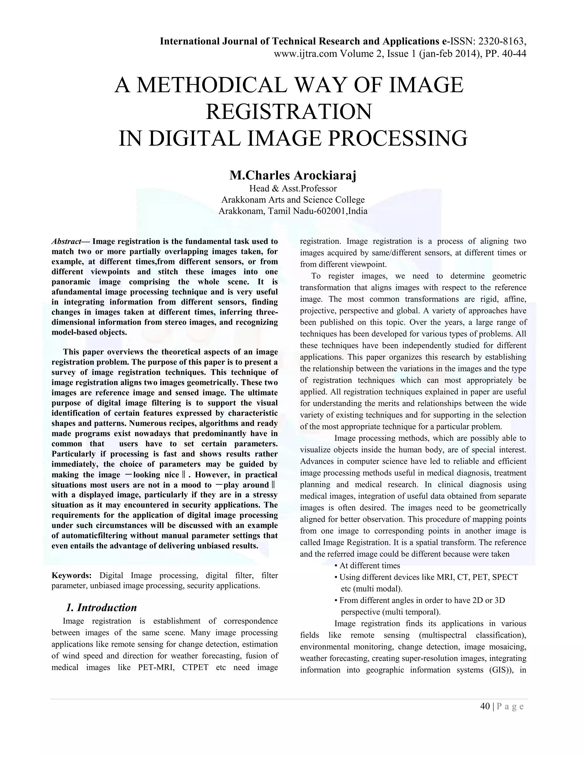
monitoring tumor growth (Figure 1), treatment verification,
comparison of the patient’s data with anatomical atlases ,in
cartography (map updating) and in computer vision (target
localization, automatic quality control).
Fig. 1 : Application of Image Registration in
MR Mammography
Figure 1 shows a maximum intensity projection of contrast
controlled MR mammography a) without registration b) with
registration. It demonstrates superior performance in
identification of cancerous lesions with the use of registration
2. Classification
P. A. Van Den Elsen, E. J. D. Pol and M. A. Viergever, had
given classification of Image registration way back in 1993 [4].
Maintz later in his survey paper [5] has given a more detailed
and augmented version of classification based on nine basic
criteria. Leszek Chmielewski, Dorota Kozinska [9] have
summarized classifications given by earlier papers [4],[6],[7],[8]
The classification is based on following features. Wan Rui,
Prof.Li Minglu [9] have also has mentioned similar
classification.
Dimensionality: 2D/2D, 2D/3D, 3D/3D Image
registrations are possible. Sometimes time could be the fourth
dimension.
Domain of transformation: It could be global or local
depending on whether the whole image or its part is to
eregistered. A transformation is called global if it applies to the
entire image and local otherwise. Optimization Procedure : The
parameters that make up the registration transformation can
either be computed directly from the available data, or
determined by finding an optimum of some function defined on
the parameter space.
Modalities Involved: Four classes of registration tasks
can be recognized based on the modalities that are involved. In
mono model applications, the images to be registered belong to
the same modality, as opposed to multimodal registration tasks,
where the images to be registered stem from two different
modalities. In modality to model and patient to modality
registration only one image is involved and the other modality is
either a model or the patient himself.
Subject: In intra-subject registration, all images
involved are from the same patient. If the registration process
involves images of different patients (or a patient and a model), it
is inter-subject registration. If one image involved is from a
patient, and the other image is created from an image information
database obtained using imaging of many subjects, then it is an
atlas registration.
Object Registration Methods : These can also be
classified according to the body parts that are involved, e.g. head,
thorax.
Interaction: Three levels of interaction can be
recognized. Automatic, where the user only supplies the
algorithm with the image data and possibly information on the
image acquisition. Interactive, where the user does the
registration himself, assisted by software supplying a visual or
numerical impression of the current transformation, and possibly
an initial transformation guess. Semi-automatic, where the
interaction is required to either initialize the algorithm, e.g., by
segmenting the data, or steer the algorithm, e.g., by rejecting or
accepting suggested registration hypotheses.
Nature of Registration Basis: Registration can be
images based or non-image based. The latter usually necessitates
the scanners to be brought in to the same physical location,and
the assumption that the patient remains motionless between both
acquisitions. Image based registration can be divided into
extrinsic and intrinsic methods. Extrinsicmethods rely on
artificial objects attached to the patient, objects which are
designed to be well visible and accurately detectable in all of the
pertinent modalities. Intrinsicmethods rely on image content
only. Registration can be based on a limited set of identified
salient points (landmarks), on the alignment of segmented binary
structures (segmentation based), mostly object surfaces, or
directly onto measures computed from the image grey values
(voxel property based).
Nature of Transformation: The transformation
applied to register the images can be categorized according to the
degrees of freedom. A rigid transformation can be defined as one
that includes only translations and rotations. If a rigid
transformation is allowed to include scaling and shearing, it is
referred to as affine. This type of transformation maps straight
lines to straight lines and preserves the parallelism between lines.
The perspective transformation differs from the affine
transformation in the sense that the parallelism of lines need not
be preserved. The fourth class consists of elastic transformations,
whichallow the mapping of straight lines to curves, and is called
curved transformation.
Type of transformation: The transformation could be
rigid, affine, projective or nonlinear.
Tightness of feature coupling: The transformation can
be interpolating (features of the objects in one image are exactly
transferred into features in the other image) or approximating.
Measure of registration Quality: Various measures
are applied depending on the data features or data itself.
Method of parameter determination: The parameters
of the transformation can be found out using direct or search
oriented methods.
3. 42 | P a g e
Subject of registration: If the two images contain the
same subject it is intra subject registration. If the subject in the
two images differs it is intersubject registration.
Type of data: It can be raw data, features extracted
from data or introduced markers in data.
Source of features: Features explicitly present in the
data are called intrinsic features where as those introduced from
outside are called as extrinsic features.
Automization level: This can be automatic or
semiautomatic depending on user intervention level.
3. Approaches to Image Registration
J.V.Chapnick, M.E.Noz, G.Q. Maguire, E.L.Kramer,
J.J.Sanger, B.A.Birnbaum, A.J.Megibow [10] have mentioned
the following approaches of image registration way back in 1993.
• Transformations using Fourier analysis
• Cross correlation approach using Fourier analysis
• Sum of squares search technique
• Eigen Value Decomposition
• Moment matching techniques
• Warping Techniques
• Procedural approach
• Anatomic Atlas
• Internal landmarks
• External Landmarks
4. ALGORITHM CLASSIFICATION
Following is the classification of image registration algorithms:
• On the basis of Intensity: Intensity based methods compare
intensity patterns in images via correlation metrics. These
methods register entire images or sub images. If sub images are
registered, center of corresponding sub images are treated as
corresponding feature point.
• On the basis of features: Feature based methods find
correspondence between image features such as points, lines and
contours. These methods establish correspondence between a
numbers of points in an image. Knowing the correspondence
between the numbers of points in an image, a transformation is
then determined to map the target image to the referenced image,
there by establishing point by point correspondence between the
referenced and target image.
• Single modality method: Single modality methods tend to
register images in the same modality acquired by the same
scanner or sensor type.
• Multi modality method: Multi modality methods tend to
register images acquired by different sensors or scanner types.
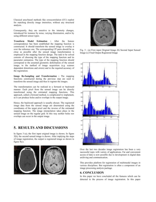
4. METHODOLOGY
Image registration essentially consists of following
steps as per Zitova and Flusser. Figure 2 illustrates the process.
• Feature detection: Salient and distinctive objects (closed-
boundary regions, edges, contours, line intersections, corners,
etc) in both reference and sensed images are detected.
• Feature matching: The correspondence between the features in
the reference and sensed image established.
• Transform model estimation: The type and parameters of the
so-called mapping functions, aligning the sensed image with the
reference image, are estimated.
• Image re-sampling and transformation: The sensed image is
transformed by means of the mapping functions.
Feature Detection : This approach is based on the extraction of
salient features /structures in the image. Significant regions, lines
or points are considered.
Region Features : The region like features can be projections of
general high contrast closed-boundary regions of an appropriate
size like water reservoirs, lakes, and buildings. The regions are
often represented by their centers of gravity, which are invariant
with respect to rotation, scaling, and skewing and stable under
random noise and gray level variation. Region features are
detected by means of segmentation methods. The accuracy of the
segmentation significantly influences the resulting registration.
Line Features : The line features can be the representations of
general line segments, object contours, coastal lines, roads or
elongated anatomic structures in medical imaging. Standard edge
detection methods, like Canny-edge detector or a detector based
on the Laplacian of Gaussian, are employed for the line feature
detection.
Point Features : The point features group consists of line
intersections, road crossings etc which are detected using the
Gabor wavelets, inflection points of curves.
Feature Matching : In the following section two major
categories, area based and feature based methods, are further be
classified into subcategories according to different methods and
advantage and disadvantage.
Area Based Methods : These methods deal with the images
without attempting to detect salient features. Windows of
predefined size o the entire image are used for the
correspondence estimation. Area based methods are preferably
applied when the images have not many prominent details and
the distinctive information is provided by gray levels/colors
rather than by local shapes and structure. From the geometric
point of view, only shift and small rotation between the images
are allowed when using area based methods. To speed up the
searching, area-based methods often employ pyramidal image
representations and sophisticated optimization algorithms.
The limitations of the area based methods originate in their basic
idea. Firstly, the rectangular window, which is most often used,
suits the registration of images which locally differ only by a
translation. If images are deformed by more complex
transformations, this type of the window is not able to cover the
same parts of the scene in the reference and sensed images.
4. Classical area-based methods like crosscorrelation (CC) exploit
for matching directly image intensities, without any structural
analysis.
Consequently, they are sensitive to the intensity changes,
introduced for instance by noise, varying illumination, and/or by
using different sensor types.
Transform Model Estimation : After the feature
correspondence has been established the mapping function is
constructed. It should transform the sensed image to overlay it
over the reference one. The corresponding CP pairs should be as
close as possible after the sensed image transformation is
employed in the mapping function design. The task to be solved
consists of choosing the type of the mapping function and its
parameter estimation. The type of the mapping function should
correspond to the assumed geometric deformation of the sensed
image, to the method of image acquisition (e.g. scanner
dependent distortions and errors) and to the required accuracy of
the registration.
Image Re-Sampling and Transformation : The mapping
functions constructed during the previous step are used to
transform the sensed image and thus to register the images.
The transformation can be realized in a forward or backward
manner. Each pixel from the sensed image can be directly
transformed using the estimated mapping functions. This
approach, called a forward method, is complicated to implement,
as it can produce holes and/or overlaps in the output image.
Hence, the backward approach is usually chosen. The registered
image data from the sensed image are determined using the
coordinates of the target pixel and the inverse of the estimated
mapping function. The image interpolation takes place in the
sensed image on the regular grid. In this way neither holes nor
overlaps can occur in the output image.
5. RESULTS AND DISCUSSIONS
In figure 3 (a), the first input original image is shown. In figure
3(b), the second sensed image is shown. After implying the steps
of image registration, the output is registered image as shown in
figure 4(c).
Fig. 3 : (a) First input Original Image (b) Second Input Sensed
Image (c) Final Output Registered Image
Over the last two decades image registration has been a very
successful topic with variety of applications. Fat and convenient
access of data is now possible due to development in digital data
archiving and communication.
This provides platform for registration of multimodal images in
various disciplines. But registration is often a component of an
image processing analysis package.
6. CONCLUSION
In this paper we have concluded all the features which can be
detected in the process of image registration. In this paper
5. 44 | P a g e
authors have presented a technique for registering the images
differ by rotation, scaling and translation. The techniques
presented can be applied a wide class of problems involving
features may be corners, edges etc. In future work we can use
various methods for detecting these features to make the process
of image registration better.
These methods may be harries corner method for detecting the
corners etc. It will help in making the technique of image
registration better as well as to reduce the errors produced in this
process of image registration.
References
[1] Manjusha P. Deshmukh & Udhav Bhosle, “A SURVEY OF
IMAGE REGISTRATION”, International Journal of Image
Processing (IJIP), Volume (5) : Issue (3) , 2011, 245-269
[2] Medha V. Wyawahare, Dr. Pradeep M. Patil, and Hemant
K.Abhyankar, “Image Registration Techniques: An overview”,
International Journal of Signal Processing, Image Processing
and Pattern Recognition Vol. 2, No.3, September 2009, 11-28
[3] D.Ruckert, L.I.Sonoda, C.Hayes,, D.L.G.Hill ,M.O. Leach
D.J.Hawkes, “Non rigid registration using free form
deformations: Application to breast MR images”, IEEE
transactions on Medical imaging, 18(8), 1999, 712-721
[4] Wan Rui, Prof.Li Minglu, “An Overview of Medical Image
Registration”, Proceedings of the Fifth International
Conference on Computational Intelligence and Multimedia
Applications (ICCIMA’03), 2003, 385-390
[5] Brown Gottesfeld L, “A survey of image Registration
Techniques”, ACM Computing surveys 24, 1992, 325-376 [6]
Barbara Zitova, Jan Flusser, “Image registration methods: a
survey”, Image and Vision Computing 21 (2003),977–1000
[7] Rafael C. Gonzalez, Richard E. Woods, Steven L.
Eddins,“Digital Image Processing Using MATLAB”, Pearson
Education.
[8] Mehfuza Holia & Dr. V.K.Thakar, “Image registration for
recovering affine transformation using Nelder Mead Simplex
method for optimization.”, International Journal of Image
Processing (IJIP) Volume(3), Issue(5), 218-228.