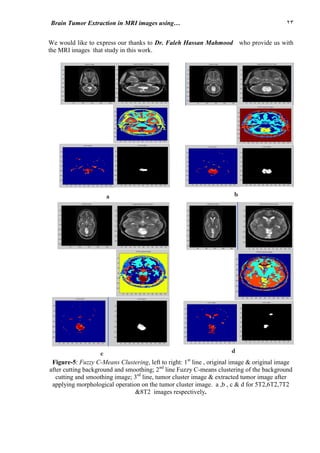
This document summarizes four techniques used to extract brain tumor regions from MRI images: 1) Gray level stretching and Sobel edge detection, 2) K-Means clustering based on location and intensity, 3) Fuzzy C-Means clustering, and 4) an adapted K-Means and Fuzzy C-Means technique. The techniques were able to successfully detect and extract brain tumors, which helps doctors identify tumor size and location. Clustering algorithms like K-Means and Fuzzy C-Means were used to segment MRI images into clusters representing different tissue types to identify tumor regions.
Geometric Correction for Braille Document Images csandit
Image processing is an important research area in computer vision. clustering is an unsupervised
study. clustering can also be used for image segmentation. there exist so many methods for image
segmentation. image segmentation plays an important role in image analysis.it is one of the first
and the most important tasks in image analysis and computer vision. this proposed system
presents a variation of fuzzy c-means algorithm that provides image clustering. the kernel fuzzy
c-means clustering algorithm (kfcm) is derived from the fuzzy c-means clustering
algorithm(fcm).the kfcm algorithm that provides image clustering and improves accuracy
significantly compared with classical fuzzy c-means algorithm. the new algorithm is called
gaussian kernel based fuzzy c-means clustering algorithm (gkfcm)the major characteristic of
gkfcm is the use of a fuzzy clustering approach ,aiming to guarantee noise insensitiveness and
image detail preservation.. the objective of the work is to cluster the low intensity in homogeneity
area from the noisy images, using the clustering method, segmenting that portion separately using
content level set approach. the purpose of designing this system is to produce better segmentation
results for images corrupted by noise, so that it can be useful in various fields like medical image
analysis, such as tumor detection, study of anatomical structure, and treatment planning.
DETECTION OF HUMAN BLADDER CANCER CELLS USING IMAGE PROCESSINGprj_publication
Bladder cancer presents a spectrum of different diatheses. A precise assessment for
individualized treatment depends on the accuracy of the initial diagnosis. In this method the
performance of the level set segmentation is subject to appropriate initialization and optimal
configuration of controlling parameters, which require substantial manual intervention. A
new fuzzy level set algorithm is proposed in this paper to facilitate medical image
segmentation. It is able to directly evolve from the initial segmentation by spatial fuzzy
clustering. The Spatial induced fuzzy c-means using pixel classification and level set
methods are utilizing dynamic variational boundaries for image segmentation. The
controlling parameters of level set evolution are also estimated from the results of clustering.
The fuzzy level set algorithm is enhanced with locally regularized evolution. Such
improvements facilitate level set manipulation and lead to more robust segmentation.
Performance evaluation of the proposed algorithm was carried on medical images
Performance Evaluation of Basic Segmented Algorithms for Brain Tumor DetectionIOSR Journals
In the field of computers segmentation of image plays a very important role. By this method the required
portion of object is traced from the image. In medical image segmentation, clustering is very famous
method . By clustering, an image is divided into a number of various groups or can also be called as clusters.
There are various methods of clustering and thresholding which have been proposed in this paper such as otsu
, region growing , K Means , fuzzy c means and Hierarchical self organizing mapping algorithm. Fuzzy c-means
(FCM) is a method of clustering which allows one piece of data to belong to two or more clusters. This method
(developed by Dunn in 1973 and improved by Bezdek in 1981) is frequently used in pattern recognition. As
process of fuzzy c mean is too slow, this drawback is then removed. In this paper by experimental analysis and
performance parameters the segmentation of hierarchical self organizing mapping method is done in a better
way as compared to other algorithms. The various parameters used for the evaluation of the performance are as follows: segmentation accuracy (Sa) , area (A), rand index (Ri),and global consistency error (Gce)
AN EFFICIENT WAVELET BASED FEATURE REDUCTION AND CLASSIFICATION TECHNIQUE FOR...ijcseit
This document proposes a technique for classifying brain MRI images to diagnose dementia using wavelet-based feature reduction and support vector machine (SVM) classification. It compares SVM trained with genetic algorithm and particle swarm optimization for feature selection and parameter optimization. Wavelet-based feature reduction is found to perform better than principal component analysis (PCA) at reducing features while retaining important information. SVM trained with particle swarm optimization achieved more accurate classification than SVM trained with genetic algorithm. The proposed method uses wavelet transforms to extract Haralick texture features from MRI images, reduces the features, and classifies the images as normal or abnormal using optimized SVM to diagnose mild or severe dementia.
This document summarizes a research paper on using a k-means clustering method to detect brain tumors in MRI images. The paper introduces brain tumors and MRI imaging. It then describes using k-means clustering for tumor segmentation, which groups similar image patterns into clusters to identify the tumor region. The paper presents results of applying k-means to two MRI images, including statistical measures of segmentation accuracy, tumor area comparison, and timing. The k-means method achieved average rand index of 0.8358, low average errors, and tumor areas close to manual segmentation in under 3 seconds, demonstrating potential for accurate and efficient brain tumor detection.
ENHANCING SEGMENTATION APPROACHES FROM FUZZY K-C-MEANS TO FUZZY-MPSO BASED LI...Christo Ananth
Christo Ananth et al. [7] discussed about the combination of Graph cut liver segmentation and Fuzzy with MPSO tumor segmentation algorithms. The system determines the elapsed time for the segmentation process. The accuracy of the proposed system is higher than the existing system. The algorithm has been successfully tested in multiple images where it has performed very well, resulting in good segmentation. It has taken high computation time for the graph cut processing algorithm. In future work, we can reduce the computation time and improves segmentation accuracy.
Classification and Segmentation of Glaucomatous Image Using Probabilistic Neu...ijsrd.com
The gradual visual field loss and there is a characteristic type of damage to the retinal nerve fiber layer associated with the progression of the disease glaucoma. Texture features within images are actively pursued for accurate and efficient glaucoma classification. Energy distribution over wavelet subband is applied to find these important texture features. In this paper, we investigate the discriminatory potential of wavelet features obtained from the Daubechies (db3), symlets (sym3), and biorthogonal (bio3.3, bio3.5, and bio3.7) wavelet filters. We propose a novel technique to extract energy signatures obtained using 2-D discrete wavelet transform, and subject these signatures to different feature ranking and feature selection strategies. Here my project aims at the use of Probabilistic Neural Network (PNN), Fuzzy C-means (FCM) and K-means helps for the detection of glaucoma disease. For this, fuzzy c-means clustering algorithm and k-means algorithm is used. Fuzzy c-means results faster and reliably good clustering when compare to k-means.
SEGMENTATION OF MAGNETIC RESONANCE BRAIN TUMOR USING INTEGRATED FUZZY K-MEANS...ijcsit
Segmentation is a process of partitioning the image into several objects. It plays a vital role in many fields
such as satellite, remote sensing, object identification, face tracking and most importantly in medical field.
In radiology, magnetic resonance imaging (MRI) is used to investigate the human body processes and
functions of organisms. In hospitals, this technique has been using widely for medical diagnosis, to find the
disease stage and follow-up without exposure to ionizing radiation.Here in this paper, we proposed a novel
MR brain image segmentation method for detecting the tumor and finding the tumor area with improved
performance over conventional segmentation techniques such as fuzzy c means (FCM), K-means and even
that of manual segmentation in terms of precision time and accuracy. Simulation performance shows that
the proposed scheme has performed superior to the existing segmentation methods.
Microscopy images segmentation algorithm based on shearlet neural networkjournalBEEI
Microscopic images are becoming important and need to be studied to know the details and how-to quantitatively evaluate decellularization. Most of the existing research focuses on deep learning-based techniques that lack simplification for decellularization. A new computational method for the segmentation microscopy images based on the shearlet neural network (SNN) has been introduced. The proposal is to link the concept of shearlets transform and neural networks into a single unit. The method contains a feed-forward neural network and uses a single hidden layer. The activation functions are depending on the standard shearlet transform. The proposed SNN is a powerful technology for segmenting an electron microscopic image that is trained without relying on the pre-information of the data. The shearlet neural networks capture the features of full accuracy and contextual information, respectively. The expected value for specific inputs is estimated by learning the functional configuration of a network for the sequence of observed value. Experimental results on the segmentation of two-dimensional microscopy images are promising and confirm the benefits of the proposed approach. Lastly, we investigate on a challenging datasets ISBI 2012 that our method (SNN) achieves superior outcomes when compared to classical and deep learning-based methods.
Optimized Neural Network for Classification of Multispectral ImagesIDES Editor
This document summarizes an article that proposes using a multiobjective particle swarm optimization (MOPSO) approach to optimize the structure of an artificial neural network for classifying multispectral satellite images. Specifically, the MOPSO is used to simultaneously select the most discriminative spectral bands from the available options and determine the optimal number of nodes in the hidden layer of the neural network. The MOPSO approach is compared to traditional classifiers like maximum likelihood classification and Euclidean classifiers. The results show that the MOPSO-optimized neural network approach provides superior performance for remote sensing image classification problems.
fMRI Segmentation Using Echo State Neural NetworkCSCJournals
This research work proposes a new intelligent segmentation technique for functional Magnetic Resonance Imaging (fMRI). It has been implemented using an Echostate Neural Network (ESN). Segmentation is an important process that helps in identifying objects of the image. Existing segmentation methods are not able to exactly segment the complicated profile of the fMRI accurately. Segmentation of every pixel in the fMRI correctly helps in proper location of tumor. The presence of noise and artifacts poses a challenging problem in proper segmentation. The proposed ESN is an estimation method with energy minimization. The estimation property helps in better segmentation of the complicated profile of the fMRI. The performance of the new segmentation method is found to be better with higher peak signal to noise ratio (PSNR) of 61 when compared to the PSNR of the existing back-propagation algorithm (BPA) segmentation method which is 57.
Hybrid Pixel-Based Method for Multimodal Medical Image Fusion Based on Integr...Dr.NAGARAJAN. S
Medical imaging plays a vital role in medical diagnosis and treatment. However, distinct imaging modality yields information only in limited domain. Studies are done for analysis information collected from distinct modalities of same patient. This led to the introduction of image fusion in the field of medicine and the progression of image fusion techniques. Image fusion is characterized as the amalgamation of significant data from numerous images and their incorporation into seldom images, generally a solitary one. This fused image will be more instructive and precise than the indi- vidual source images that have been utilized, and the resultant fused image comprises paramount information. The main objective of image fusion is to incorporate all the essential data from source images which would be pertinent and comprehensible for human and machine recognition. Image fusion is the strategy of combining images from distinct modalities into a single image [1]. The resultant image is utilized in variety of applications such as medical diagnosis, identification of tumor and surgery treatment [2]. Before fusing images from two distinct modalities, it is essential to preserve the features so that the fused image is free from inconsistencies or artifacts in the output.
Classification of MR medical images Based Rough-Fuzzy KMeansIOSRJM
The document summarizes a proposed algorithm for classifying MR medical images using Rough-Fuzzy K-Means (FRKM). It begins with an introduction to the challenges of medical image classification and a literature review of previous techniques. It then provides background on rough set theory, fuzzy set theory, and K-means clustering. The proposed FRKM algorithm is described as using rough set theory for feature selection and dimensionality reduction, followed by a K-means clustering with probabilities assigned based on rough set approximations to classify ambiguous areas. Experimental results show the FRKM approach achieves 94.4% accuracy, higher than other techniques.
Brain Image Fusion using DWT and Laplacian Pyramid Approach and Tumor Detecti...INFOGAIN PUBLICATION
Image fusion is the process of combining important information from two or more images into a single image. The resulting image will be more enhanced than any of the input pictures. The idea of combining multiple image modalities to furnish a single, more enhanced image is well established, special fusion methods have been proposed in literature. This paper is based on image fusion using laplacian pyramid and Discreet Wavelet Transform (DWT) methods. This system uses an easy and effective algorithm for multi-focus image fusion which uses fusion rules to create fused image. Subsequently, the fused image is obtained by applying inverse discreet wavelet transform. After fused image is obtained, watershed segmentation algorithm is applied to detect the tumor part in fused image.
Ijeee 16-19-a novel approach to brain tumor classification using wavelet and ...Kumar Goud
This document presents a novel approach for classifying brain tumors using magnetic resonance images (MRIs). The proposed technique uses two stages: 1) discrete wavelet transform for dimensionality reduction and feature extraction, and 2) probabilistic neural network (PNN) for classification. MRIs of benign and malignant brain tumors were collected and preprocessed using discrete wavelet transform to extract features. A PNN classifier was then trained on these features to classify tumors as benign or malignant. The technique aims to provide an automated brain tumor classification method using artificial intelligence.
HIGH RESOLUTION MRI BRAIN IMAGE SEGMENTATION TECHNIQUE USING HOLDER EXPONENTijsc
Image segmentation is a technique to locate certain objects or boundaries within an image. Image
segmentation plays a crucial role in many medical imaging applications. There are many algorithms and
techniques have been developed to solve image segmentation problems. Spectral pattern is not sufficient in
high resolution image for image segmentation due to variability of spectral and structural information.
Thus the spatial pattern or texture techniques are used. Thus the concept of Holder Exponent for
segmentation of high resolution medical image is an efficient image segmentation technique. The proposed
method is implemented in Matlab and verified using various kinds of high resolution medical images. The
experimental results shows that the proposed image segmentation system is efficient than the existing
segmentation systems.
Brain Tumor Extraction from T1- Weighted MRI using Co-clustering and Level Se...CSCJournals
The aim of the paper is to propose effective technique for tumor extraction from T1-weighted magnetic resonance brain images with combination of co-clustering and level set methods. The co-clustering is the effective region based segmentation technique for the brain tumor extraction but have a drawback at the boundary of tumors. While, the level set without re-initialization which is good edge based segmentation technique but have some drawbacks in providing initial contour. Therefore, in this paper the region based co-clustering and edge-based level set method are combined through initially extracting tumor using co-clustering and then providing the initial contour to level set method, which help in cancelling the drawbacks of co-clustering and level set method. The data set of five patients, where one slice is selected from each data set is used to analyze the performance of the proposed method. The quality metrics analysis of the proposed method is proved much better as compared to level set without re-initialization method.
K Mean and Fuzzy Clustering Algorithm Predicated Brain Tumor Segmentation And...IRJET Journal
This document discusses using K-means and fuzzy clustering algorithms to segment and estimate the area of predicted brain tumors in MRI images. It begins with an introduction to brain tumors and MRI imaging. Then, it describes the related work on brain tumor detection and classification using techniques like artificial neural networks. The proposed method is outlined as applying K-means clustering initially for segmentation followed by fuzzy C-means clustering for more precise tumor shape extraction. Results from applying the algorithms to images are presented and analyzed, showing the processing time, estimated tumor area, and other metrics. The conclusion is that K-means works well for mass tumors while fuzzy C-means is better for segmenting malignant tumors to precisely estimate the shape and position.
Fully Automatic Method for 3D T1-Weighted Brain Magnetic Resonance Images Seg...CSCJournals
In the domain of medical imaging, accurate segmentation of brain MR images is of interest for many brain disorders. However, due to several factors such noise, imaging artefacts, intrinsic tissue variation and partial volume effects, tissue segmentation remains a challenging task. So, in this paper, a full automatic method for segmentation of brain MR images is presented. The method consists of four steps segmentation procedure. First, noise removing by median filtering is done; second segmentation of brain/non-brain tissue is performed by using a Threshold Morphologic Brain Extraction method (TMBE). Then initial centroids estimation by gray level histogram analysis based is executed. Finally, Fuzzy C-means Algorithm is used for MRI tissue segmentation. The efficiency of the proposed method is demonstrated by extensive segmentation experiments using simulated and real MR images.
1. Self-organizing maps (SOM) are an unsupervised learning algorithm that transform high-dimensional data into lower dimensions for visualization while preserving topological properties.
2. The SOM network has an input layer fully connected to an output layer arranged in a grid, with each node containing a weight vector of the same dimension as inputs.
3. During training, the best matching unit (BMU) and its neighbors on the grid have their weight vectors adjusted to better match the input based on their distance from the BMU, with learning rates decreasing over time.
This document summarizes a research paper on using bilateral symmetry analysis to detect brain tumors from MRI images. It begins by introducing the problem of brain tumor detection and importance of asymmetry analysis. It then describes the proposed algorithm which involves defining a bilateral symmetry axis between the two brain hemispheres and detecting any regions of asymmetry that could indicate a tumor. The algorithm uses edge detection techniques to find the symmetry axis. Performance is evaluated on sample patient data and results show the method can successfully identify tumor locations and sizes. In conclusion, analyzing bilateral symmetry is an effective approach for automated brain tumor detection from MRI images.
Abstract
This paper proposes a survey on the classification techniques of lung nodules. We have the different classifications about the nodules in the lungs. It contains the different methods of classification, segmentation and detection techniques. Malignant cell presented in the lungs named , nodules are classified for the treatment processes. Thresholding and Robust segmentation techniques are used in the segmentation process and the feature set is used for classification. Low Dose CT(Computed Tomography) images are applied. This survey has the information about the efficient techniques which are all used for the nodule classification. In these days lung cancer is the dangerous dead disease in the world, So we need to have the knowledge of that cancer. In starting stages the micro nodules are then formed into a cancer cell. Among the cancer affected population about 20% of the people are dead due to lung cancer. If nodules are found in a starting stage, we can be extend the lifetime of the patient. The main process of this paper involves with the nodule classification and segmentation process of the lung nodules. Here we taken the different procedures involved with nodule detections. CT is the most appropriate imaging technique to obtain anatomical information about lung nodules and the surrounding structures. Here we taken the Low Dose CT(LDCT) images for operations. This paper has the various approaches of the nodule classification. In this survey different techniques are presented which are used for detection and classification of the nodules in the lungs. By differentiating the nodules from the anatomical parts of the lungs, the nodules are identified.
Keywords: PLSA, Robust Segmentation and Partitioning.
FUZZY SEGMENTATION OF MRI CEREBRAL TISSUE USING LEVEL SET ALGORITHMAM Publications
The current study investigated a median filter with the fuzzy level set method to propose fuzzy segmentation of magnetic resonance imaging (MRI) cerebral tissue images. An MRI image was used as an input image. A median filter and fuzzy c-means (FCM) clustering were utilized to remove image noise and create image clusters, respectively. The image clusters showed initial and final cluster centers. The level set method was then used for segmentation after separating and extracting white matter from gray matter. Fuzzy c-means was sensitive to the choice of the initial cluster center. Improper center selection caused the method to produce suboptimal solutions. The proposed algorithm was successfully utilized to segment MRI cerebral tissue images. The algorithm efficiently performed segmentation of test MRI cerebral tissue images compared with algorithms proposed in previous studies.
Development of algorithm for identification of maligant growth in cancer usin...IJECEIAES
The precise identification and characterization of small pulmonary nodules at low-dose CT is a necessary requirement for the completion of valuable lung cancer screening. It is compulsory to develop some automated tool, in order to detect pulmonary nodules at low dose ct at the beginning stage itself. The various algorithms had been proposed earlier by many researchers within the past, but the accuracy of prediction is usually a challenging task. During this work, a man-made neural networ based methodology is proposed to seek out the irregular growth of lung tissues. Higher probability of detection is taken as a goal to urge an automatic tool, with great accuracy. The best feature sets derived from Haralick Gray level co occurrence Matrix and used because the dimension reduction way for feeding neural network. During this work, a binary Binary classifier neural network has been proposed to spot the traditional images out of all the images. The potential of the proposed neural network has been quantitatively computed using confusion matrix and located in terms of accuracy.
The document provides a literature review of different clustering techniques. It begins by defining clustering and its applications. It then categorizes and describes several clustering methods including hierarchical (BIRCH, CURE, CHAMELEON), partitioning (k-means, k-medoids), density-based (DBSCAN, OPTICS, DENCLUE), grid-based (CLIQUE, STING, MAFIA), and model-based (RBMN, SOM) methods. For each method, it discusses the algorithm, advantages, disadvantages and time complexity. The document aims to provide an overview of various clustering techniques for classification and comparison.
International Journal of Research in Engineering and Science is an open access peer-reviewed international forum for scientists involved in research to publish quality and refereed papers. Papers reporting original research or experimentally proved review work are welcome. Papers for publication are selected through peer review to ensure originality, relevance, and readability.
SVM Classifiers at it Bests in Brain Tumor Detection using MR Imagesijtsrd
This paper presents some case study frameworks to limelight SVM classifiers as most efficient one compared to existing classifiers like Otsu, k-means and fuzzy c-means. In general, Computed Tomography (CT) and Magnetic Resonance Imaging (MR) are more dominant imaging technique for any brain lesions detection like brain tumor, Alzheimer' disease and so on. MR imaging takes a lead technically for imaging medical images due to its possession of large spatial resolution and provides better contrast for the soft tissues like white matter (WM), gray matter (GM) and cerebrospinal fluid (CSF). The usual method used for classification of lesions in brain images consists of pre-processing, feature extraction, feature reduction and classification. Early detection of the tumor region without much time lapse in computation can be achieved by using efficient SVM classifier model. Brain tumor grade classifications with the assistance of morphologically selected features are extracted and tumor classification is attained using SVM classifier. The assessment of SVM classifications are evaluated through metrics termed as sensitivity, exactness and accuracy of segmentation. These measures are then compared with existing methods to exhibit the SVM classifier as significant classifier model. Dr. R Manjunatha Prasad | Roopa B S"SVM Classifiers at it Bests in Brain Tumor Detection using MR Images" Published in International Journal of Trend in Scientific Research and Development (ijtsrd), ISSN: 2456-6470, Volume-2 | Issue-5 , August 2018, URL: http://www.ijtsrd.com/papers/ijtsrd18372.pdf http://www.ijtsrd.com/engineering/electronics-and-communication-engineering/18372/svm-classifiers-at-it-bests-in-brain-tumor-detection-using-mr-images/dr-r-manjunatha-prasad
GAUSSIAN KERNEL BASED FUZZY C-MEANS CLUSTERING ALGORITHM FOR IMAGE SEGMENTATIONcscpconf
Image processing is an important research area in computer vision. clustering is an unsupervised study. clustering can also be used for image segmentation. there exist so many methods for image segmentation. image segmentation plays an important role in image analysis.it is one of the first and the most important tasks in image analysis and computer vision. this proposed system presents a variation of fuzzy c-means algorithm that provides image clustering. the kernel fuzzy
c-means clustering algorithm (kfcm) is derived from the fuzzy c-means clustering algorithm(fcm).the kfcm algorithm that provides image clustering and improves accuracy significantly compared with classical fuzzy c-means algorithm. the new algorithm is called gaussian kernel based fuzzy c-means clustering algorithm (gkfcm)the major characteristic of gkfcm is the use of a fuzzy clustering approach ,aiming to guarantee noise insensitiveness and image detail preservation.. the objective of the work is to cluster the low intensity in homogeneity area from the noisy images, using the clustering method, segmenting that portion separately using content level set approach. the purpose of designing this system is to produce better segmentation results for images corrupted by noise, so that it can be useful in various fields like medical image analysis, such as tumor detection, study of anatomical structure, and treatment planning.
GAUSSIAN KERNEL BASED FUZZY C-MEANS CLUSTERING ALGORITHM FOR IMAGE SEGMENTATIONcsandit
Image processing is an important research area in computer vision. clustering is an unsupervised
study. clustering can also be used for image segmentation. there exist so many methods for image
segmentation. image segmentation plays an important role in image analysis.it is one of the first
and the most important tasks in image analysis and computer vision. this proposed system
presents a variation of fuzzy c-means algorithm that provides image clustering. the kernel fuzzy
c-means clustering algorithm (kfcm) is derived from the fuzzy c-means clustering
algorithm(fcm).the kfcm algorithm that provides image clustering and improves accuracy
significantly compared with classical fuzzy c-means algorithm. the new algorithm is called
gaussian kernel based fuzzy c-means clustering algorithm (gkfcm)the major characteristic of
gkfcm is the use of a fuzzy clustering approach ,aiming to guarantee noise insensitiveness and
image detail preservation.. the objective of the work is to cluster the low intensity in homogeneity
area from the noisy images, using the clustering method, segmenting that portion separately using
content level set approach. the purpose of designing this system is to produce better segmentation
results for images corrupted by noise, so that it can be useful in various fields like medical image
analysis, such as tumor detection, study of anatomical structure, and treatment planning.
Comparison of Image Segmentation Algorithms for Brain Tumor DetectionIJMTST Journal
This paper deals with the implementation of Simple Algorithms for detection of size and shape of tumor in brain using MRI images. Generally, CT scan or MRI that is directed into intracranial cavity produces a complete image of brain. This image is visually examined by the physician for detection & diagnosis of brain tumor. However this method of detection resists the accurate determination of stage & size of tumor. To avoid that, this project uses computer aided method for segmentation (detection) of brain tumor by applying Fuzzy C-Means, K-Means, Gaussian Kernel and Pillar K-means algorithms. This segmentation process includes a new mechanism for clustering the elements of high-resolution images in order to improve precision and reduce computation time. The system applies FCM, Gaussian kernel and K-means clustering to the image later optimized by Pillar Algorithm. It designates the initial centroids’ positions by calculating the Euclidian distance metric between each data point and all previous centroids. Then it selects data points which have the maximum distance as new initial centroids. This algorithm distributes all initial centroids according to the maximum accumulated distance metric. In addition, it also reduces the time for analysis. At the end of the process the tumor is extracted from the MRI image and its exact position and the shape is also determined. This paper evaluates the proposed approach for Brain tumor detection by comparing with K-means, Fuzzy C means, Gaussian Kernel and manually segmented algorithms. The experimental results clarify the effectiveness of proposed approach to improve the segmentation quality in aspects of precision and computational time.
MALIGNANT AND BENIGN BRAIN TUMOR SEGMENTATION AND CLASSIFICATION USING SVM WI...sipij
In this article a method is proposed for segmentation and classification of benign and malignant tumor slices in brain Computed Tomography (CT) images. In this study image noises are removed using median and wiener filter and brain tumors are segmented using Support Vector Machine (SVM). Then a two-level discrete wavelet decomposition of tumor image is performed and the approximation at the second level is obtained to replace the original image to be used for texture analysis. Here, 17 features are extracted that 6 of them are selected using Student’s t-test. Dominant gray level run length and gray level co-occurrence texture features are used for SVM training. Malignant and benign tumors are classified using SVM with kernel width and Weighted kernel width (WSVM) and k-Nearest Neighbors (k-NN) classifier. Classification accuracy of classifiers are evaluated using 10 fold cross validation method. The segmentation results are
also compared with the experienced radiologist ground truth. The experimental results show that the proposed WSVM classifier is able to achieve high classification accuracy effectiveness as measured by sensitivity and specificity.
A Wavelet Based Automatic Segmentation of Brain Tumor in CT Images Using Opti...CSCJournals
This document summarizes a research paper that proposes a new method for automatically segmenting brain tumors in CT images. The method uses a combination of wavelet-based texture features extracted from discrete wavelet transformed sub-bands. These features are optimized using genetic algorithms and used to train probabilistic neural network and feedforward neural network classifiers to segment tumors. The proposed method is evaluated on brain CT images and shown to outperform existing segmentation methods.
This document presents a comparative study of two segmentation methods - k-means clustering and fuzzy c-means clustering with genetic algorithm - for detecting brain tumors in MRI images. K-means clustering is used to segment MRI images into clusters and identify tumor regions. Fuzzy c-means clustering with genetic algorithm aims to improve upon k-means by eliminating over-segmentation issues and providing faster, more efficient clustering results. The experimental results indicate fuzzy c-means performs better than k-means for brain tumor segmentation. The document also reviews several other related works applying techniques like edge detection and probabilistic neural networks to segment brain tumors from MRI scans.
Lec13: Clustering Based Medical Image Segmentation MethodsUlaş Bağcı
Clustering – K-means
– FCM (fuzzyc-means)
– SMC (simple membership based clustering) – AP(affinity propagation)
– FLAB(fuzzy locally adaptive Bayesian)
– Spectral Clustering Methods
ShapeModeling – M-reps – Active Shape Models (ASM) – Oriented Active Shape Models (OASM) – Application in anatomy recognition and segmentation – Comparison of ASM and OASM ActiveContour(Snake) • LevelSet • Applications Enhancement, Noise Reduction, and Signal Processing • MedicalImageRegistration • MedicalImageSegmentation • MedicalImageVisualization • Machine Learning in Medical Imaging • Shape Modeling/Analysis of Medical Images Deep Learning in Radiology Fuzzy Connectivity (FC) – Affinity functions • Absolute FC • Relative FC (and Iterative Relative FC) • Successful example applications of FC in medical imaging • Segmentation of Airway and Airway Walls using RFC based method Energy functional – Data and Smoothness terms • GraphCut – Min cut – Max Flow • ApplicationsinRadiologyImages
Performance Evaluation of Basic Segmented Algorithms for Brain Tumor DetectionIOSR Journals
This document evaluates and compares the performance of various segmentation algorithms for detecting brain tumors in MRI images, including hierarchical self-organizing mapping (HSOM), region growing, Otsu, K-means, and fuzzy C-means. It finds that HSOM performs best according to evaluation metrics like segmentation accuracy, Rand index, global consistency error, and variation of information. HSOM is able to segment brain tumor images with higher accuracy and consistency compared to other algorithms like region growing, Otsu, K-means and fuzzy C-means.
International Journal of Computational Engineering Research(IJCER) is an intentional online Journal in English monthly publishing journal. This Journal publish original research work that contributes significantly to further the scientific knowledge in engineering and Technology
MAGNETIC RESONANCE BRAIN IMAGE SEGMENTATIONVLSICS Design
Segmentation of tissues and structures from medical images is the first step in many image analysis applications developed for medical diagnosis. With the growing research on medical image segmentation, it is essential to categorize the research outcomes and provide researchers with an overview of the existing segmentation techniques in medical images. In this paper, different image segmentation methods applied on magnetic resonance brain images are reviewed. The selection of methods includes sources from image processing journals, conferences, books, dissertations and thesis. The conceptual details of the methods are explained and mathematical details are avoided for simplicity. Both broad and detailed categorizations of reviewed segmentation techniques are provided. The state of art research is provided with emphasis on developed techniques and image properties used by them. The methods defined are not always mutually independent. Hence, their inter relationships are also stated. Finally, conclusions are drawn summarizing commonly used techniques and their complexities in application.
This document provides a review of different techniques for segmenting brain MRI images to detect tumors. It compares the K-means and Fuzzy C-means clustering algorithms. K-means is an exclusive clustering algorithm that groups data points into distinct clusters, while Fuzzy C-means is an overlapping clustering algorithm that allows data points to belong to multiple clusters. The document finds that Fuzzy C-means requires more time for brain tumor detection compared to other methods like hierarchical clustering or K-means. It also reviews related work applying these clustering algorithms to segment brain MRI images.
Human brain is the most complex structure where identifying the tumor like diseases are extremely challenging because differentiating the components of a brain is complex. In this paper, pillar k-means algorithm is used for segmentation of brain tumor from magnetic resonance image (MRI).Generally, the brain tumor is detected by radiologist through analysis of MR images which takes longer time. The pillar k-means algorithm’s experimental results clarify the effectiveness of our approach to improve the segmentation quality, accuracy, and computational time. Classify, the tumor from the brain MR images using Bayesian classification.
1) The document presents an integrated technique for detecting brain tumors in MRI images that combines modified texture-based region growing segmentation and edge detection.
2) The technique first performs pre-processing on MRI images, then uses modified texture-based region growing to segment regions. It then applies edge detection to extract the tumor region.
3) Experimental results show the integrated technique provides more accurate tumor detection compared to individual segmentation methods and manual segmentation.
Automatic Diagnosis of Abnormal Tumor Region from Brain Computed Tomography I...ijcseit
The research work presented in this paper is to achieve the tissue classification and automatically
diagnosis the abnormal tumor region present in Computed Tomography (CT) images using the wavelet
based statistical texture analysis method. Comparative studies of texture analysis method are performed
for the proposed wavelet based texture analysis method and Spatial Gray Level Dependence Method
(SGLDM). Our proposed system consists of four phases i) Discrete Wavelet Decomposition (ii)
Feature extraction (iii) Feature selection (iv) Analysis of extracted texture features by classifier. A
wavelet based statistical texture feature set is derived from normal and tumor regions. Genetic Algorithm
(GA) is used to select the optimal texture features from the set of extracted texture features. We construct
the Support Vector Machine (SVM) based classifier and evaluate the performance of classifier by
comparing the classification results of the SVM based classifier with the Back Propagation Neural network
classifier(BPN). The results of Support Vector Machine (SVM), BPN classifiers for the texture analysis
methods are evaluated using Receiver Operating Characteristic (ROC) analysis. Experimental results
show that the classification accuracy of SVM is 96% for 10 fold cross validation method. The system
has been tested with a number of real Computed Tomography brain images and has achieved satisfactory
results.
Primary challenges are the identification, segmentation, and extraction of the afflicted area from the scanning of magnetic resonance. However, it is a time-consuming and tiresome for clinical specialists. In this paper,
an automated brain tumor system is proposed. The proposed system employs hybrid image processing techniques such as contrast correction, histogram normalization, thresholding techniques, arithmetic, and morphological operations to quarantine nearby organs and other tissue from the brain for improving the localization of the affected region. At first, the skull stripping process is proposed to segregate the non-designated regions to extract the designated brain regions. Those resultant brain region images are further subjected to discover the brain tumor. The planned scheme is studied on the magnetic resonance (MR) images with the use of T1, T2, T1c, and fluid-attenuated inversion recovery (FLAIR). The proposed hybrid method employed. The results reveal that the proposed method is quite efficient to extract the tumor region. The accuracy rate for segmentation and separation of area of interest in brain tumor reached to 95%. Finally, the significance of the proposed procedure is confirmed using the real image clinical dataset got from ten patients were diagnosed as begin, malignant, and metastatic brain tumors in Al-Yarmouk and Baghdad teaching hospital in Baghdad, Iraq.
Mammogram image segmentation using rough clusteringeSAT Journals
This document discusses using rough clustering algorithms for mammogram image segmentation. It proposes using Rough K-Means clustering on Haralick texture features extracted from mammogram images. The Rough K-Means algorithm is compared to traditional K-Means and Fuzzy C-Means using metrics like mean square error and root mean square error. Preliminary results found that Rough K-Means produced better segmentation results than the other methods. The document provides background on rough set theory, image segmentation, feature extraction, and different clustering algorithms that can be used.
IJRET : International Journal of Research in Engineering and Technology is an international peer reviewed, online journal published by eSAT Publishing House for the enhancement of research in various disciplines of Engineering and Technology. The aim and scope of the journal is to provide an academic medium and an important reference for the advancement and dissemination of research results that support high-level learning, teaching and research in the fields of Engineering and Technology. We bring together Scientists, Academician, Field Engineers, Scholars and Students of related fields of Engineering and Technology.
SEGMENTATION AND CLASSIFICATION OF BRAIN TUMOR CT IMAGES USING SVM WITH WEIGH...csandit
In this article a method is proposed for segmentation and classification of benign and malignant
tumor slices in brain Computed Tomography (CT) images. In this study image noises are
removed using median and wiener filter and brain tumors are segmented using Support Vector
Machine (SVM). Then a two-level discrete wavelet decomposition of tumor image is performed
and the approximation at the second level is obtained to replace the original image to be used
for texture analysis. Here, 17 features are extracted that 6 of them are selected using Student’s
t-test. Dominant gray level run length and gray level co-occurrence texture features are used for
SVM training. Malignant and benign tumors are classified using SVM with kernel width and
Weighted kernel width (WSVM) and k-Nearest Neighbors (k-NN) classifier. Classification
accuracy of classifiers are evaluated using 10 fold cross validation method. The segmentation
results are also compared with the experienced radiologist ground truth. The experimental
results show that the proposed WSVM classifier is able to achieve high classification accuracy
effectiveness as measured by sensitivity and specificity.
SEGMENTATION AND CLASSIFICATION OF BRAIN TUMOR CT IMAGES USING SVM WITH WEIGH...
vol.4.1.2.july.13
1. Brain Tumor Extraction in MRI images using
Clustering and Morphological Operations
Techniques
S.M. Ali1
, Loay Kadom Abood2
, and Rabab Saadoon Abdoon3
1
Remote Sensing Unit - college of science - University of Baghdad- Iraq,
2
Department of Computer Science-College of Science- University of Baghdad-
Iraq, 3
Department of Physics - College of Science-University of Babylon-Iraq
e-mail: Sensing.remote@gmail.com, loayka@yahoo.com, sr614@ymail.com
Abstract
In this paper, Magnetic Resonance Images,T2 weighted modality , have
been pre-processed by bilateral filter to reduce the noise and maintaining
edges among the different tissues. Four different techniques with
morphological operations have been applied to extract the tumor region.
These were: Gray level stretching and Sobel edge detection, K-Means
Clustering technique based on location and intensity, Fuzzy C-Means
Clustering, and An Adapted K-Means clustering technique and Fuzzy C-
Means technique. The area of the extracted tumor regions has been
calculated. The present work showed that the four implemented techniques
can successfully detect and extract the brain tumor and thereby help doctors
in identifying tumor's size and region.
Keywords: MRI, Brain Tumor, Clustering, Morphological Operations, K-Means and
FCM
1 Introduction
Imaging is an essential aspect of medical science to visualize the anatomical
structures of the human body. Several new complex multidimensional digital
images of physiological structures can be processed and manipulated to help
visualize hidden diagnostic features that are otherwise difficult or impossible to
identify using planar imaging methods [1]. Magnetic Resonance Imaging (MRI)
is a significant technique for examining the human body, it helps to clarify and
distinguish the neural architecture of the human brain. It is unharmed method of
obtaining images of the specific structure in the human body. MRI scanner
employs magnetic field and radio waves to generate exhaustive images of the
human brain, its data is most relevant in the studies of a head, specifically, for
tracking the size of brain tumor and other brain related problems. It helps for
early detection of intracranial tumors and precise estimation of tumor boundaries.
Analytical, MRI scan can also been used to assess the maturity of the central
nervous system and diagnose malformations. The resonance is also crucial for
2. 13 Brain Tumor Extraction in MRI images using…
imaging of vascular changes. Using this method of diagnostic, imaging allows
obtaining information about aneurysms and accompanying symptoms. It is also
helps in showing seditious changes of the central nervous system and gives
accurate assessment of the degree of brain atrophy . The automatic classification
of brain's MRI can thus be used to identify regions having various brain diseases
like cerebro-vascular, Alzheimer, brain tumor, inflammatory, etc [2].
The segmented MR images used in the medical diagnostic process depends on
a combination of two, often conflicting, requirements; i.e. the removal of the
unnecessary information present in the original MR images and the maintenance
of the significant details in the resulting segmented images. MRI segmentation
methods are usually evaluate based on their ability to differentiate: i) between
cerebro-spinal fluid (CSF), white matter, and gray matter, and ii) between normal
tissues and abnormalities. Many segmentation techniques have been proposed in
the recent years, which were used for segmentation of brain tissues from MRI,
are classical pattern recognition methods, rule-based systems, image analysis
methods, crisp and fuzzy clustering procedures, feed-forward neural networks,
fuzzy reasoning, geometric models to determine lesion boundaries, connected
component analysis, deterministic annealing, atlas based methods and contouring
approaches[3]. Lots of researches have been performed for the segmentation of
MR brain images to detect and extract tumor regions from these images. Some of
these related works regarding the segmentation of brain tissues using clustering
and other methods can be found in [3]-[8].
2 Image segmentation
Image segmentation methods can be classified into three categories: edge-based
methods, region-based methods, and pixel-based methods. For Brain
segmentation, two types of segmentation techniques have been adopted in the
literature; i.e. region detection methods and boundary detection methods.
Mostly, the existing methods are dedicated for specific objects. The K-means
clustering technique is a pixel-based method, it is one of the most simple
techniques, it's complexity is relatively lower than other region-based or edge-
based methods. Furthermore, K-means clustering is suitable for biomedical
image segmentation as the number of clusters is usually known for images of
particular regions of the human anatomy. Combined with the existing methods
and aiming to get better results, it is useful to take soft segmentation methods
into account. In soft segmentation, pixels are classified into different classes with
various degrees of uncertainty which are specified by functions. The larger the
value of function for a specific pixel, the larger the possibility that this pixel
belongs to that cluster. The fuzzy C-means (FCM) clustering algorithm is soft
segmentation method, and has aroused comprehensive attention. There have been
many different families of fuzzy clustering algorithms proposed, for instance see
[1] and [5].
3 Clustering
Clustering is the process of grouping feature vectors into classes in the self-
organizing mode. Let {x(q): q = 1,…,Q} be a set of Q feature vectors. Each
feature vector x(q)= (x1(q), …, xN (q)) has N components. The process of
clustering is to assign the Q feature vectors into K clusters {c(k): k = 1, …, K},
3. 14 S. M. Ali, Loay Kadom Abood, and Rabab Saadoon Abdoon
usually by the minimum distance assignment principle. Choosing the
representation of cluster centers (or prototypes) is crucial to the clustering.
Feature vectors that are farther away from the cluster center should not have as
much weight to those are close. These more distant feature vectors are outliers
usually caused by errors in one or more measurements or a deviation in the
processes that formed the object. The simplest weighting method is arithmetic
averaging; it adds all feature vectors in a cluster and takes the average as
prototype. Because of its simplicity, it is still widely used in the clustering
initialization. The arithmetic averaging gives the central located feature vectors
the same weights as outliers. To lower the influence of the outliers, median
vectors are used in some proposed algorithms. To be more immune to outliers
and more representatives, the fuzzy weighted average is introduced to represent
prototypes [1]:
}:{
)()(
kqq
q
nqk
k
n XWZ
(1)
Rather than a Boolean value "1-True" (means it belongs to the cluster), or 0-
False (does not belong),
The weight Wqk in equation (1) represents partial membership to a cluster. It is
called a fuzzy weight. There are different means to generate fuzzy weights.
One way of generating fuzzy weights is the reciprocal of distance [1]; i.e.
)0Dif1(W,
1
qkqk
qk
qk
D
W
(2)
The earlier fuzzy clustering algorithms; when the distance between the feature
vector and the prototype is large, the weight is small, and it is large when the
distance is small. Using Gaussian functions to generate fuzzy weights is the most
natural way for clustering. It is not only immune to outliers but also provides
appropriate weighting for more centrally and densely located vectors. It is used
in the fuzzy clustering and fuzzy merging (FCFM) algorithm [1].
4 K-Means Clustering
It is one of the simplest unsupervised learning algorithms to solve the well
known clustering problem. The procedure follows a simple and easy way to
classify a given data set through a certain number of clusters (assume k clusters)
fixed a priori. Occasionally the extracted features that used affect the clustering
method response. In this work, an adaptive K-Means Clustering algorithm is
proposed, the intensity and the location distance from the center of the skull
is used. The nature of the skull creature reflect a Centro or near Centro symmetry
with organized tissues layer alike; which can be defined by a distance, to
segment MRI images of brain in order to detect the tumor, since the
detection of brain tumor through MRI images can provide the valuable outlook
and accuracy of earlier brain tumor detection.
4. 15 Brain Tumor Extraction in MRI images using…
5 Fuzzy Clustering Algorithms
The fuzzy C-means (FCM) is widely used method like the K-means algorithm
[9]. It aim is minimizing an objective function. It is more preferable than the K-
means because in the K-means the feature vectors of a data's set is partitioned
into hard clusters, and the feature vector can exactly be a member of one cluster
only, while the fuzzy C-means relax the condition by allowing the feature vector
to have multiple membership grades to multiple clusters. Suppose the data set
with known clusters and a data point which is close to both clusters but also
equidistant to them. Fuzzy clustering gracefully copes with such dilemmas by
assigning this data point equal but partial memberships to both clusters; i.e. the
point may belong to both clusters with some degree of membership grades varies
from 0 to 1[6]. It uses reciprocal distance to compute fuzzy weights. It computes
the cluster's center using Gaussian weights, uses large initial prototypes, and adds
processes of elimination, clustering and merging. The FCM algorithm was
introduced by J. C. Bezdek [10], using weights that minimize the total weighted
mean-square error, i.e.;
K
k
K
k
kq
qk
k
qk ZXWZWJ
1
2
1
)()()(
)(,( (3)
qfor eachW
K
k
qk ,1Where
1
(4)
K
k
)(p
qk
)(p
qk
qk
)
D
(
)
D
(
W
1
1
1
2
1
1
2
1
1
and
(5)
The FCM allows each feature vector to belong to every cluster with a fuzzy truth
value (between 0 and 1), which is computed using Equation (5). The algorithm
assigns a feature vector to a cluster according to the maximum weight of the
feature vector over all clusters [1] .
5.1 K-Means Based Fuzzy C-Mean Clustering
It is well known that the output of K-Means algorithm depends hardly on the
initial seeds number as well as the final clusters number. Therefore to avoid such
obstacle K-Means based FCM is suggested. The idea behind this suggestion is to
supply the K-Means with well defined clusters centers based on optimal
calculation instead of random ones. In addition to that it is well known that the
fuzzy C-Mean algorithm assign probability for each point to be classified rather
than deterministic class assignment by K-means; therefore one can switch form
probability to deterministic by this algorithm.
6 Bilateral Filters
In this work the bilateral filter that introduced by Manduchi et al. (1998) [11],
has been adopted. It performs nonlinear smoothing on image to reduce the noise
5. 16 S. M. Ali, Loay Kadom Abood, and Rabab Saadoon Abdoon
and retaining the edge information. Nonlinear smoothing is performed by
combining the geometric and intensity similarity of pixels. The filtering
operation is given by[11]
N
Nn
N
Nm
N
Nn
N
Nm
g
b
mnyxW
mynxImnyxW
yxI
),,,(
),(),,,(
),( (6)
If Ig(x , y) be a grayscale image having values in the range [0, 1] , Ib(x, y) will be
the bilateral filtered version of Ig(x, y). This equation is simply a normalized
weighted average of a neighborhood of (2N + 1) by (2N + 1) pixels around the
pixel location (x, y). The weight W(x, y, n, m) is computed by multiplying the
following two factors [11]:
),,,(),,,(),,,( mnyxWmnyxWmnyxW rs (7)
Where: Ws (x, y, n, m) is the geometric weight factor. It is based on the Euclidean
distance between the center pixel (x, y) and the (x − n, y − m) pixel as [11]:
]
2
)()(
exp[),,,(
2
22
z
s
mynx
mnyxW
(8)
The second weight Wr(x, y, n, m) is based on the grayscale intensity distance
between the values at (x, y) and (x − n, y − m). Again , it is based on the
Euclidean distance between intensity values as [11] :
]
2
)),(),((
exp[),,,(
2
2
r
gg
r
mynxIyxI
mnyxW
(9)
For discarding noise terms without disturbing object boundaries, the Ib function
should be normalized by W(x, y, n, m).
7 Morphological Operations
Morphological operators have been used in the field of image processing and are
known for their robust performance in preserving the shape of a signal, while
suppressing the noise. Image morphology provides a way to incorporate
neighborhood and distance information into algorithms. The basic idea in
mathematical morphology is to convolve an image with a given mask (known as
the structuring element) and to binaries the result of the convolution using a
given function. Choice of convolution mask and binarization function depends
on the particular morphological operator being used. Shrinking or expanding a
binary image based on iterative neighborhood transformations or a
“mathematical morphology” as applied by G. Matheron and J. Serra [12] allows
processing of an image based on its shape. Morphological operations may be
viewed as shape filters which remove information from an image based on the
shape of objects in the image, and how they relate to the shape of the filter
retaining only the information of interest in the image. There are two basic
6. 17 Brain Tumor Extraction in MRI images using…
morphological operators: erosion and dilation, opening and closing are two
derived operations in terms of erosion and dilation [13].
8 Material and Datasets
The samples of images adopted in this work have been supplied by AL-Shiek
Zayed Hospital. They have been obtained with 1.5 Tesla magnetic resonance,
MRI device (Siemens, syngo fast view, standard viewing tool for the Digital
Imaging and Communications for Medicine (DICOM) standard was created by
the National Electrical Manufacturers Association (NEMA) to aid the
distribution and viewing of medical images. The used samples of MRI were 4-
slices for T2-wieghted axial orientation (5, 6, 7, and 8) for a patient of an
abnormal case, named as 5T2, 6T2, 7T2&8T2 images. Each image has size
equals to 166 × 276 pixels per slice (spatial resolution 1mm), with slice thickness
of 5mm. The reason behind the selection of these images belongs to the
distinguishable appearance of the tumor which is the important requirement in
this work, because our techniques can be applied on images with tumor of high
intensity rather than other regions or tissues in the brain.
9 Methodology
The processes involved in this work can be summarized by the following block
diagram, shown in fig-1:
Figure-1: Block Diagram of the proposed work.
Preprocessing (a)
Background Cutting
Preprocessing (b)
Smoothing using
Bilateral Filter
FCM CLUSTERING K-MEANS
CLUSTERING BASED
ON FCM CLUSTER
K-MEANS CLUSTERING
BASED ON INTENSITY
& POSITION
GRAY LEVEL
STRETCHING &
MORPHOLOGICAL
OPERATIONS
GRAY LEVEL
STRETCHING &
MORPHOLOGICAL
OPERATIONS
TUMOR REGION
EXTRACTION
COMPUTING AREA OF
TUMOR REGION
Input MRI
Image
7. 18 S. M. Ali, Loay Kadom Abood, and Rabab Saadoon Abdoon
10 Experiments and Results
The proposed techniques are applied on images with tumor of high intensity
rather than other regions or tissues in the brain.
Preprocessing Stage: included;
a- Automatically cutting the background of the images.
b- Implementing bilateral filter to smooth images.
Image segmentation follows the preprocessing operation, utilizing the four
mentioned techniques; i.e.
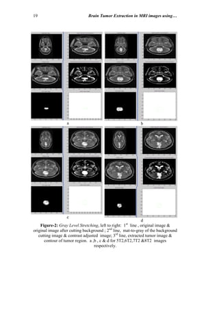
10.1 Gray Level Stretching; includes
a. Gray Level Stretching: performing contrast adjustment to stretch the gray
level of the input image from the range [0.3-0.7] to the range [0 1].
b. Morphological Operation: after converting the image into binary form by
choosing threshold value (depending on the image intensity), many
morphological operations have been applied using structural element of 'disk-
shape', of 6-pixels diameter, these operations are:
1-Erosion: applied on the binary image.
2-Dilation: applied on the resultant image from the previous step.
The dilated image then convolutes with the input reduced intensity image (0.03
of its original intensity value).
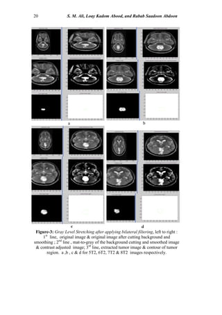
c. Edge Detection
In this step, the Sobel operator is implemented on the resultant image from the
previous steps, followed by filling process to represent the final image of the
tumor. The last step involves contouring the tumor region, are illustrated in figs-
2&3.
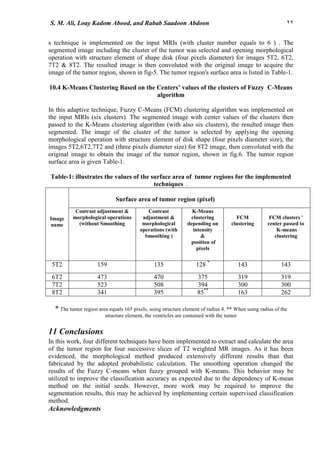
d. Surface Area of the Tumor Region:
The last step was computing the surface area of the tumor region in pixel unit, as
listed Table-1. All the above processes have been applied without smoothing the
original image; the results are shown in fig-2.
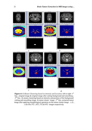
10.2 K-Means Clustering Based on Intensity and Location:
In this technique, the K-Means clustering algorithm was implemented on the
input MRIs (with six clusters). The segmented image included the cluster of the
tumor then selected, using opening morphological operation with structure
element of shape disk (five pixels diameter), and the resulted image then
convoluted with the original image to acquire the image of the tumor region,
shown in fig-4. The tumor region's surface area is presented in Table-1.
8. 19 Brain Tumor Extraction in MRI images using…
a b
c
d
Figure-2: Gray Level Stretching, left to right: 1st
line , original image &
original image after cutting background ; 2nd
line, mat-to-gray of the background
cutting image & contrast adjusted image; 3rd
line, extracted tumor image &
contour of tumor region. a ,b , c & d for 5T2,6T2,7T2 &8T2 images
respectively.
9. 20 S. M. Ali, Loay Kadom Abood, and Rabab Saadoon Abdoon
c d
a b
Figure-3: Gray Level Stretching after applying bilateral filtering, left to right :
1st
line, original image & original image after cutting background and
smoothing ; 2nd
line , mat-to-gray of the background cutting and smoothed image
& contrast adjusted image; 3rd
line, extracted tumor image & contour of tumor
region. a ,b , c & d for 5T2, 6T2, 7T2 & 8T2 images respectively.
10. 21 Brain Tumor Extraction in MRI images using…
d
a b
c
Figure-4: K-Means Clustering based on intensity and Location, left to right: 1st
line , original image & original image after cutting background and smoothing ;
2nd
line , K-means clustering based on intensity and position of the background
cutting and smoothing image & tumor cluster image ; 3rd
line ,extracted tumor
image after applying morphological operation on the tumor cluster image . a ,b ,
c & d for 5T2 ,6T2 ,7T2 & 8T2 images respectively.
11. 22S. M. Ali, Loay Kadom Abood, and Rabab Saadoon Abdoon
s technique is implemented on the input MRIs (with cluster number equals to 6 ) . The
segmented image including the cluster of the tumor was selected and opening morphological
operation with structure element of shape disk (four pixels diameter) for images 5T2, 6T2,
7T2 & 8T2. The resulted image is then convoluted with the original image to acquire the
image of the tumor region, shown in fig-5. The tumor region's surface area is listed in Table-1.
10.4 K-Means Clustering Based on the Centers’ values of the clusters of Fuzzy C-Means
algorithm
In this adaptive technique, Fuzzy C-Means (FCM) clustering algorithm was implemented on
the input MRIs (six clusters). The segmented image with center values of the clusters then
passed to the K-Means clustering algorithm (with also six clusters), the resulted image then
segmented. The image of the cluster of the tumor is selected by applying the opening
morphological operation with structure element of disk shape (four pixels diameter size), the
images 5T2,6T2,7T2 and (three pixels diameter size) for 8T2 image, then convoluted with the
original image to obtain the image of the tumor region, shown in fig.6. The tumor region
surface area is given Table-1.
Table-1: illustrates the values of the surface area of tumor regions for the implemented
techniques .
Surface area of tumor region (pixel)
FCM clusters '
center passed to
K-means
clustering
FCM
clustering
K-Means
clustering
depending on
intensity
&
position of
pixels
Contrast
adjustment &
morphological
operations (with
Smoothing )
Contrast adjustment &
morphological operations
(without Smoothing
Image
name
143143128 *
1351595T2
3193193754704736T2
3003003945085237T2
26216385**
3953418T2
* The tumor region area equals 165 pixels, using structure element of radius 4. ** When using radius of the
structure element, the ventricles are contained with the tumor
11 Conclusions
In this work, four different techniques have been implemented to extract and calculate the area
of the tumor region for four successive slices of T2 weighted MR images. As it has been
evidenced, the morphological method produced extensively different results than that
fabricated by the adopted probabilistic calculation. The smoothing operation changed the
results of the Fuzzy C-means when fuzzy grouped with K-means. This behavior may be
utilized to improve the classification accuracy as expected due to the dependency of K-mean
method on the initial seeds. However, more work may be required to improve the
segmentation results, this may be achieved by implementing certain supervised classification
method.
Acknowledgments
12. 22Brain Tumor Extraction in MRI images using…
a b
c d
We would like to express our thanks to Dr. Faleh Hassan Mahmood who provide us with
the MRI images that study in this work.
Figure-5: Fuzzy C-Means Clustering, left to right: 1st
line , original image & original image
after cutting background and smoothing; 2nd
line Fuzzy C-means clustering of the background
cutting and smoothing image; 3rd
line, tumor cluster image & extracted tumor image after
applying morphological operation on the tumor cluster image. a ,b , c & d for 5T2,6T2,7T2
&8T2 images respectively.
13. 22S. M. Ali, Loay Kadom Abood, and Rabab Saadoon Abdoon
a b
d
Figure-6: K-Means Clustering based on the centers’ values of the clusters of Fuzzy C-Means
algorithm, left to right: 1st
line, original image & original image after cutting background and
smoothing ; 2nd
line, Fuzzy C-means clustering of the background cutting and smoothing
image & K-means clustering of the background cutting and smoothing image based on
clusters' centers values of FCM algorithm; 3rd
line, tumor cluster image & extracted tumor
image after applying morphological operation on the tumor cluster image . a, b, c& d for 5T2,
6T2, 7T2 & 8T2 images respectively. .
c
14. 22Brain Tumor Extraction in MRI images using…
References
[1]. Hari Prasath S.P, G.Kharmega Sundararaj, A.Jayachandran ,(2012) ,"
Brain Tumor Segmentation of Contrast Material Applied MRI
Using Enhanced Fuzzy C-Means Clustering," International Journal of
Engineering and Innovative Technology (IJEIT) Volume 1, Issue 2.
[2].Parveen Lehana , Swapna Devi Satnam Singh , Pawanesh Abrol, Saleem
Khan, Sandeep Arya, (2012), "Investigations of the MRI Images using
Aura Transformation ," Signal & Image Processing : An International
Journal (SIPIJ) Vol.3, No.1.
[3].S. Javeed Hussain , T. Satya Savithri, P.V. Sree Devi,
(2012),"Segmentation of Tissues in Brain MRI Images using Dynamic
Neuro-Fuzzy Technique," International Journal of Soft Computing and
Engineering (IJSCE) ISSN: 2231-2307, Volume-1, Issue-6.
[4].K. S. Angel Viji & J. Jayakumari, (2012), "Performance Evaluation of
Standard Image Segmentation Methods and Clustering Algorithms for
Segmentation of MRI Brain Tumor Images," European Journal of
Scientific Research ISSN 1450-216X Vol.79 No.2 , pp.166-179 .
[5].Sarbani Datta & Dr. Monisha Chakraborty, (2011),"Brain Tumor
Detection from Pre-Processed MR Images using Segmentation
Techniques," IJCA Special Issue on “2nd National Conference-
Computing, Communication and Sensor Network” CCSN.
[6].S.R Kannan, (2005) "Segmentation of MRI Using New Unsupervised
Fuzzy C-Mean Algorithm", ICGSTGVIP Journal. Vol. 5. No.2.
[7].Rajesh C. Patil and A. S. Bhalchandra, (2011), "Brain Tumor Extraction
from MRI Images Using MATLAB," ISSN: 2277-9477, Volume 2, Issue 1.
[8].Sudipta Roy and Samir K. Bandyopadhyay, (2012), "Detection and
Quantification of Brain Tumor from MRI of Brain and it’s Symmetric
Analysis," ICT Journal., Volume 2 No.6.
[9].A. Struyf, Mia Hubert, and Peter J. Rousseeuw, (1997) "Integrating
Robust Clustering Techniques in S-PLUS," Computational Statistics &
Data Analysis, Volume 26, Issue 1, 6 November 1997, Pages 17–37
[10]. F.M. Enzinger and S.W. Weiss, (1995)," Malignant vascular tumors, in:
Soft Tissue Tumors," 3rd
ed., Mosby, St. Louis, MO, pp. 655–677.
[11]. TOMASI, C. and MANDUCHI, R., (1998), “Bilateral filtering for gray
and color images,” In Proceedings of the International Conference on
Computer Vision, pp. 839–846.
[12]. Serra J., (1982), “Image Analysis and Mathematical Morphology,”
London Academic Press .
[13]. Asma'a Abbas Ajwad Al-Tamimy , (2005), "Artificial Intelligence for
Magnetic Resonence Image (MRI) Recognition ", M.Sc. Thesis in Medical
Engineering.