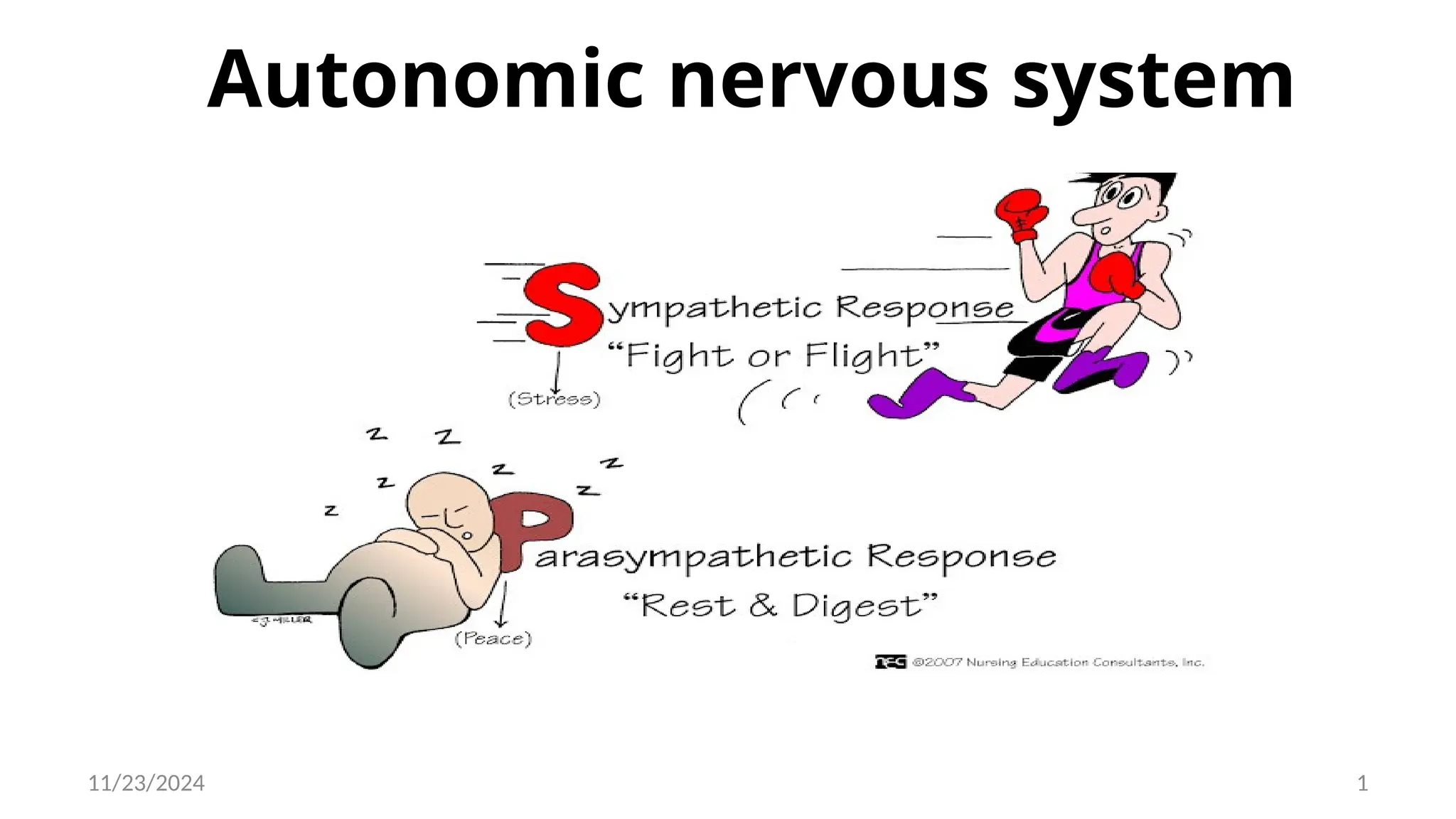
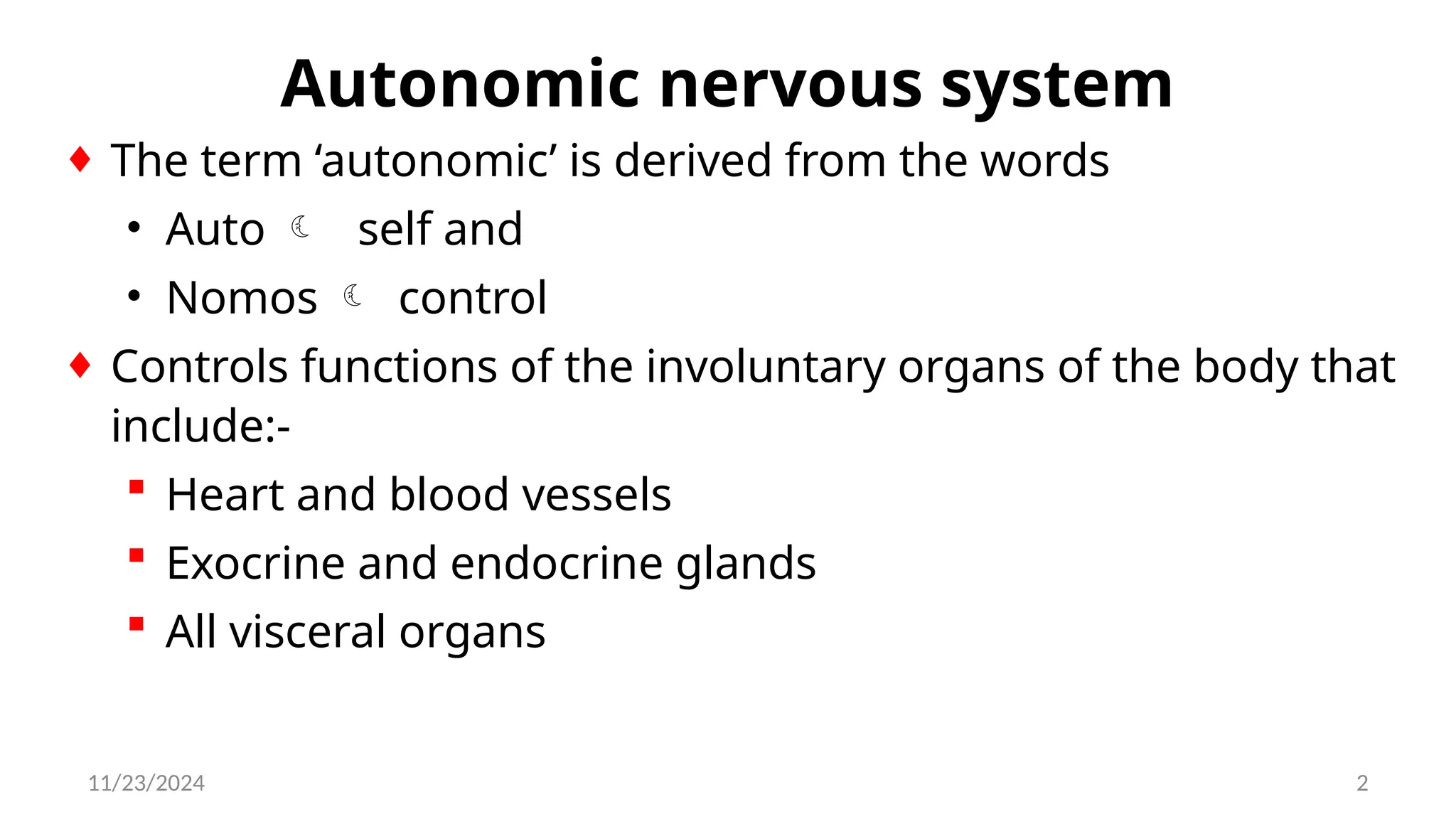
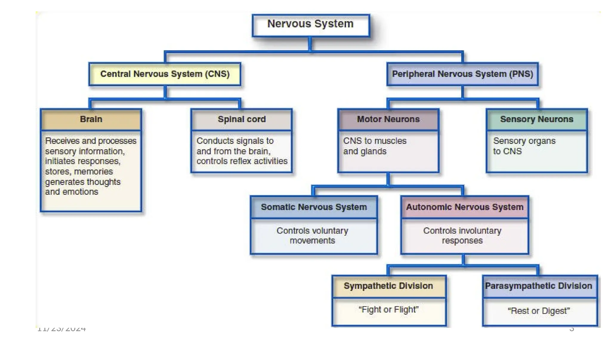
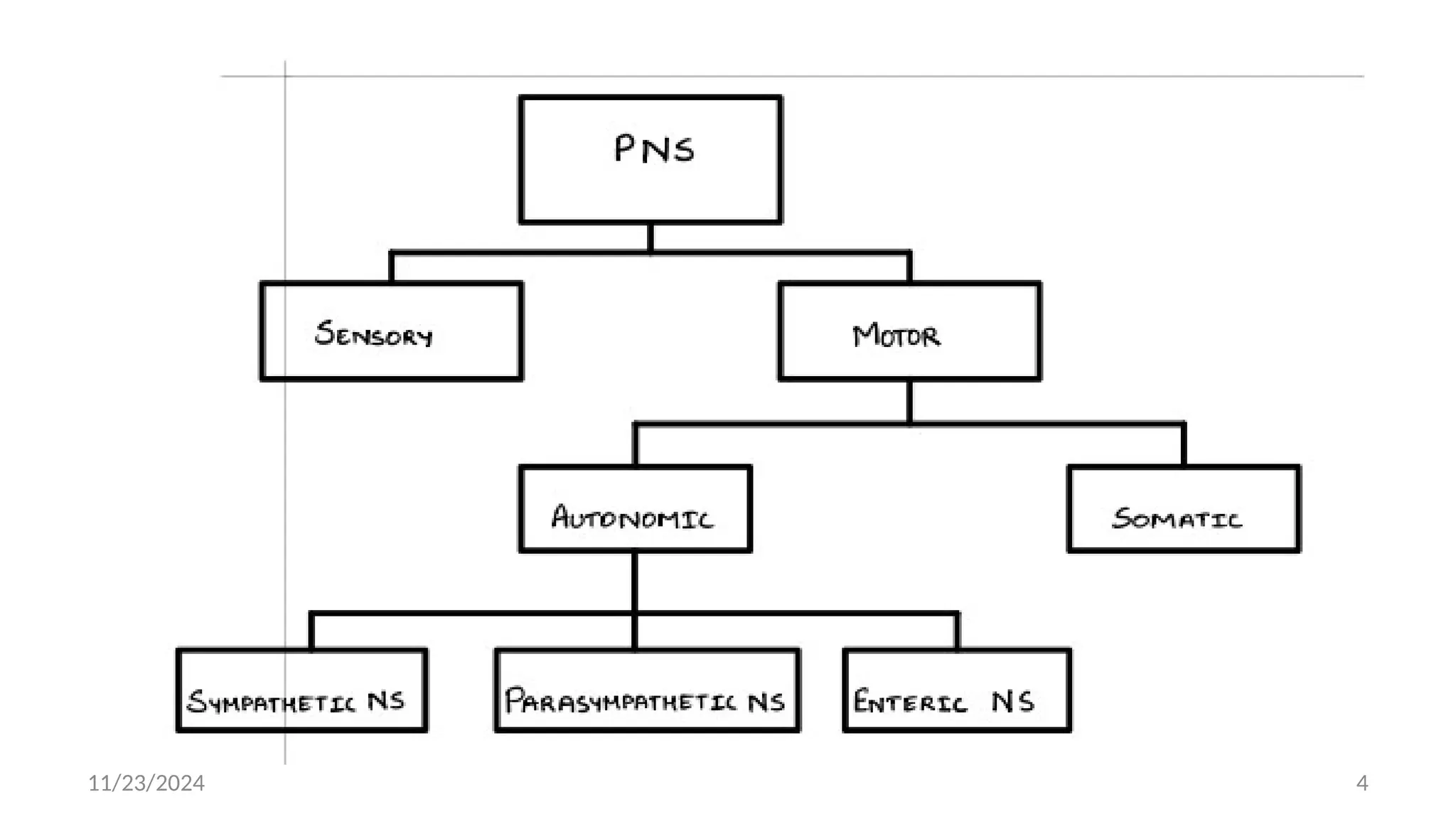
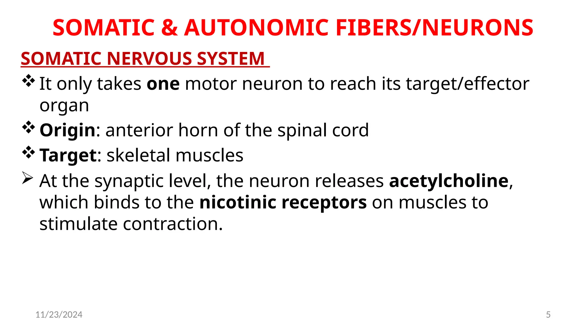
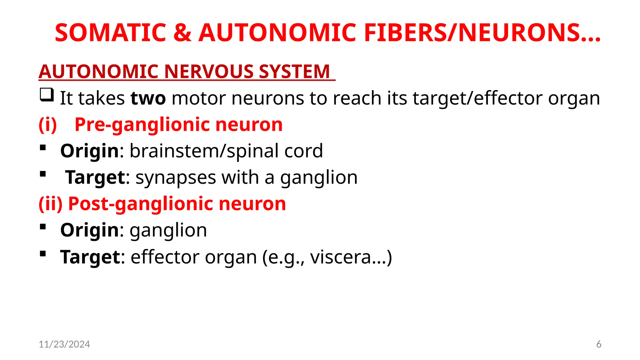
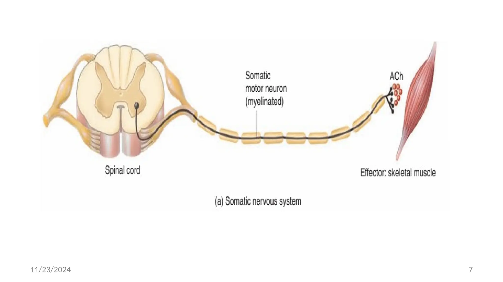
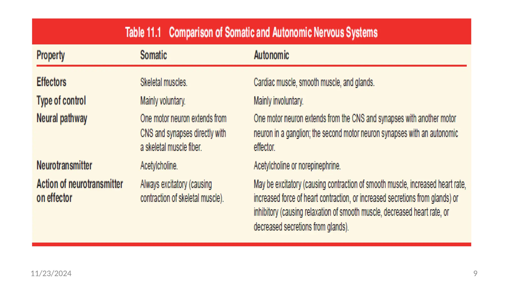
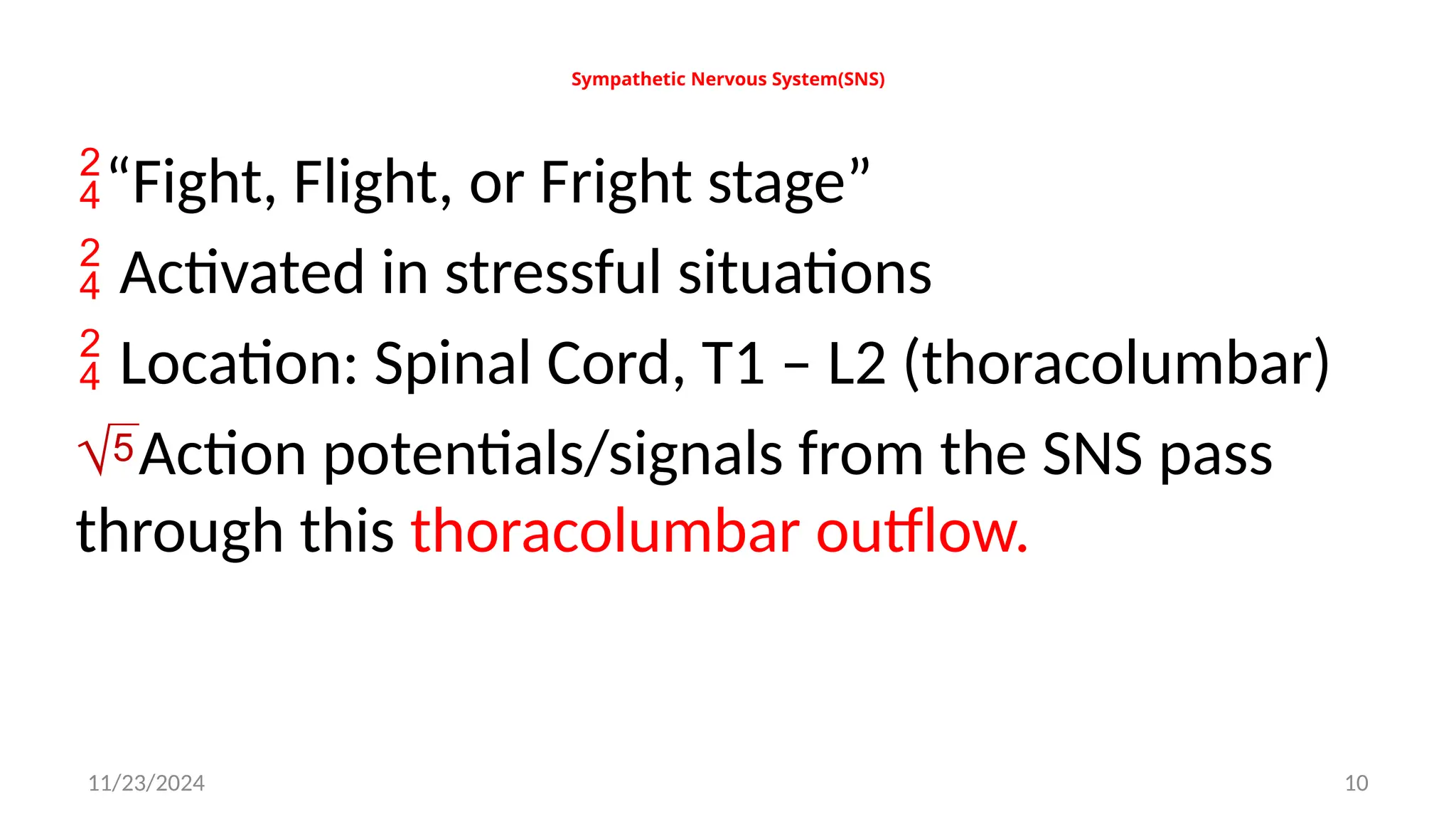
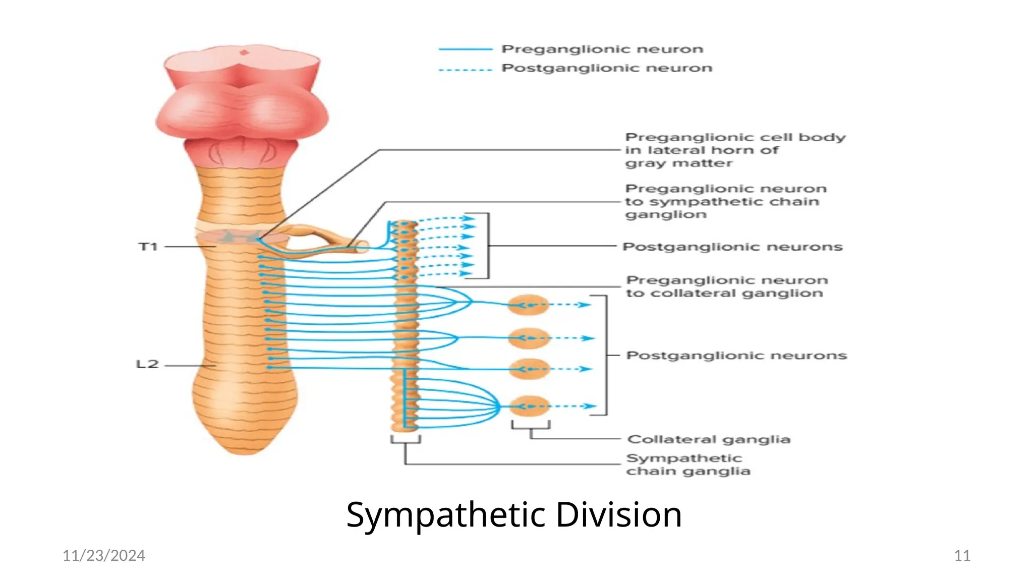
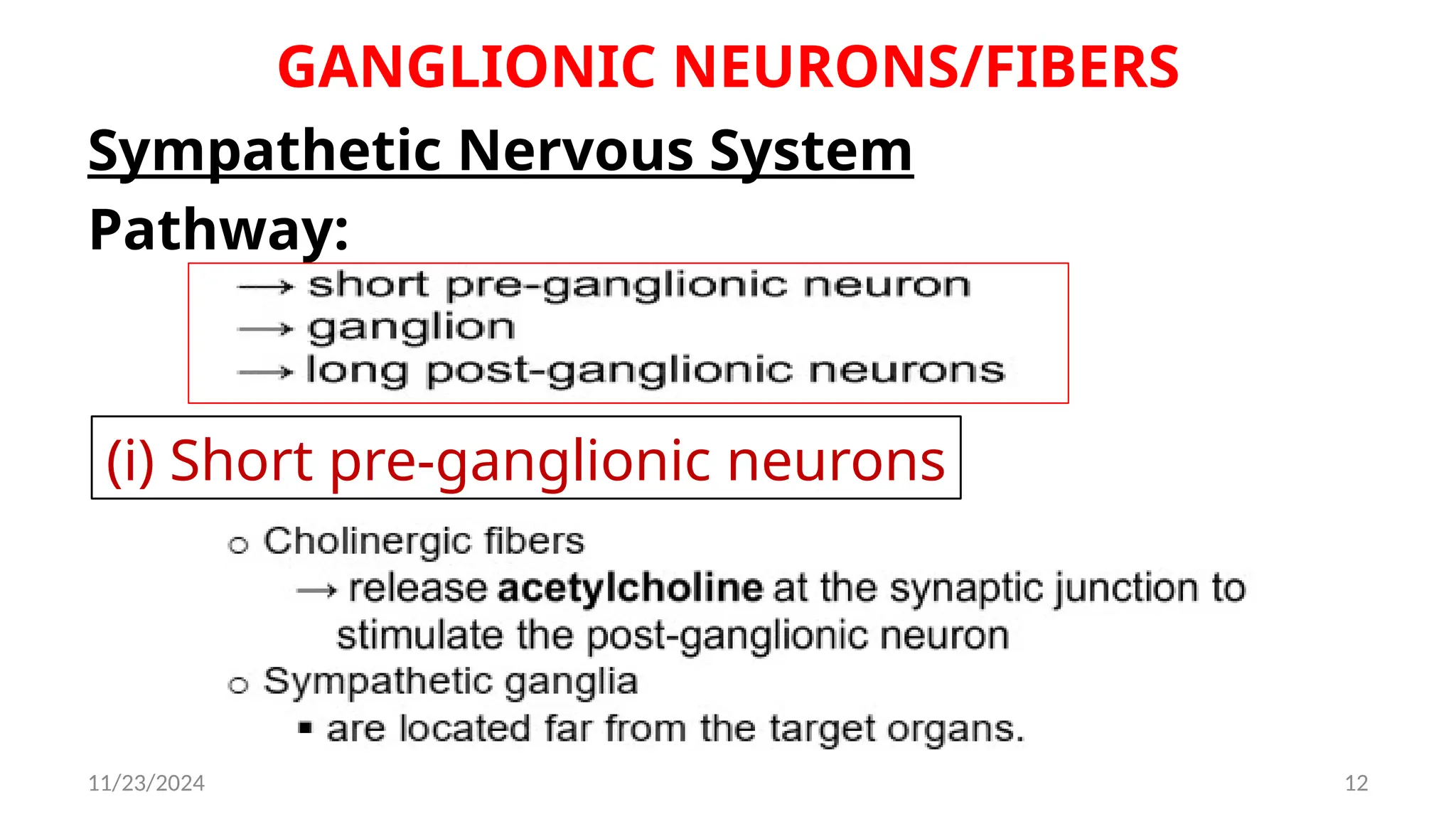
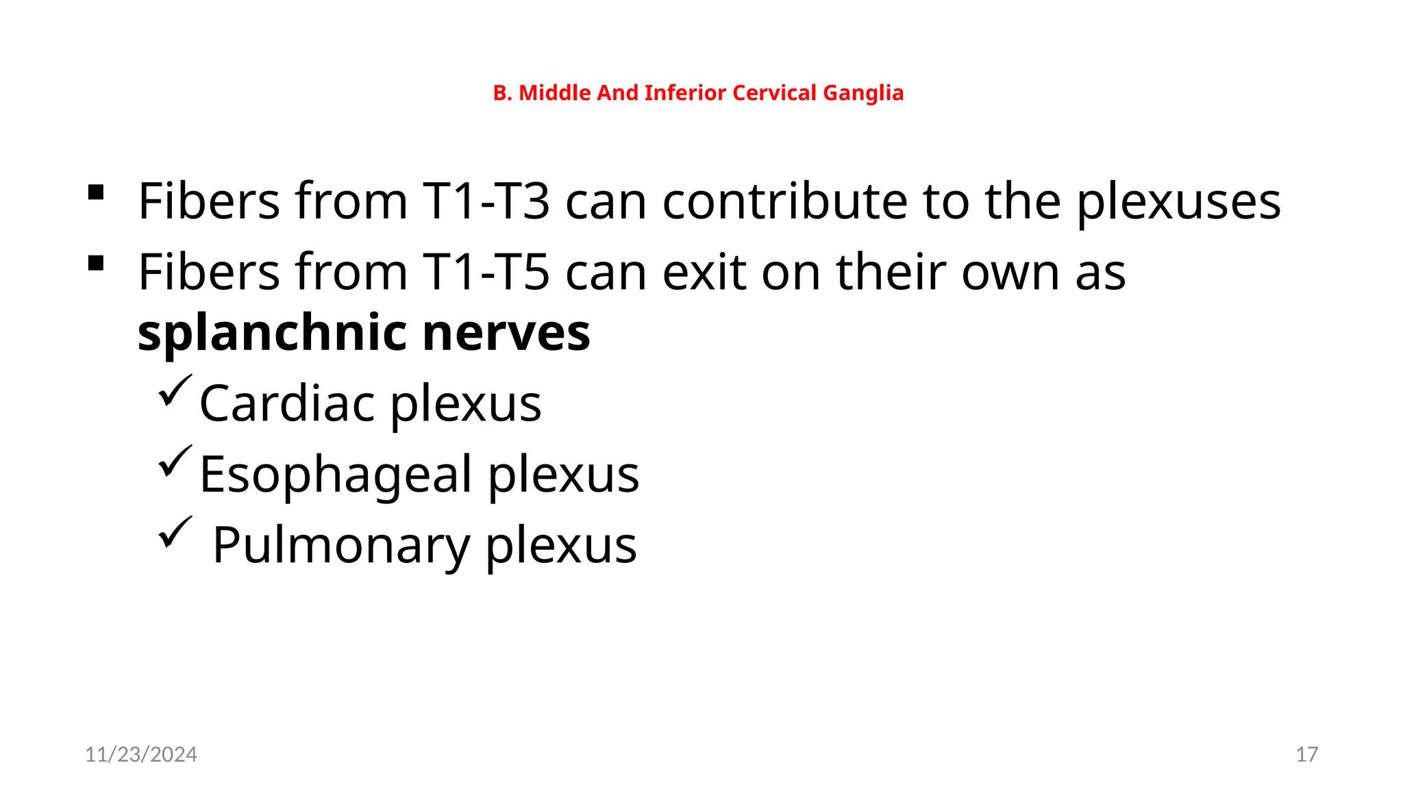
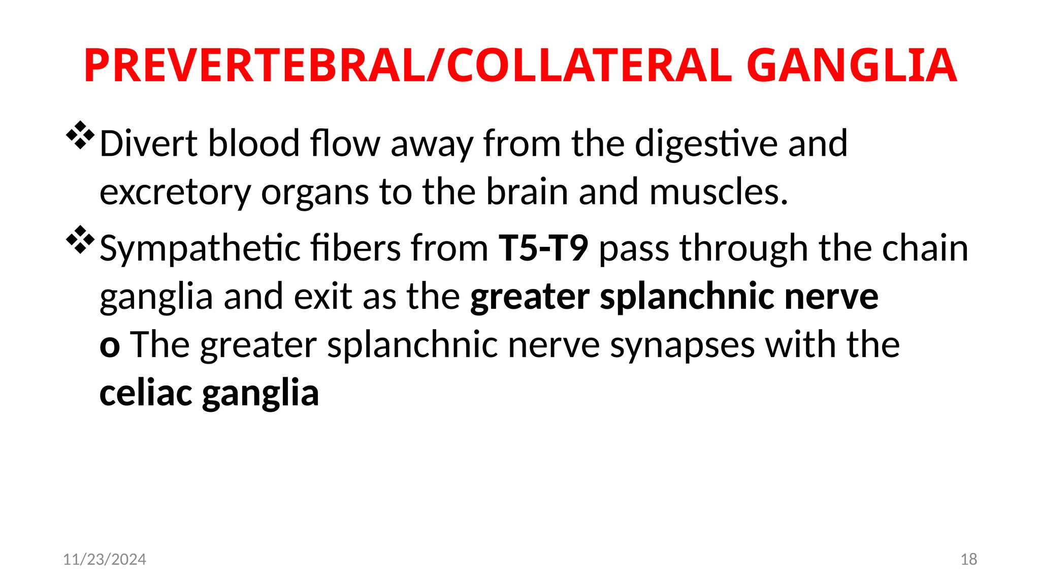
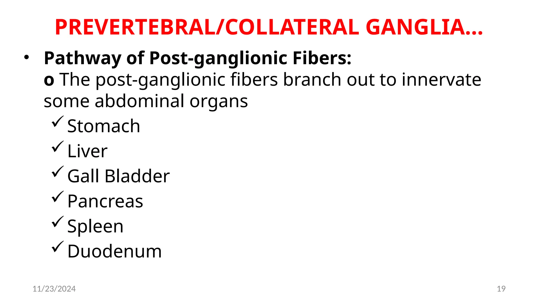
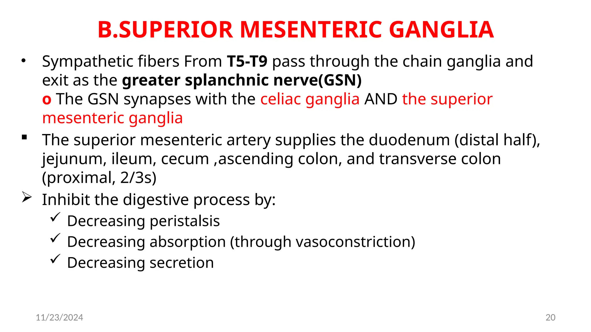
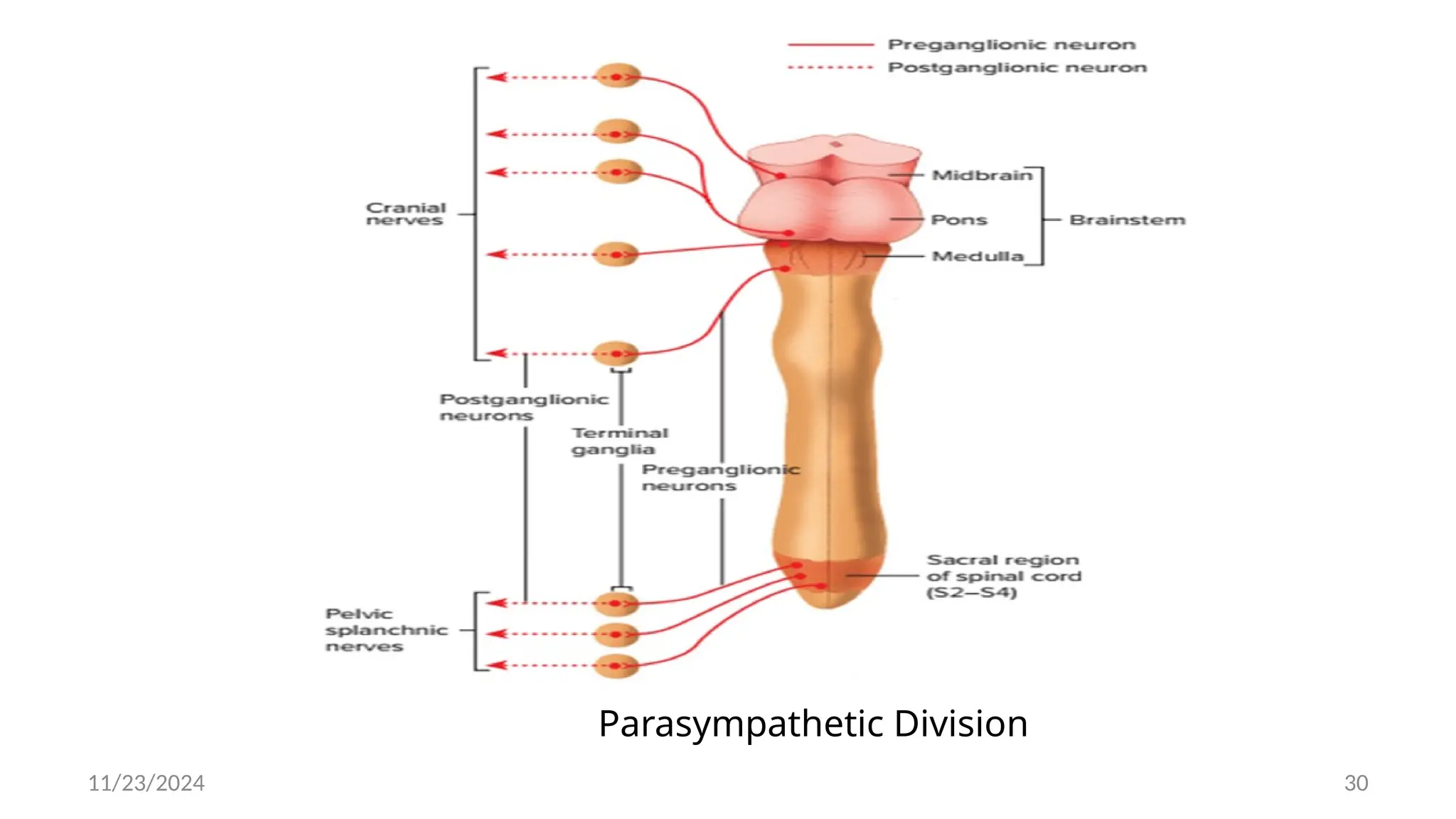
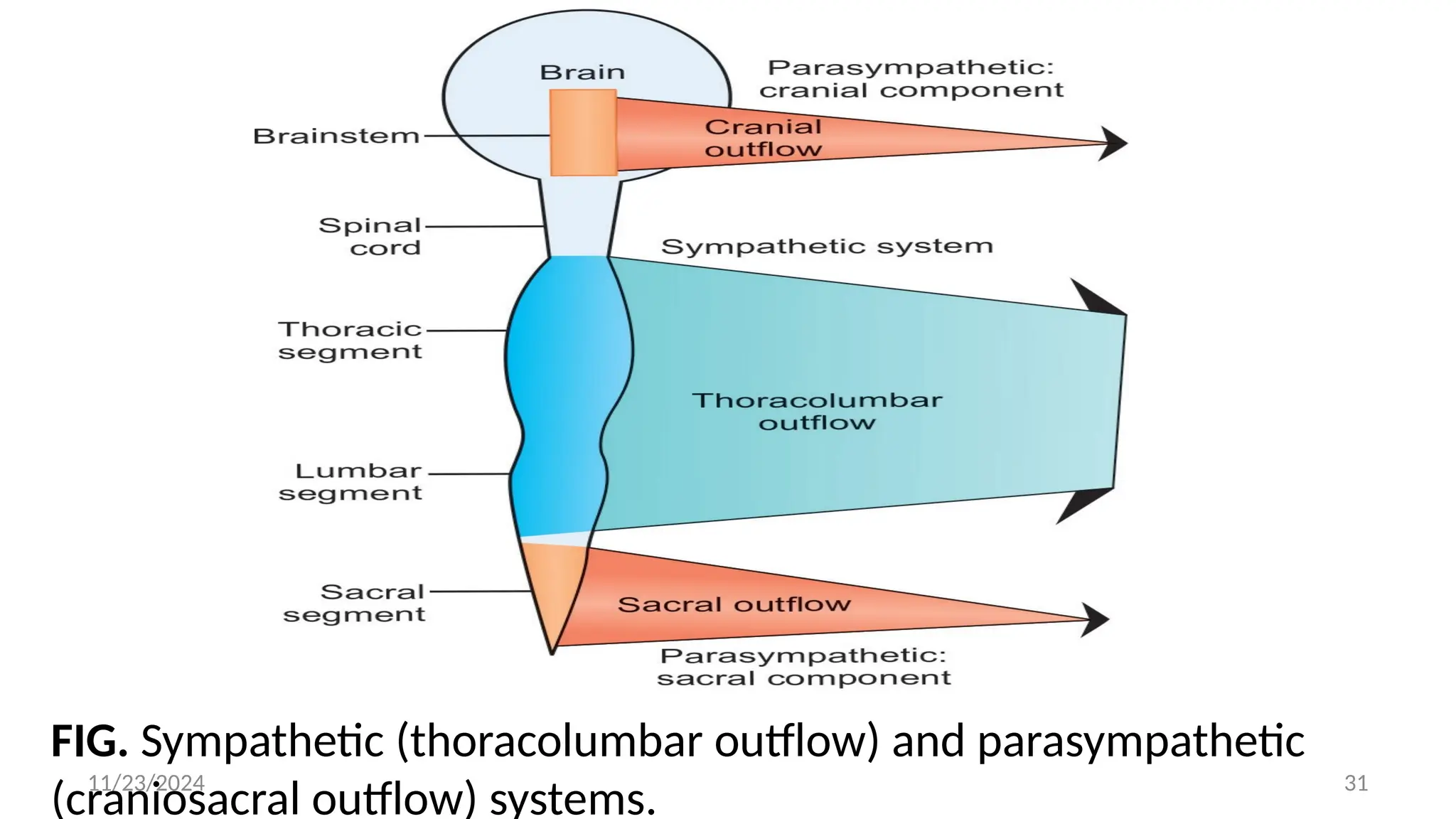
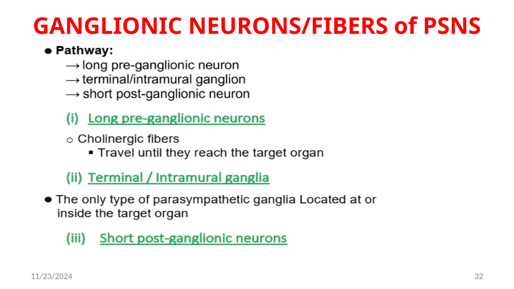
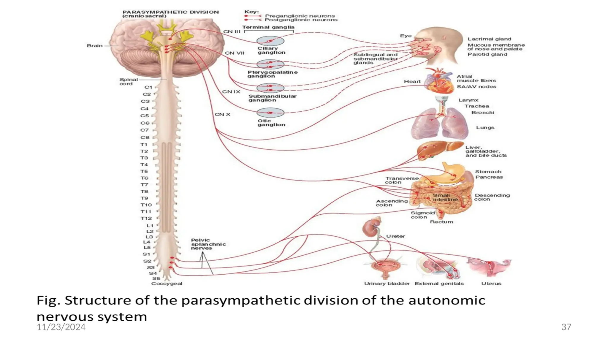
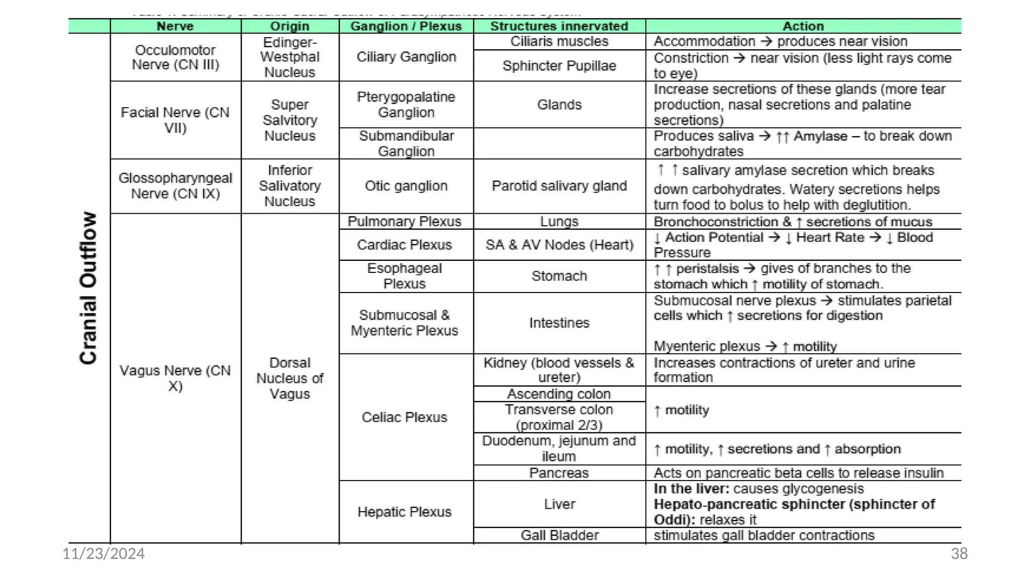
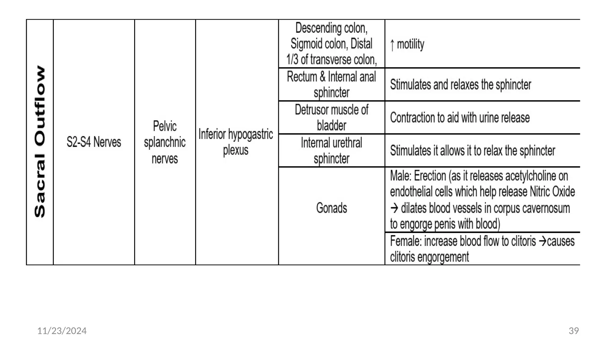
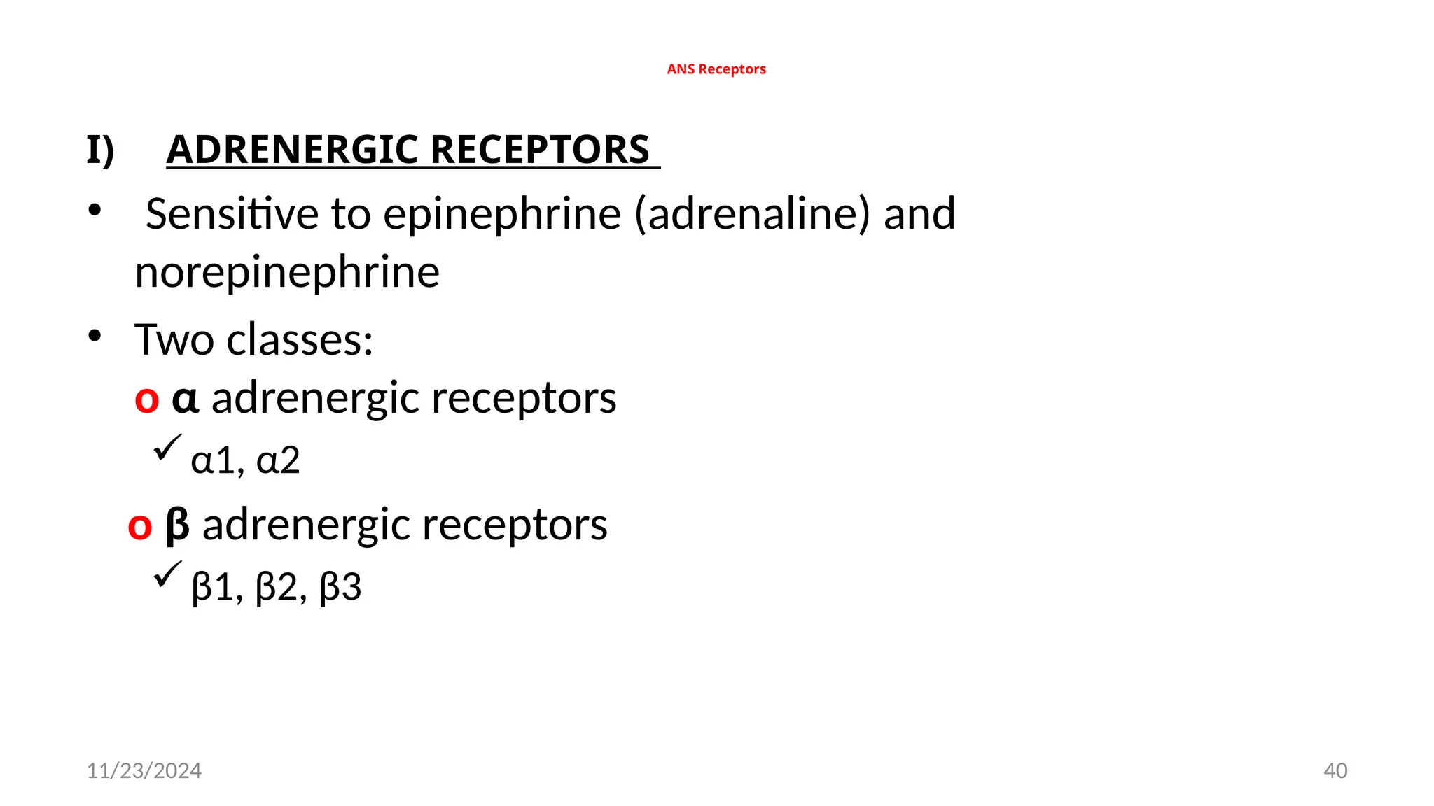
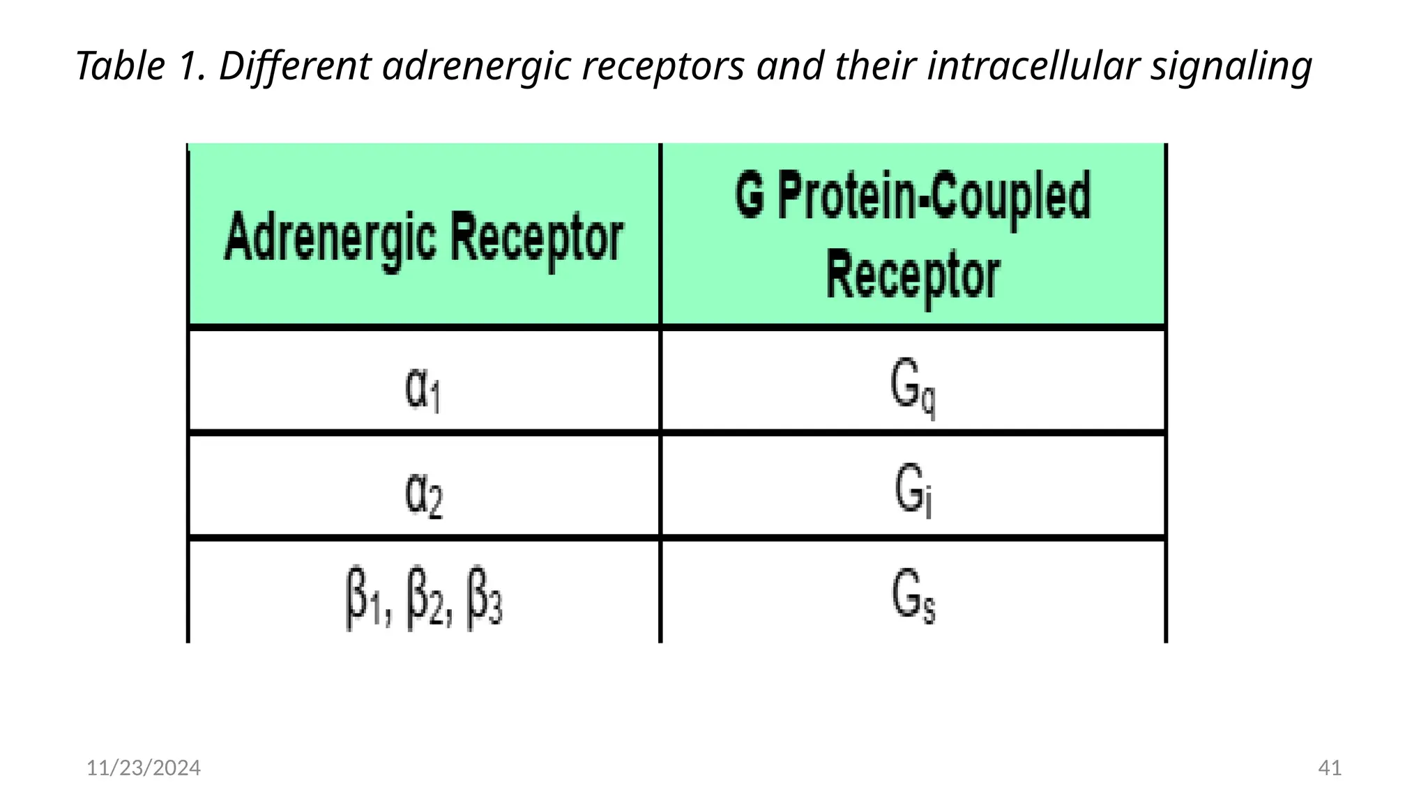
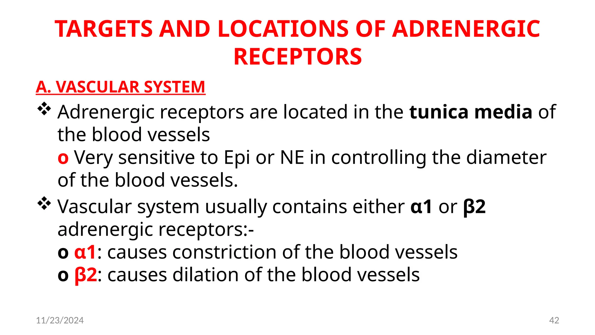
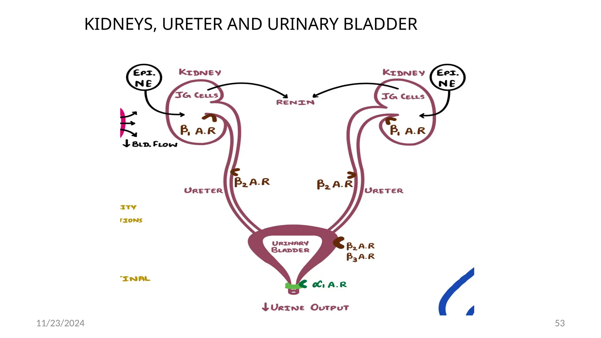
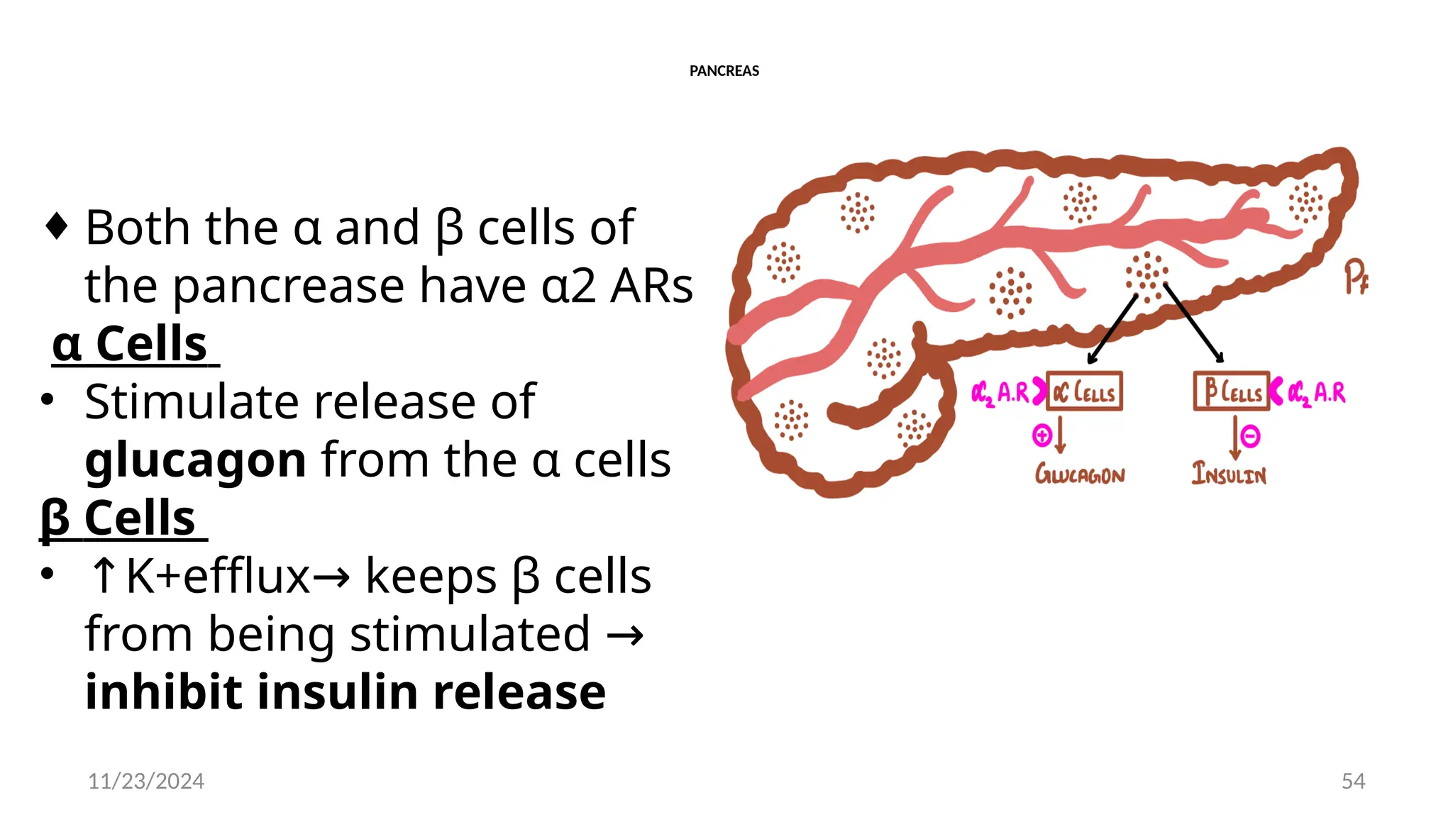
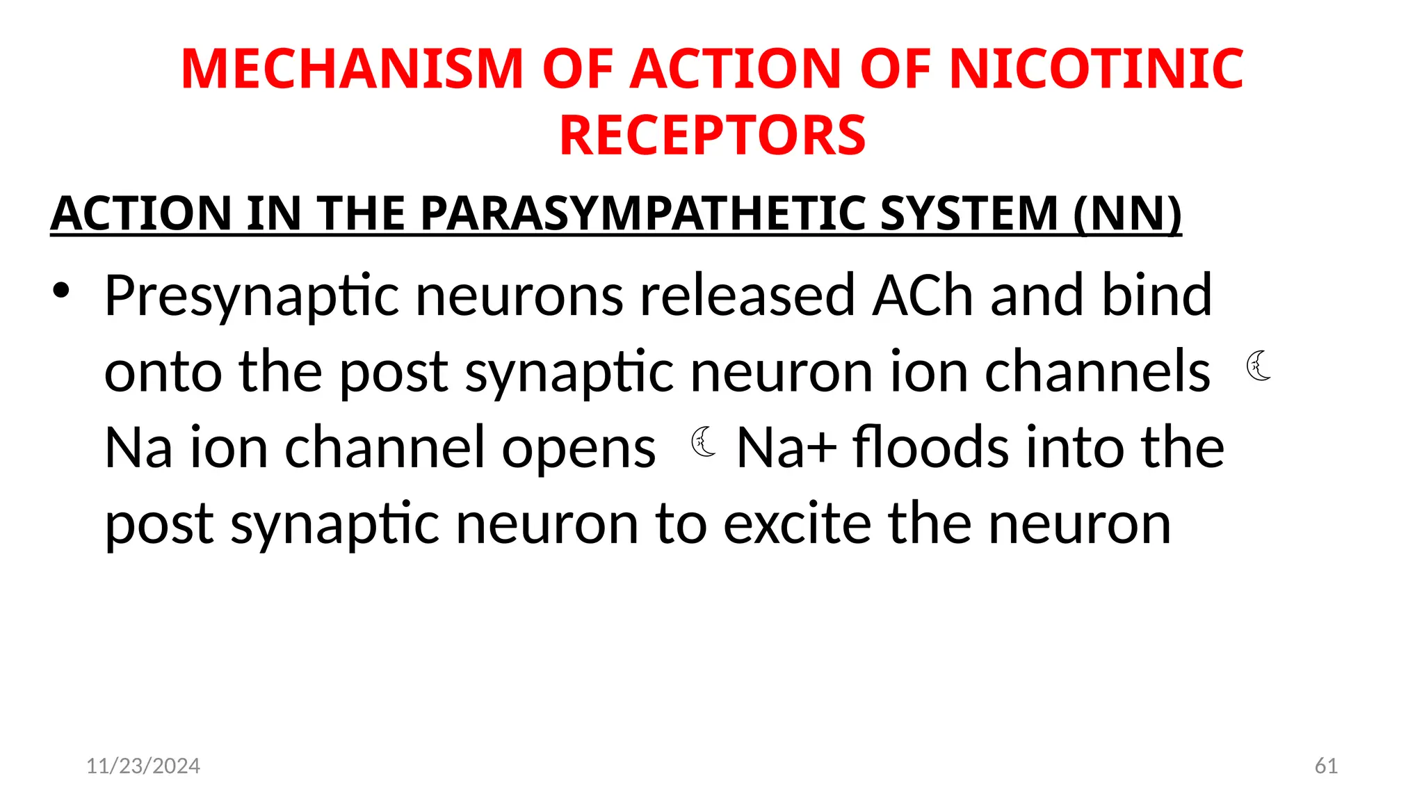
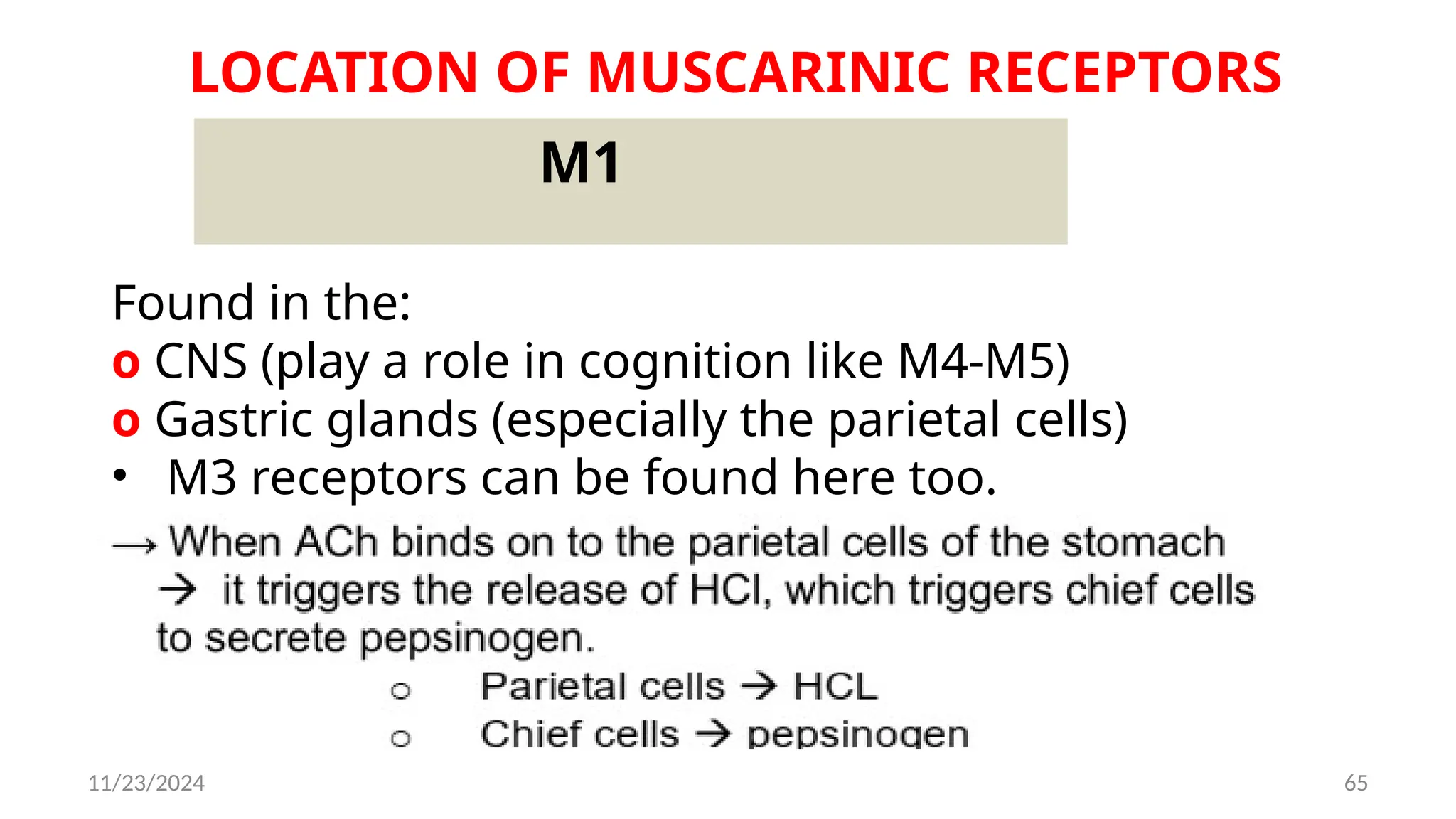
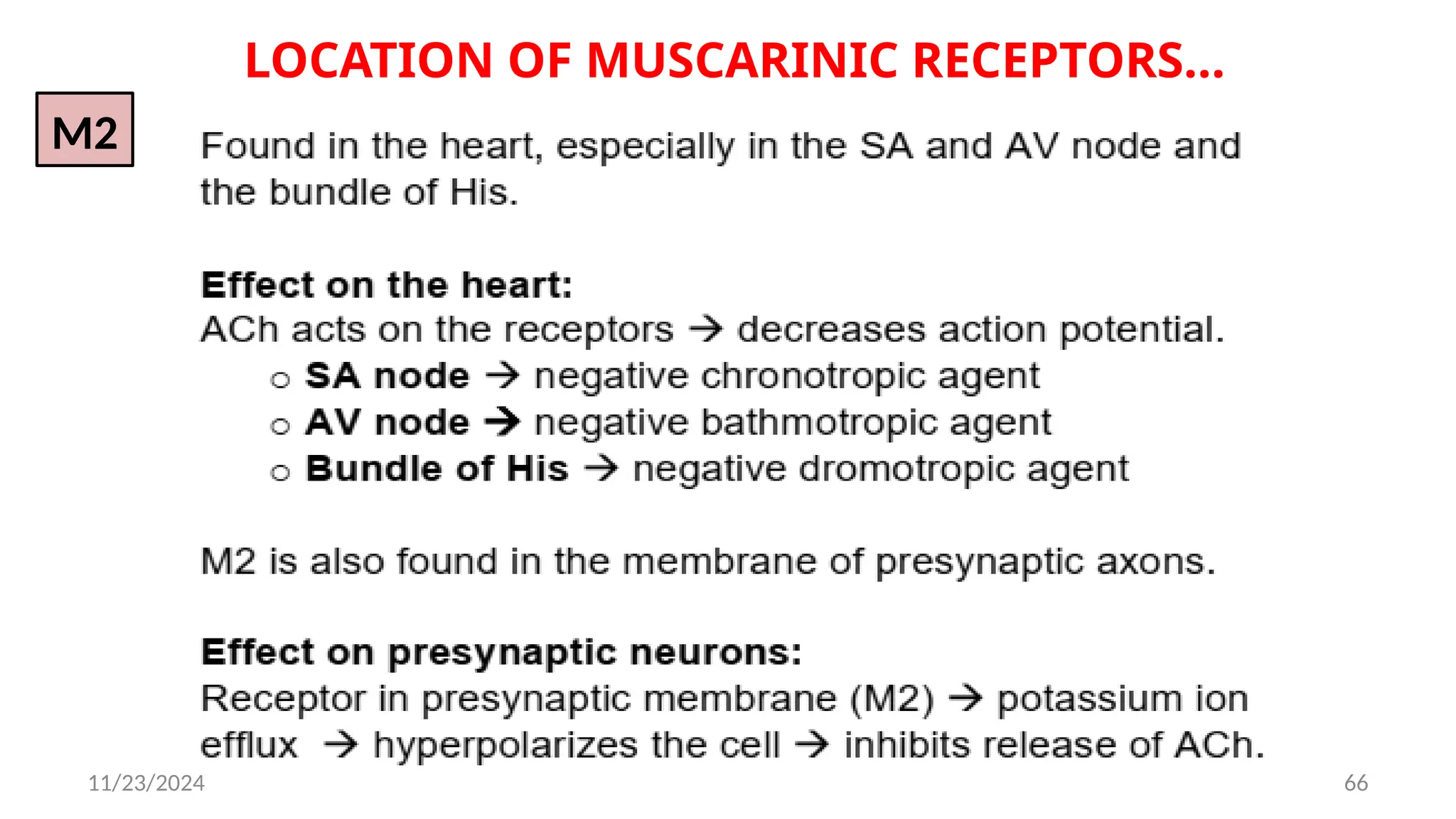
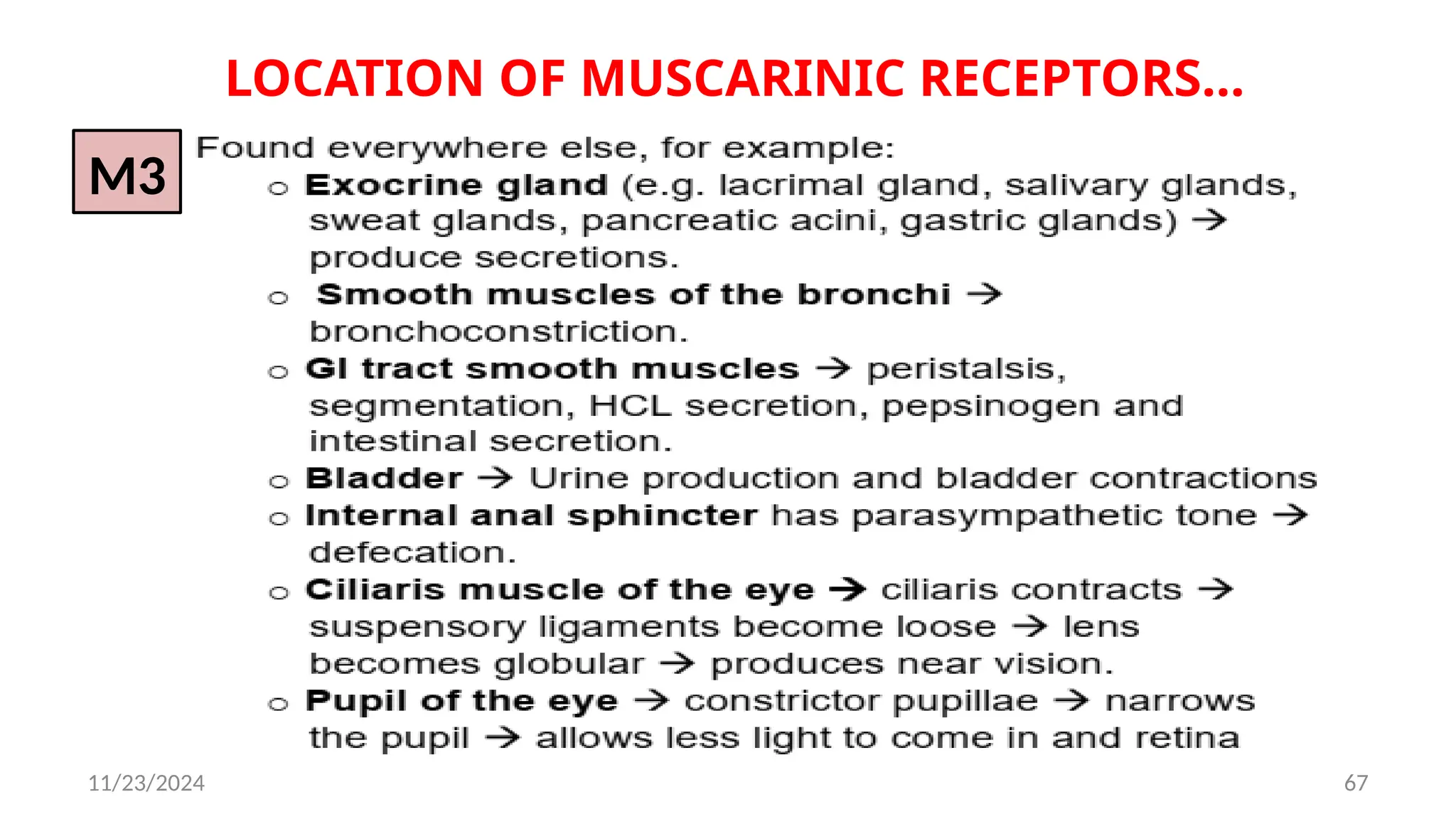
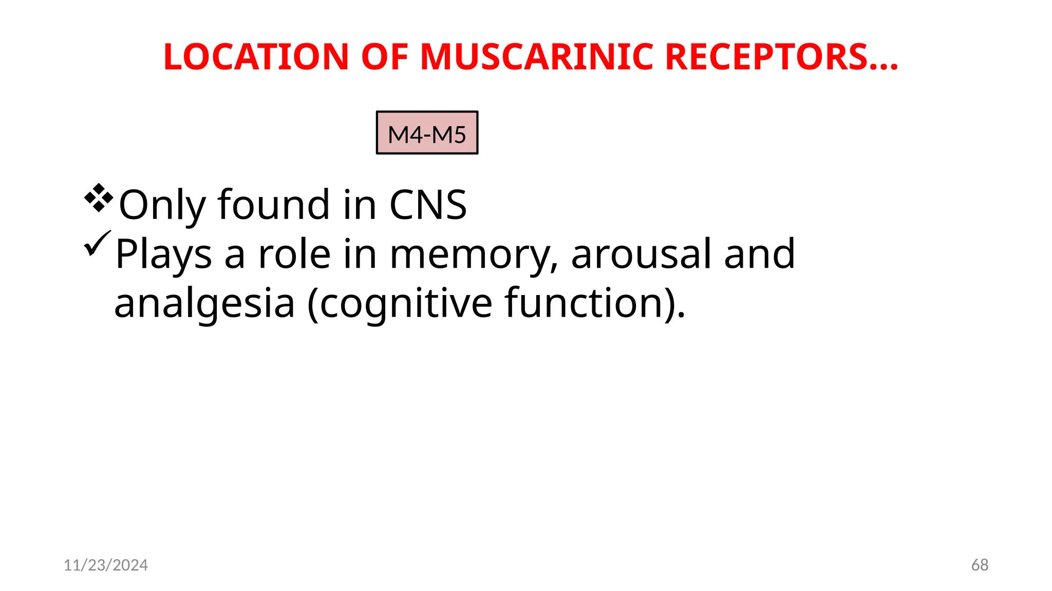
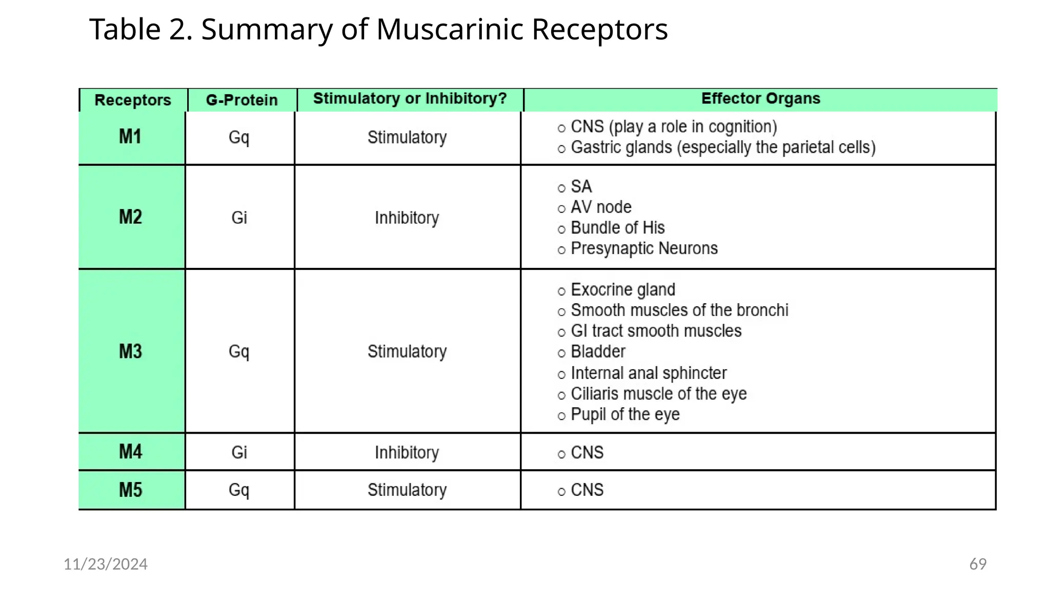
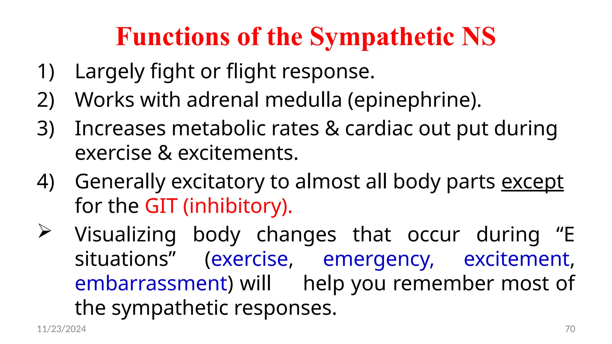
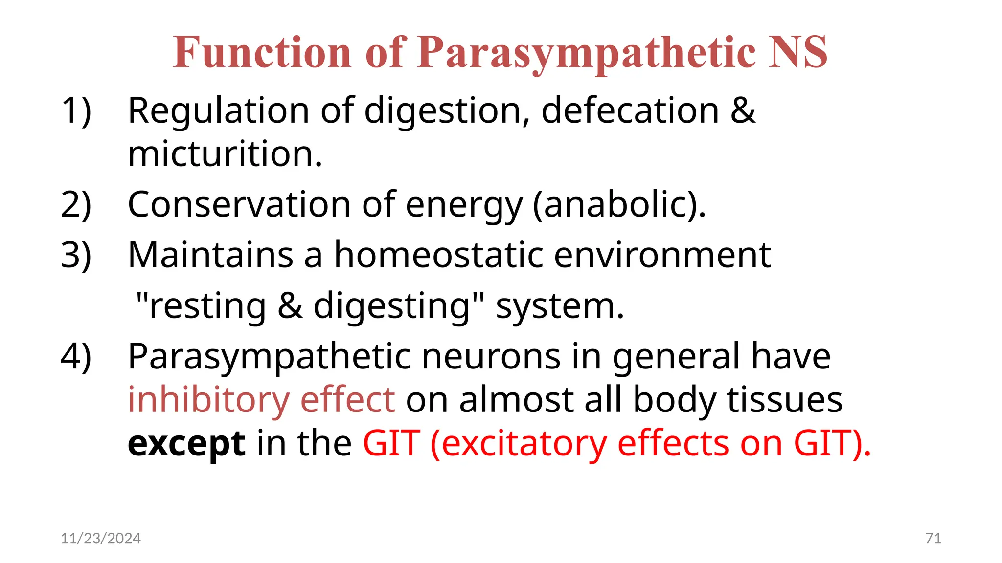
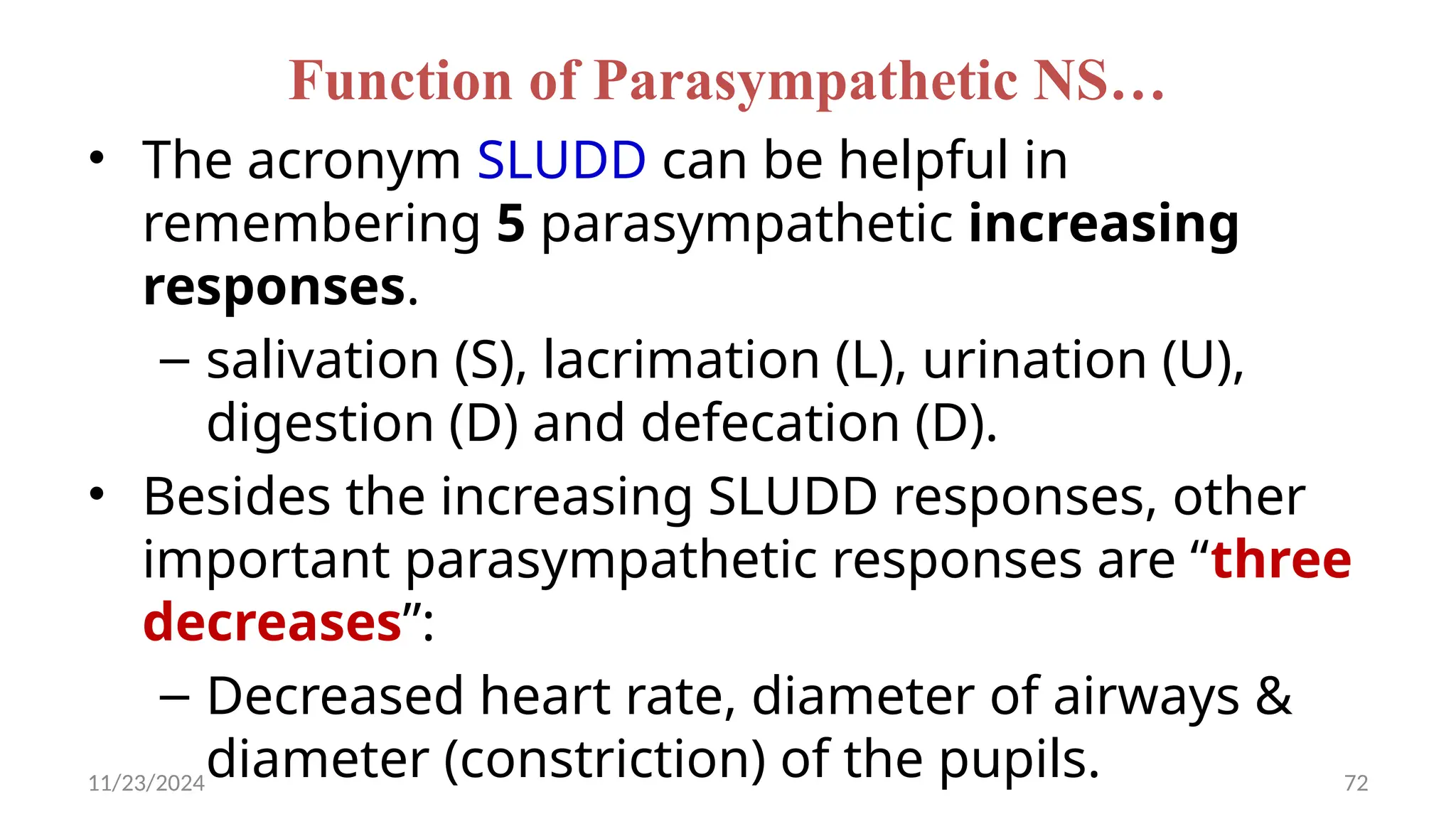
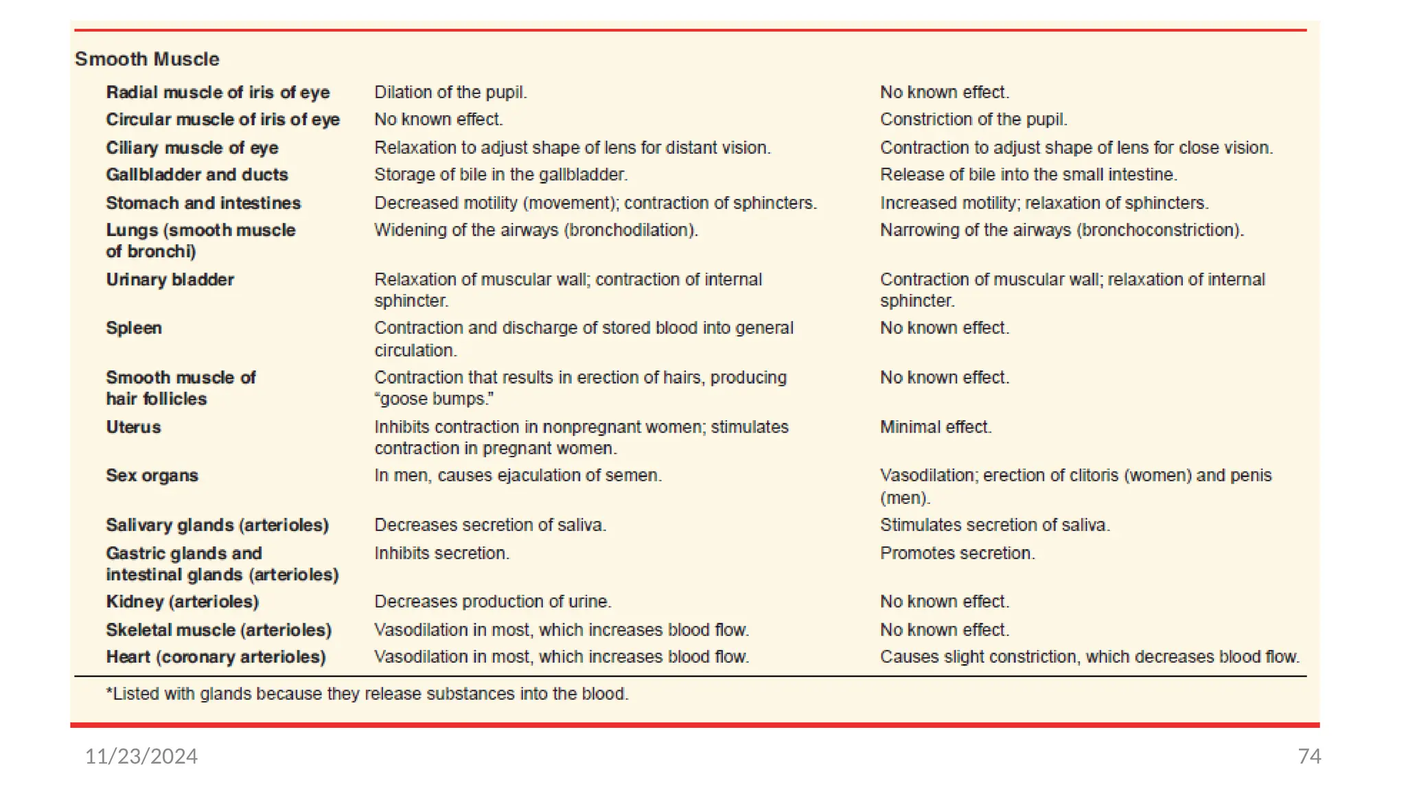
11/23/2024 1
Autonomic nervous system
11/23/2024 2
Autonomic nervous system
¨ The term ‘autonomic’ is derived from the words
• Auto  self and
• Nomos  control
¨ Controls functions of the involuntary organs of the body that
include:-
 Heart and blood vessels
 Exocrine and endocrine glands
 All visceral organs
11/23/2024 3
11/23/2024 4
11/23/2024 5
SOMATIC & AUTONOMIC FIBERS/NEURONS
SOMATIC NERVOUS SYSTEM
It only takes one motor neuron to reach its target/effector
organ
Origin: anterior horn of the spinal cord
Target: skeletal muscles
 At the synaptic level, the neuron releases acetylcholine,
which binds to the nicotinic receptors on muscles to
stimulate contraction.
11/23/2024 6
SOMATIC & AUTONOMIC FIBERS/NEURONS…
AUTONOMIC NERVOUS SYSTEM
 It takes two motor neurons to reach its target/effector organ
(i) Pre-ganglionic neuron
 Origin: brainstem/spinal cord
 Target: synapses with a ganglion
(ii) Post-ganglionic neuron
 Origin: ganglion
 Target: effector organ (e.g., viscera...)
11/23/2024 7
11/23/2024 8
11/23/2024 9
11/23/2024 10
Sympathetic Nervous System(SNS)
“Fight, Flight, or Fright stage”
 Activated in stressful situations
 Location: Spinal Cord, T1 – L2 (thoracolumbar)
Action potentials/signals from the SNS pass
through this thoracolumbar outflow.
11/23/2024 11
Sympathetic Division
11/23/2024 12
GANGLIONIC NEURONS/FIBERS
Sympathetic Nervous System
Pathway:
(i) Short pre-ganglionic neurons
11/23/2024 13
GANGLIONIC NEURONS/FIBERS…
(ii) Long post-ganglionic neurons
 Adrenergic fibers - release norepinephrine at
the synaptic junction
11/23/2024 14
SYMPATHETIC NERVOUS SYSTEM…
CHAIN/PARAVERTEBRAL GANGLIA
 Sympathetic fibers from the upper thoracic spinal cord
extend to the cervical region to synapse with the
cervical ganglia:
o Superior cervical ganglia
o Middle cervical ganglia
o Inferior cervical ganglia
o Stellate ganglia
 These cervical ganglia provide sympathetic efferent
innervation to the face and head.
11/23/2024 15
SYMPATHETIC NERVOUS SYSTEM…
A.SUPERIOR CERVICAL GANGLIA
• Pathway of Pre-ganglionic Fibers: Sympathetic fibers from T1-
T3 pass through the chain ganglia then extend superiorly to
synapse with the superior cervical ganglia
 Pathway of Post-ganglionic Fibers:
o Join/Contribute to spinal nerves to supply:
 Arrector pili
 Blood vessels
 Sudoriferous sweat glands
 Contribute branches to the heart, lungs, and
esophagus
 Form carotid plexus
11/23/2024 16
SUPERIOR CERVICAL GANGLIA…
¨ The carotid plexus branches into three to
innervate:
1. Dilator pupillae
2. Ciliary muscle (ciliaris)-- Contracts to make the
lens flat (accommodation)
3. Salivary glands
• Decreasing salivary output
• Increases mucin
4. Lacrimal glands
11/23/2024 17
B. Middle And Inferior Cervical Ganglia
 Fibers from T1-T3 can contribute to the plexuses
 Fibers from T1-T5 can exit on their own as
splanchnic nerves
Cardiac plexus
Esophageal plexus
 Pulmonary plexus
11/23/2024 18
PREVERTEBRAL/COLLATERAL GANGLIA
Divert blood flow away from the digestive and
excretory organs to the brain and muscles.
Sympathetic fibers from T5-T9 pass through the chain
ganglia and exit as the greater splanchnic nerve
o The greater splanchnic nerve synapses with the
celiac ganglia
11/23/2024 19
PREVERTEBRAL/COLLATERAL GANGLIA…
• Pathway of Post-ganglionic Fibers:
o The post-ganglionic fibers branch out to innervate
some abdominal organs
Stomach
Liver
Gall Bladder
Pancreas
Spleen
Duodenum
11/23/2024 20
B.SUPERIOR MESENTERIC GANGLIA
• Sympathetic fibers From T5-T9 pass through the chain ganglia and
exit as the greater splanchnic nerve(GSN)
o The GSN synapses with the celiac ganglia AND the superior
mesenteric ganglia
 The superior mesenteric artery supplies the duodenum (distal half),
jejunum, ileum, cecum ,ascending colon, and transverse colon
(proximal, 2/3s)
 Inhibit the digestive process by:
 Decreasing peristalsis
 Decreasing absorption (through vasoconstriction)
 Decreasing secretion
11/23/2024 21
C. ADRENAL MEDULLA
• Pathway of Pre-ganglionic Fibers: The greater
splanchnic nerve synapses with the chromaffin cells of
the adrenal medulla
o Example of an intramural ganglion
o Exception to the usual two-neuron structure of the
sympathetic nervous system
• Pathway of Post-ganglionic Fibers
o The chromaffin cells release norepinephrine and
epinephrine to the general circulation, causing a
diffuse, systemic effect
11/23/2024 22
D.AORTICORENAL GANGLIA
o Sympathetic fibers from T10-T11 pass through the chain
ganglia and exit as the lesser splanchnic nerve.
o The lesser splanchnic nerve synapses with the aorticorenal
ganglia.
o The post-ganglionic fibers innervate the kidney and
ureter
 Decrease urine production (by decreasing blood
flow to the kidneys)
 JGC release renin, which triggers the angiotensin II cascade to
increases blood pressure
11/23/2024 23
E.RENAL PLEXUS
• Pre-ganglionic Fibers: Sympathetic fibers from T12
pass through the chain ganglia and synapse with diffuse
ganglion cells (renal plexus)
• Post-ganglionic Fibers: The post-ganglionic fibers enter
the kidney to:
o Support the aorticorenal ganglion fibers in decreasing
urine production in the kidney.
o Decrease peristalsis of the ureter
11/23/2024 24
F.INFERIOR MESENTERIC GANGLION
• Pathway of Pre-ganglionic Fibers:
o Sympathetic fibers from L1-L2 (sometimes L3) pass
through the chain ganglia and exit as lumbar
splanchnic nerves
o The lumbar splanchnic nerves innervate the inferior
mesenteric ganglion
Pathway of Post-ganglionic Fibers:
 The inferior mesenteric artery supplies the transverse
colon (distal 1/3), descending colon, sigmoid colon,
and upper rectum---decrease peristalsis and
defecation
11/23/2024 25
G. SUPERIOR HYPOGASTRIC PLEXUS
 Sympathetic fibers from T12-L2 (sometimes L3) pass
through the chain
ganglia and synapse with the superior hypogastric
plexus.
 The postganglionic fibers go to the internal urethral
sphincter(-1AR) and detrusor (-2 and -3
receptor)
11/23/2024 26
H. INFERIOR HYPOGASTRIC PLEXUS
o Sympathetic fibers from T10-L2 pass through the
chain ganglia and exit around the sacral region as the
sacral splanchnic nerves
o The sacral splanchnic nerves synapse with the
inferior hypogastric plexus
• The postganglionic fibers of this plexus innervate
the gonads
11/23/2024 27
INFERIOR HYPOGASTRIC PLEXUS…
1. Males
 The postganglionic fibers of the inferior hypogastric
plexus innervate the penis and scrotum
o Initiate ejaculation
o Stimulate prostate gland to produce seminal fluid
2. Females
 The postganglionic fibers innervate the uterus and
vagina.
11/23/2024 28
Fig. Structure of the sympathetic division of the autonomic nervous system
11/23/2024 29
Parasympathetic Nervous System(PSNS)
✹“Rest and Digest stage”
✹ Location: Brainstem (CN III, VII, IX, X) and
Spinal Cord, S2-S4 (craniosacral)
✹ Action potentials/signals from the PSNS pass
through this craniosacral outflow
11/23/2024 30
Parasympathetic Division
11/23/2024 31
FIG. Sympathetic (thoracolumbar outflow) and parasympathetic
(craniosacral outflow) systems.
11/23/2024 32
GANGLIONIC NEURONS/FIBERS of PSNS
11/23/2024 33
OCCULOMOTOR NERVE - CN III
• Long preganglionic PS fibers outwards to ciliary ganglion
postganglionic fibers
pierce through sclera and into the ciliary muscle.
Action:
1. Ciliaris muscles contract zonules become relax
lens becomes globular (accommodation) produces near
vision
2. Sphincter pupillae  constricts  near vision
(less light rays come to eye)
11/23/2024 34
FACIAL NERVE – CN VII
¨ Super Salvitory Nucleus long preganglionic axons 
synapses to two ganglions:
¨ 1. pterygopalatine ganglion Postganglionic fibers
stimulates glandular structures like lacrimal, nasal and
palatine glands
2. submandibular ganglion  Postganglionic
stimulate glandular structures like submandibular and
sublingual glands
 ↑↑ production of saliva (↑↑ Amylase – to break down
carbohydrates)
11/23/2024 35
GLOSSOPHARYNGEAL NERVE - CN IX
• Long preganglionic axons from Inferior Salvitory
Nucleus  synapse with otic ganglionPost ganglionic
fibers innervate the parotid salivary gland
Action:
• ↑↑ Salivary amylase secretion which breaks down
carbohydrates.
• Watery secretions helps turn food to bolus to help with
deglutition.
11/23/2024 36
VAGUS NERVE – CN X
Dorsal Nucleus of Vaguslong preganglionic axons
 synapse with:
1. Pulmonary Plexus Lungs
2. Cardiac Plexus  Heart
3. Esophageal Plexus  Stomach
4. Submucosal & Myenteric PlexusIntestines
5. Celiac Plexus  Kidney & Intestines
6. Hepatic Plexus Liver & Gall Bladder
11/23/2024 37
11/23/2024 38
11/23/2024 39
11/23/2024 40
ANS Receptors
I) ADRENERGIC RECEPTORS
• Sensitive to epinephrine (adrenaline) and
norepinephrine
• Two classes:
o α adrenergic receptors
α1, α2
o β adrenergic receptors
β1, β2, β3
11/23/2024 41
Table 1. Different adrenergic receptors and their intracellular signaling
11/23/2024 42
TARGETS AND LOCATIONS OF ADRENERGIC
RECEPTORS
A. VASCULAR SYSTEM
 Adrenergic receptors are located in the tunica media of
the blood vessels
o Very sensitive to Epi or NE in controlling the diameter
of the blood vessels.
 Vascular system usually contains either α1 or β2
adrenergic receptors:-
o α1: causes constriction of the blood vessels
o β2: causes dilation of the blood vessels
11/23/2024 43
TARGETS AND LOCATIONS OF ADRENERGIC
RECEPTORS…
1. Blood Flow to the Integumentary System
Vascular smooth muscles of the skin contain α1
adrenergic receptors.
o ↓Blood flow to the skin → palish skin appearance
o Arrector pili muscle contracts → hair stands up
11/23/2024 44
Blood Flow to the Kidneys
• Blood vessels to the kidneys have α1 receptors →
blood flow blood filtered
↓ → ↓ → urine
↓
Blood Flow to the GIT
• Splanchnic circulation has α1 adrenergic
receptors blood flow digestion,
→ ↓ → ↓
absorption and secretions.
11/23/2024 45
11/23/2024 46
Blood Flow to the Skeletal Muscles
Vascular smooth muscles of the skeletal muscles contain
β2 adrenergic receptors
o Dilate the blood vessels blood flow
→ ↑ → ↑perfusion to
the muscles
Blood Flow to the Heart
 Under metabolic control
 The ANS DOES NOT have a significant effect on the
coronary circulation
11/23/2024 47
Blood Flow to the CNS
• The blood flow of the CNS is NOT dependent on the
autonomic nervous system
• Therefore, vascular system to the CNS contains α1 and
β2 adrenergic receptors.
 Myogenic mechanism
The dilation or constriction of the blood vessels
depends on BP
 ↑BP = constriction
 ↓BP = dilation
11/23/2024 48
B. EYES
(1) Ciliaris Muscle
• Contains β2 adrenergic receptors
• Ciliaris muscles relax → ciliary zonules tighten → lens
flatten → far vision
(2) Dilator Pupillae
• Contains α1 adrenergic receptors
• Contraction of dilator pupillae → pupil dilation → ↑light
rays hitting the retina → far vision
11/23/2024 49
11/23/2024 50
(C) SALIVARY GLAND
• Contains β2 adrenergic receptors
o Switches salivary secretions into making
↓Water and electrolyte-rich saliva
↑Mucin proteins
↑Different salivary enzymes
End result: thicker saliva
11/23/2024 51
Fig. Adrenergic receptors on the salivary gland.
11/23/2024 52
D. HEART
 SA node, AV node, Bundle of His and the
contractile myocardium contains β1 adrenergic
receptors.
o ↑APs→ ↑HR → ↑CO → ↑BP
o ↑Ca2+ loading → ↑contractility → ↑SV →
↑CO → ↑BP
11/23/2024 53
KIDNEYS, URETER AND URINARY BLADDER
11/23/2024 54
PANCREAS
¨ Both the α and β cells of
the pancrease have α2 ARs
α Cells
• Stimulate release of
glucagon from the α cells
β Cells
• ↑K+efflux keeps β cells
→
from being stimulated →
inhibit insulin release
11/23/2024 55
LIVER
11/23/2024 56
RESPIRATORY SYSTEM
(1) Bronchial Smooth Muscles
• Contain β2 ARs
o Stimulate relaxation of the smooth
muscles bronchial diameter
→ ↑ →
ventilation perfusion
↑ → ↑ →
blood O2
↑
(2) Mast Cells
• Contain β2 ARs
o Inhibit release of histamines →
bronchial arteries constrict blood
→ ↓
flow secretions in seromucous
→ ↓
glands open patent airway
→ →
ventilation
↑
11/23/2024 57
CHOLINERGIC RECEPTORS
¨ Are mainly specific for the PSNS.
¨ Respond to acetylcholine (ACh) release at the
preganglionic and postganglionic fibers.
o Nicotinic Receptors
o Muscarinic Receptors
 All preganglionic neurons are cholinergic and release
ACh, which interacts at nicotinic receptors on the
postganglionic neurons.
 Most postganglionic neurons of the parasympathetic
division are also cholinergic.
11/23/2024 58
CHOLINERGIC RECEPTORS…
 Receptors for ACh in the effector organs are
muscarinic receptors rather than nicotinic
receptors.
o Ach released from preganglionic neurons of the
PS division activates nicotinic receptors.
o ACh released from postganglionic neurons of the
PS division activates muscarinic receptors.
11/23/2024 59
Nicotinic Receptors
• Respond to ACh and nicotine (an Ach agonist).
• They are ligand gated ion channels.
• Types:
o Nn found in autonomic ganglia & CNS.
o Nm  located in NMJ , specific to skeletal
muscles.
11/23/2024 60
Muscarinic receptors
G-protein coupled receptors or serpentine
receptors.
o M1
o M2
o M3
o M4
o M5
11/23/2024 61
MECHANISM OF ACTION OF NICOTINIC
RECEPTORS
ACTION IN THE PARASYMPATHETIC SYSTEM (NN)
• Presynaptic neurons released ACh and bind
onto the post synaptic neuron ion channels 
Na ion channel opens Na+ floods into the
post synaptic neuron to excite the neuron
11/23/2024 62
MOA OF NICOTINIC RECEPTORS…
11/23/2024 63
MOA OF NICOTINIC RECEPTORS…
ACTION IN THE SOMATIC NERVOUS SYSTEM (NM)
• Nicotinic receptors at the neuromuscular
junction (Nm) have delta, alpha, gamma
chains
11/23/2024 64
MUSCARINIC RECEPTORS
 Muscarinic receptors either stimulate or inhibit the target
organs.
o M1 (+)
o M2 (-)
o M3 (+)
o M4 (-)
o M5 (+)
 (+)  stimulates the organ, Gq coupled receptor.
 (-)  inhibits the organ, Gi coupled receptor.
11/23/2024 65
LOCATION OF MUSCARINIC RECEPTORS
M1
Found in the:
o CNS (play a role in cognition like M4-M5)
o Gastric glands (especially the parietal cells)
• M3 receptors can be found here too.
11/23/2024 66
LOCATION OF MUSCARINIC RECEPTORS…
M2
11/23/2024 67
LOCATION OF MUSCARINIC RECEPTORS…
M3
11/23/2024 68
LOCATION OF MUSCARINIC RECEPTORS…
M4-M5
Only found in CNS
Plays a role in memory, arousal and
analgesia (cognitive function).
11/23/2024 69
Table 2. Summary of Muscarinic Receptors
11/23/2024 70
Functions of the Sympathetic NS
1) Largely fight or flight response.
2) Works with adrenal medulla (epinephrine).
3) Increases metabolic rates & cardiac out put during
exercise & excitements.
4) Generally excitatory to almost all body parts except
for the GIT (inhibitory).
 Visualizing body changes that occur during “E
situations” (exercise, emergency, excitement,
embarrassment) will help you remember most of
the sympathetic responses.
11/23/2024 71
Function of Parasympathetic NS
1) Regulation of digestion, defecation &
micturition.
2) Conservation of energy (anabolic).
3) Maintains a homeostatic environment
"resting & digesting" system.
4) Parasympathetic neurons in general have
inhibitory effect on almost all body tissues
except in the GIT (excitatory effects on GIT).
11/23/2024 72
Function of Parasympathetic NS…
• The acronym SLUDD can be helpful in
remembering 5 parasympathetic increasing
responses.
– salivation (S), lacrimation (L), urination (U),
digestion (D) and defecation (D).
• Besides the increasing SLUDD responses, other
important parasympathetic responses are “three
decreases”:
– Decreased heart rate, diameter of airways &
diameter (constriction) of the pupils.
11/23/2024 73
11/23/2024 74

3.ANS and their side effect and important

  • 1.
  • 2.
    11/23/2024 2 Autonomic nervoussystem ¨ The term ‘autonomic’ is derived from the words • Auto  self and • Nomos  control ¨ Controls functions of the involuntary organs of the body that include:-  Heart and blood vessels  Exocrine and endocrine glands  All visceral organs
  • 3.
  • 4.
  • 5.
    11/23/2024 5 SOMATIC &AUTONOMIC FIBERS/NEURONS SOMATIC NERVOUS SYSTEM It only takes one motor neuron to reach its target/effector organ Origin: anterior horn of the spinal cord Target: skeletal muscles  At the synaptic level, the neuron releases acetylcholine, which binds to the nicotinic receptors on muscles to stimulate contraction.
  • 6.
    11/23/2024 6 SOMATIC &AUTONOMIC FIBERS/NEURONS… AUTONOMIC NERVOUS SYSTEM  It takes two motor neurons to reach its target/effector organ (i) Pre-ganglionic neuron  Origin: brainstem/spinal cord  Target: synapses with a ganglion (ii) Post-ganglionic neuron  Origin: ganglion  Target: effector organ (e.g., viscera...)
  • 7.
  • 8.
  • 9.
  • 10.
    11/23/2024 10 Sympathetic NervousSystem(SNS) “Fight, Flight, or Fright stage”  Activated in stressful situations  Location: Spinal Cord, T1 – L2 (thoracolumbar) Action potentials/signals from the SNS pass through this thoracolumbar outflow.
  • 11.
  • 12.
    11/23/2024 12 GANGLIONIC NEURONS/FIBERS SympatheticNervous System Pathway: (i) Short pre-ganglionic neurons
  • 13.
    11/23/2024 13 GANGLIONIC NEURONS/FIBERS… (ii)Long post-ganglionic neurons  Adrenergic fibers - release norepinephrine at the synaptic junction
  • 14.
    11/23/2024 14 SYMPATHETIC NERVOUSSYSTEM… CHAIN/PARAVERTEBRAL GANGLIA  Sympathetic fibers from the upper thoracic spinal cord extend to the cervical region to synapse with the cervical ganglia: o Superior cervical ganglia o Middle cervical ganglia o Inferior cervical ganglia o Stellate ganglia  These cervical ganglia provide sympathetic efferent innervation to the face and head.
  • 15.
    11/23/2024 15 SYMPATHETIC NERVOUSSYSTEM… A.SUPERIOR CERVICAL GANGLIA • Pathway of Pre-ganglionic Fibers: Sympathetic fibers from T1- T3 pass through the chain ganglia then extend superiorly to synapse with the superior cervical ganglia  Pathway of Post-ganglionic Fibers: o Join/Contribute to spinal nerves to supply:  Arrector pili  Blood vessels  Sudoriferous sweat glands  Contribute branches to the heart, lungs, and esophagus  Form carotid plexus
  • 16.
    11/23/2024 16 SUPERIOR CERVICALGANGLIA… ¨ The carotid plexus branches into three to innervate: 1. Dilator pupillae 2. Ciliary muscle (ciliaris)-- Contracts to make the lens flat (accommodation) 3. Salivary glands • Decreasing salivary output • Increases mucin 4. Lacrimal glands
  • 17.
    11/23/2024 17 B. MiddleAnd Inferior Cervical Ganglia  Fibers from T1-T3 can contribute to the plexuses  Fibers from T1-T5 can exit on their own as splanchnic nerves Cardiac plexus Esophageal plexus  Pulmonary plexus
  • 18.
    11/23/2024 18 PREVERTEBRAL/COLLATERAL GANGLIA Divertblood flow away from the digestive and excretory organs to the brain and muscles. Sympathetic fibers from T5-T9 pass through the chain ganglia and exit as the greater splanchnic nerve o The greater splanchnic nerve synapses with the celiac ganglia
  • 19.
    11/23/2024 19 PREVERTEBRAL/COLLATERAL GANGLIA… •Pathway of Post-ganglionic Fibers: o The post-ganglionic fibers branch out to innervate some abdominal organs Stomach Liver Gall Bladder Pancreas Spleen Duodenum
  • 20.
    11/23/2024 20 B.SUPERIOR MESENTERICGANGLIA • Sympathetic fibers From T5-T9 pass through the chain ganglia and exit as the greater splanchnic nerve(GSN) o The GSN synapses with the celiac ganglia AND the superior mesenteric ganglia  The superior mesenteric artery supplies the duodenum (distal half), jejunum, ileum, cecum ,ascending colon, and transverse colon (proximal, 2/3s)  Inhibit the digestive process by:  Decreasing peristalsis  Decreasing absorption (through vasoconstriction)  Decreasing secretion
  • 21.
    11/23/2024 21 C. ADRENALMEDULLA • Pathway of Pre-ganglionic Fibers: The greater splanchnic nerve synapses with the chromaffin cells of the adrenal medulla o Example of an intramural ganglion o Exception to the usual two-neuron structure of the sympathetic nervous system • Pathway of Post-ganglionic Fibers o The chromaffin cells release norepinephrine and epinephrine to the general circulation, causing a diffuse, systemic effect
  • 22.
    11/23/2024 22 D.AORTICORENAL GANGLIA oSympathetic fibers from T10-T11 pass through the chain ganglia and exit as the lesser splanchnic nerve. o The lesser splanchnic nerve synapses with the aorticorenal ganglia. o The post-ganglionic fibers innervate the kidney and ureter  Decrease urine production (by decreasing blood flow to the kidneys)  JGC release renin, which triggers the angiotensin II cascade to increases blood pressure
  • 23.
    11/23/2024 23 E.RENAL PLEXUS •Pre-ganglionic Fibers: Sympathetic fibers from T12 pass through the chain ganglia and synapse with diffuse ganglion cells (renal plexus) • Post-ganglionic Fibers: The post-ganglionic fibers enter the kidney to: o Support the aorticorenal ganglion fibers in decreasing urine production in the kidney. o Decrease peristalsis of the ureter
  • 24.
    11/23/2024 24 F.INFERIOR MESENTERICGANGLION • Pathway of Pre-ganglionic Fibers: o Sympathetic fibers from L1-L2 (sometimes L3) pass through the chain ganglia and exit as lumbar splanchnic nerves o The lumbar splanchnic nerves innervate the inferior mesenteric ganglion Pathway of Post-ganglionic Fibers:  The inferior mesenteric artery supplies the transverse colon (distal 1/3), descending colon, sigmoid colon, and upper rectum---decrease peristalsis and defecation
  • 25.
    11/23/2024 25 G. SUPERIORHYPOGASTRIC PLEXUS  Sympathetic fibers from T12-L2 (sometimes L3) pass through the chain ganglia and synapse with the superior hypogastric plexus.  The postganglionic fibers go to the internal urethral sphincter(-1AR) and detrusor (-2 and -3 receptor)
  • 26.
    11/23/2024 26 H. INFERIORHYPOGASTRIC PLEXUS o Sympathetic fibers from T10-L2 pass through the chain ganglia and exit around the sacral region as the sacral splanchnic nerves o The sacral splanchnic nerves synapse with the inferior hypogastric plexus • The postganglionic fibers of this plexus innervate the gonads
  • 27.
    11/23/2024 27 INFERIOR HYPOGASTRICPLEXUS… 1. Males  The postganglionic fibers of the inferior hypogastric plexus innervate the penis and scrotum o Initiate ejaculation o Stimulate prostate gland to produce seminal fluid 2. Females  The postganglionic fibers innervate the uterus and vagina.
  • 28.
    11/23/2024 28 Fig. Structureof the sympathetic division of the autonomic nervous system
  • 29.
    11/23/2024 29 Parasympathetic NervousSystem(PSNS) ✹“Rest and Digest stage” ✹ Location: Brainstem (CN III, VII, IX, X) and Spinal Cord, S2-S4 (craniosacral) ✹ Action potentials/signals from the PSNS pass through this craniosacral outflow
  • 30.
  • 31.
    11/23/2024 31 FIG. Sympathetic(thoracolumbar outflow) and parasympathetic (craniosacral outflow) systems.
  • 32.
  • 33.
    11/23/2024 33 OCCULOMOTOR NERVE- CN III • Long preganglionic PS fibers outwards to ciliary ganglion postganglionic fibers pierce through sclera and into the ciliary muscle. Action: 1. Ciliaris muscles contract zonules become relax lens becomes globular (accommodation) produces near vision 2. Sphincter pupillae  constricts  near vision (less light rays come to eye)
  • 34.
    11/23/2024 34 FACIAL NERVE– CN VII ¨ Super Salvitory Nucleus long preganglionic axons  synapses to two ganglions: ¨ 1. pterygopalatine ganglion Postganglionic fibers stimulates glandular structures like lacrimal, nasal and palatine glands 2. submandibular ganglion  Postganglionic stimulate glandular structures like submandibular and sublingual glands  ↑↑ production of saliva (↑↑ Amylase – to break down carbohydrates)
  • 35.
    11/23/2024 35 GLOSSOPHARYNGEAL NERVE- CN IX • Long preganglionic axons from Inferior Salvitory Nucleus  synapse with otic ganglionPost ganglionic fibers innervate the parotid salivary gland Action: • ↑↑ Salivary amylase secretion which breaks down carbohydrates. • Watery secretions helps turn food to bolus to help with deglutition.
  • 36.
    11/23/2024 36 VAGUS NERVE– CN X Dorsal Nucleus of Vaguslong preganglionic axons  synapse with: 1. Pulmonary Plexus Lungs 2. Cardiac Plexus  Heart 3. Esophageal Plexus  Stomach 4. Submucosal & Myenteric PlexusIntestines 5. Celiac Plexus  Kidney & Intestines 6. Hepatic Plexus Liver & Gall Bladder
  • 37.
  • 38.
  • 39.
  • 40.
    11/23/2024 40 ANS Receptors I)ADRENERGIC RECEPTORS • Sensitive to epinephrine (adrenaline) and norepinephrine • Two classes: o α adrenergic receptors α1, α2 o β adrenergic receptors β1, β2, β3
  • 41.
    11/23/2024 41 Table 1.Different adrenergic receptors and their intracellular signaling
  • 42.
    11/23/2024 42 TARGETS ANDLOCATIONS OF ADRENERGIC RECEPTORS A. VASCULAR SYSTEM  Adrenergic receptors are located in the tunica media of the blood vessels o Very sensitive to Epi or NE in controlling the diameter of the blood vessels.  Vascular system usually contains either α1 or β2 adrenergic receptors:- o α1: causes constriction of the blood vessels o β2: causes dilation of the blood vessels
  • 43.
    11/23/2024 43 TARGETS ANDLOCATIONS OF ADRENERGIC RECEPTORS… 1. Blood Flow to the Integumentary System Vascular smooth muscles of the skin contain α1 adrenergic receptors. o ↓Blood flow to the skin → palish skin appearance o Arrector pili muscle contracts → hair stands up
  • 44.
    11/23/2024 44 Blood Flowto the Kidneys • Blood vessels to the kidneys have α1 receptors → blood flow blood filtered ↓ → ↓ → urine ↓ Blood Flow to the GIT • Splanchnic circulation has α1 adrenergic receptors blood flow digestion, → ↓ → ↓ absorption and secretions.
  • 45.
  • 46.
    11/23/2024 46 Blood Flowto the Skeletal Muscles Vascular smooth muscles of the skeletal muscles contain β2 adrenergic receptors o Dilate the blood vessels blood flow → ↑ → ↑perfusion to the muscles Blood Flow to the Heart  Under metabolic control  The ANS DOES NOT have a significant effect on the coronary circulation
  • 47.
    11/23/2024 47 Blood Flowto the CNS • The blood flow of the CNS is NOT dependent on the autonomic nervous system • Therefore, vascular system to the CNS contains α1 and β2 adrenergic receptors.  Myogenic mechanism The dilation or constriction of the blood vessels depends on BP  ↑BP = constriction  ↓BP = dilation
  • 48.
    11/23/2024 48 B. EYES (1)Ciliaris Muscle • Contains β2 adrenergic receptors • Ciliaris muscles relax → ciliary zonules tighten → lens flatten → far vision (2) Dilator Pupillae • Contains α1 adrenergic receptors • Contraction of dilator pupillae → pupil dilation → ↑light rays hitting the retina → far vision
  • 49.
  • 50.
    11/23/2024 50 (C) SALIVARYGLAND • Contains β2 adrenergic receptors o Switches salivary secretions into making ↓Water and electrolyte-rich saliva ↑Mucin proteins ↑Different salivary enzymes End result: thicker saliva
  • 51.
    11/23/2024 51 Fig. Adrenergicreceptors on the salivary gland.
  • 52.
    11/23/2024 52 D. HEART SA node, AV node, Bundle of His and the contractile myocardium contains β1 adrenergic receptors. o ↑APs→ ↑HR → ↑CO → ↑BP o ↑Ca2+ loading → ↑contractility → ↑SV → ↑CO → ↑BP
  • 53.
    11/23/2024 53 KIDNEYS, URETERAND URINARY BLADDER
  • 54.
    11/23/2024 54 PANCREAS ¨ Boththe α and β cells of the pancrease have α2 ARs α Cells • Stimulate release of glucagon from the α cells β Cells • ↑K+efflux keeps β cells → from being stimulated → inhibit insulin release
  • 55.
  • 56.
    11/23/2024 56 RESPIRATORY SYSTEM (1)Bronchial Smooth Muscles • Contain β2 ARs o Stimulate relaxation of the smooth muscles bronchial diameter → ↑ → ventilation perfusion ↑ → ↑ → blood O2 ↑ (2) Mast Cells • Contain β2 ARs o Inhibit release of histamines → bronchial arteries constrict blood → ↓ flow secretions in seromucous → ↓ glands open patent airway → → ventilation ↑
  • 57.
    11/23/2024 57 CHOLINERGIC RECEPTORS ¨Are mainly specific for the PSNS. ¨ Respond to acetylcholine (ACh) release at the preganglionic and postganglionic fibers. o Nicotinic Receptors o Muscarinic Receptors  All preganglionic neurons are cholinergic and release ACh, which interacts at nicotinic receptors on the postganglionic neurons.  Most postganglionic neurons of the parasympathetic division are also cholinergic.
  • 58.
    11/23/2024 58 CHOLINERGIC RECEPTORS… Receptors for ACh in the effector organs are muscarinic receptors rather than nicotinic receptors. o Ach released from preganglionic neurons of the PS division activates nicotinic receptors. o ACh released from postganglionic neurons of the PS division activates muscarinic receptors.
  • 59.
    11/23/2024 59 Nicotinic Receptors •Respond to ACh and nicotine (an Ach agonist). • They are ligand gated ion channels. • Types: o Nn found in autonomic ganglia & CNS. o Nm  located in NMJ , specific to skeletal muscles.
  • 60.
    11/23/2024 60 Muscarinic receptors G-proteincoupled receptors or serpentine receptors. o M1 o M2 o M3 o M4 o M5
  • 61.
    11/23/2024 61 MECHANISM OFACTION OF NICOTINIC RECEPTORS ACTION IN THE PARASYMPATHETIC SYSTEM (NN) • Presynaptic neurons released ACh and bind onto the post synaptic neuron ion channels  Na ion channel opens Na+ floods into the post synaptic neuron to excite the neuron
  • 62.
    11/23/2024 62 MOA OFNICOTINIC RECEPTORS…
  • 63.
    11/23/2024 63 MOA OFNICOTINIC RECEPTORS… ACTION IN THE SOMATIC NERVOUS SYSTEM (NM) • Nicotinic receptors at the neuromuscular junction (Nm) have delta, alpha, gamma chains
  • 64.
    11/23/2024 64 MUSCARINIC RECEPTORS Muscarinic receptors either stimulate or inhibit the target organs. o M1 (+) o M2 (-) o M3 (+) o M4 (-) o M5 (+)  (+)  stimulates the organ, Gq coupled receptor.  (-)  inhibits the organ, Gi coupled receptor.
  • 65.
    11/23/2024 65 LOCATION OFMUSCARINIC RECEPTORS M1 Found in the: o CNS (play a role in cognition like M4-M5) o Gastric glands (especially the parietal cells) • M3 receptors can be found here too.
  • 66.
    11/23/2024 66 LOCATION OFMUSCARINIC RECEPTORS… M2
  • 67.
    11/23/2024 67 LOCATION OFMUSCARINIC RECEPTORS… M3
  • 68.
    11/23/2024 68 LOCATION OFMUSCARINIC RECEPTORS… M4-M5 Only found in CNS Plays a role in memory, arousal and analgesia (cognitive function).
  • 69.
    11/23/2024 69 Table 2.Summary of Muscarinic Receptors
  • 70.
    11/23/2024 70 Functions ofthe Sympathetic NS 1) Largely fight or flight response. 2) Works with adrenal medulla (epinephrine). 3) Increases metabolic rates & cardiac out put during exercise & excitements. 4) Generally excitatory to almost all body parts except for the GIT (inhibitory).  Visualizing body changes that occur during “E situations” (exercise, emergency, excitement, embarrassment) will help you remember most of the sympathetic responses.
  • 71.
    11/23/2024 71 Function ofParasympathetic NS 1) Regulation of digestion, defecation & micturition. 2) Conservation of energy (anabolic). 3) Maintains a homeostatic environment "resting & digesting" system. 4) Parasympathetic neurons in general have inhibitory effect on almost all body tissues except in the GIT (excitatory effects on GIT).
  • 72.
    11/23/2024 72 Function ofParasympathetic NS… • The acronym SLUDD can be helpful in remembering 5 parasympathetic increasing responses. – salivation (S), lacrimation (L), urination (U), digestion (D) and defecation (D). • Besides the increasing SLUDD responses, other important parasympathetic responses are “three decreases”: – Decreased heart rate, diameter of airways & diameter (constriction) of the pupils.
  • 73.
  • 74.