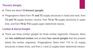
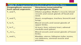
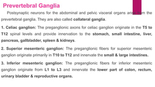
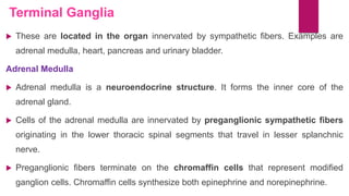
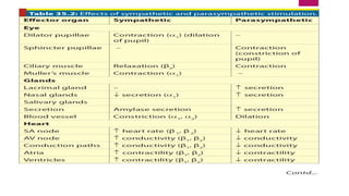
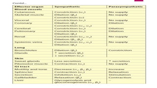
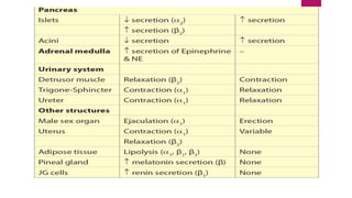
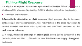
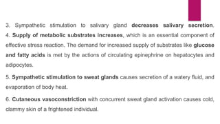
The document provides an overview of the sympathetic and parasympathetic nervous systems. It discusses the structural organization and functions of each system. The sympathetic system is activated during fight or flight responses and increases heart rate, blood pressure, respiration and mobilizes energy stores. It is organized with cell bodies in the spinal cord that project to ganglia. The parasympathetic system counteracts the sympathetic responses and is organized with cell bodies in the brainstem and sacral cord that project to target organs. Both systems use acetylcholine as a neurotransmitter but target different receptor types to produce their effects.
The autonomic nervous system (ANS), formerly the vegetative nervous system, is a division of the peripheral nervous system that supplies smooth muscle and glands, and thus influences the function of internal organs. The autonomic nervous system is a control system that acts largely unconsciously and regulates bodily functions, such as the heart rate, digestion, respiratory rate, pupillary response, urination, and sexual arousal.
Unit IV -
Peripheral nervous system
Classification of peripheral nervous system: Structure and functions of
sympathetic and parasympathetic nervous system.
Origin and functions of spinal and cranial nerves.
Unit IV -
Peripheral nervous system
Classification of peripheral nervous system: Structure and functions of
sympathetic and parasympathetic nervous system.
Origin and functions of spinal and cranial nerves.
Integrating Ayurveda into Parkinson’s Management: A Holistic ApproachAyurveda ForAll
Explore the benefits of combining Ayurveda with conventional Parkinson's treatments. Learn how a holistic approach can manage symptoms, enhance well-being, and balance body energies. Discover the steps to safely integrate Ayurvedic practices into your Parkinson’s care plan, including expert guidance on diet, herbal remedies, and lifestyle modifications.
Local Advanced Lung Cancer: Artificial Intelligence, Synergetics, Complex Sys...Oleg Kshivets
Overall life span (LS) was 1671.7±1721.6 days and cumulative 5YS reached 62.4%, 10 years – 50.4%, 20 years – 44.6%. 94 LCP lived more than 5 years without cancer (LS=2958.6±1723.6 days), 22 – more than 10 years (LS=5571±1841.8 days). 67 LCP died because of LC (LS=471.9±344 days). AT significantly improved 5YS (68% vs. 53.7%) (P=0.028 by log-rank test). Cox modeling displayed that 5YS of LCP significantly depended on: N0-N12, T3-4, blood cell circuit, cell ratio factors (ratio between cancer cells-CC and blood cells subpopulations), LC cell dynamics, recalcification time, heparin tolerance, prothrombin index, protein, AT, procedure type (P=0.000-0.031). Neural networks, genetic algorithm selection and bootstrap simulation revealed relationships between 5YS and N0-12 (rank=1), thrombocytes/CC (rank=2), segmented neutrophils/CC (3), eosinophils/CC (4), erythrocytes/CC (5), healthy cells/CC (6), lymphocytes/CC (7), stick neutrophils/CC (8), leucocytes/CC (9), monocytes/CC (10). Correct prediction of 5YS was 100% by neural networks computing (error=0.000; area under ROC curve=1.0).
micro teaching on communication m.sc nursing.pdfAnurag Sharma
Microteaching is a unique model of practice teaching. It is a viable instrument for the. desired change in the teaching behavior or the behavior potential which, in specified types of real. classroom situations, tends to facilitate the achievement of specified types of objectives.
Basavarajeeyam is a Sreshta Sangraha grantha (Compiled book ), written by Neelkanta kotturu Basavaraja Virachita. It contains 25 Prakaranas, First 24 Chapters related to Rogas& 25th to Rasadravyas.
Tom Selleck Health: A Comprehensive Look at the Iconic Actor’s Wellness Journeygreendigital
Tom Selleck, an enduring figure in Hollywood. has captivated audiences for decades with his rugged charm, iconic moustache. and memorable roles in television and film. From his breakout role as Thomas Magnum in Magnum P.I. to his current portrayal of Frank Reagan in Blue Bloods. Selleck's career has spanned over 50 years. But beyond his professional achievements. fans have often been curious about Tom Selleck Health. especially as he has aged in the public eye.
Follow us on: Pinterest
Introduction
Many have been interested in Tom Selleck health. not only because of his enduring presence on screen but also because of the challenges. and lifestyle choices he has faced and made over the years. This article delves into the various aspects of Tom Selleck health. exploring his fitness regimen, diet, mental health. and the challenges he has encountered as he ages. We'll look at how he maintains his well-being. the health issues he has faced, and his approach to ageing .
Early Life and Career
Childhood and Athletic Beginnings
Tom Selleck was born on January 29, 1945, in Detroit, Michigan, and grew up in Sherman Oaks, California. From an early age, he was involved in sports, particularly basketball. which played a significant role in his physical development. His athletic pursuits continued into college. where he attended the University of Southern California (USC) on a basketball scholarship. This early involvement in sports laid a strong foundation for his physical health and disciplined lifestyle.
Transition to Acting
Selleck's transition from an athlete to an actor came with its physical demands. His first significant role in "Magnum P.I." required him to perform various stunts and maintain a fit appearance. This role, which he played from 1980 to 1988. necessitated a rigorous fitness routine to meet the show's demands. setting the stage for his long-term commitment to health and wellness.
Fitness Regimen
Workout Routine
Tom Selleck health and fitness regimen has evolved. adapting to his changing roles and age. During his "Magnum, P.I." days. Selleck's workouts were intense and focused on building and maintaining muscle mass. His routine included weightlifting, cardiovascular exercises. and specific training for the stunts he performed on the show.
Selleck adjusted his fitness routine as he aged to suit his body's needs. Today, his workouts focus on maintaining flexibility, strength, and cardiovascular health. He incorporates low-impact exercises such as swimming, walking, and light weightlifting. This balanced approach helps him stay fit without putting undue strain on his joints and muscles.
Importance of Flexibility and Mobility
In recent years, Selleck has emphasized the importance of flexibility and mobility in his fitness regimen. Understanding the natural decline in muscle mass and joint flexibility with age. he includes stretching and yoga in his routine. These practices help prevent injuries, improve posture, and maintain mobilit
Knee anatomy and clinical tests 2024.pdfvimalpl1234
This includes all relevant anatomy and clinical tests compiled from standard textbooks, Campbell,netter etc..It is comprehensive and best suited for orthopaedicians and orthopaedic residents.
These lecture slides, by Dr Sidra Arshad, offer a quick overview of the physiological basis of a normal electrocardiogram.
Learning objectives:
1. Define an electrocardiogram (ECG) and electrocardiography
2. Describe how dipoles generated by the heart produce the waveforms of the ECG
3. Describe the components of a normal electrocardiogram of a typical bipolar lead (limb II)
4. Differentiate between intervals and segments
5. Enlist some common indications for obtaining an ECG
6. Describe the flow of current around the heart during the cardiac cycle
7. Discuss the placement and polarity of the leads of electrocardiograph
8. Describe the normal electrocardiograms recorded from the limb leads and explain the physiological basis of the different records that are obtained
9. Define mean electrical vector (axis) of the heart and give the normal range
10. Define the mean QRS vector
11. Describe the axes of leads (hexagonal reference system)
12. Comprehend the vectorial analysis of the normal ECG
13. Determine the mean electrical axis of the ventricular QRS and appreciate the mean axis deviation
14. Explain the concepts of current of injury, J point, and their significance
Study Resources:
1. Chapter 11, Guyton and Hall Textbook of Medical Physiology, 14th edition
2. Chapter 9, Human Physiology - From Cells to Systems, Lauralee Sherwood, 9th edition
3. Chapter 29, Ganong’s Review of Medical Physiology, 26th edition
4. Electrocardiogram, StatPearls - https://www.ncbi.nlm.nih.gov/books/NBK549803/
5. ECG in Medical Practice by ABM Abdullah, 4th edition
6. Chapter 3, Cardiology Explained, https://www.ncbi.nlm.nih.gov/books/NBK2214/
7. ECG Basics, http://www.nataliescasebook.com/tag/e-c-g-basics
2. FUNCTIONAL OVERVIEW
The Sympathetic System
When the body requires support to meet emergency conditions and stressful
situations, sympathetic system provides the desirable assistance.
Sympathetic system also helps to control normal functions of the body such
as regulation of blood pressure, respiration, metabolism etc.
The cell bodies of sympathetic system are located in the thoracic and
abdominal segments of the spinal cord and sympathetic ganglia are present
as a chain close to the vertebral column.
This system is also termed as the thoracolumbar division of the ANS for the
location of the ganglia and cell bodies of neurons at thoracic and lumbar
segments of spinal cord
3. The Parasympathetic System
The component of ANS that works by the side of the sympathetic system is the
parasympathetic division of ANS. It has a reciprocal influence on organ functions
to that of sympathetic influence.
The cell bodies are found in the brainstem cranial nerve nuclei (cranial
component), and in the most caudal part of the spinal cord (spinal or sacral
component).
Hence, it is also termed as craniosacral division of ANS.
The Enteric Nervous System
The enteric nervous system (ENS) is the local neural network in GI system which
has strong anatomical and physiological link with ANS. Therefore, ENS is
considered as the ‘Third division of ANS’.
4.
5. The Sympathetic System
Sympathetic Neuron
Preganglionic Neurons
Cell bodies of preganglionic neurons of the sympathetic division are located in the
intermediolateral horn of the thoracic (T1 to T12) and upper lumbar (L1 to L3)
segments of spinal cord.
The preganglionic neurons come out of the spinal cord via ventral roots. After the
merger of dorsal and ventral roots, spinal nerve emerges.
Sympathetic preganglionic axons leave the spinal nerve via the white rami
communicantes and enter the paravertebral sympathetic ganglia, which is an
interconnected chain located on both sides of the vertebral column.
6. Postganglionic Neurons
Postganglionic neurons for somatic structures such as sweat glands, piloerector
muscles, cutaneous blood vessels and blood vessels of skeletal muscles leave the
paravertebral ganglion in the gray rami communicantes and reenter the spinal
nerve to supply the target tissues.
Postganglionic neurons to head, heart and lungs originate in the cervical or
upper thoracic paravertebral ganglia and proceed to the organs as separate
nerves, for example the cardiac nerve to the heart.
7. Paravertebral Ganglia: Cervical Ganglia:
SUPERIOR CERVICAL GANGLION: The superior cervical ganglion provides
sympathetic fibers that innervate the structures in the head.
These sympathetic fibers travel in the perivascular plexus along the carotid arteries
and innervate radial muscle of the iris that causes dilation of the pupil, supply
Muller’s muscle that assists in elevating the eyelid, and innervate lacrimal and
salivary glands.
MIDDLE & INFERIOR CERVICAL GANGLIA: The middle and inferior cervical
ganglia innervate structures in the chest, including the trachea, esophagus, heart
and lungs.
8.
9. Thoracic Ganglia
There are about 12 thoracic ganglia.
Preganglionic fibers from T1 and T2 supply structures in head and neck, from
T3 and T4 supply thoracic viscera, from T5 to T9 supply structures in upper
limb, and from T6 to T12 supply upper abdominal viscera.
Lumbar & Sacral Ganglia
There are three lumbar ganglia for three lumbar segments. However, there
are two additional lumbar and at least four sacral ganglia that are present
below the lumbar segments. Preganglionic fibers from T10 to L2 supply
structures in lower limbs, and from L1 and L2 supply lower abdominal viscera
10.
11. Prevertebral Ganglia
Postsynaptic neurons for the abdominal and pelvic visceral organs arise from the
prevertebral ganglia. They are also called collateral ganglia.
1. Celiac ganglion: The preganglionic axons for celiac ganglion originate in the T5 to
T12 spinal levels and provide innervation to the stomach, small intestine, liver,
pancreas, gallbladder, spleen & kidneys.
2. Superior mesenteric ganglion: The preganglionic fibers for superior mesenteric
ganglion originate primarily in T10 to T12 and innervate the small & large intestines.
3. Inferior mesenteric ganglion: The preganglionic fibers for inferior mesenteric
ganglion originate from L1 to L3 and innervate the lower part of colon, rectum,
urinary bladder & reproductive organs.
12. Terminal Ganglia
These are located in the organ innervated by sympathetic fibers. Examples are
adrenal medulla, heart, pancreas and urinary bladder.
Adrenal Medulla
Adrenal medulla is a neuroendocrine structure. It forms the inner core of the
adrenal gland.
Cells of the adrenal medulla are innervated by preganglionic sympathetic fibers
originating in the lower thoracic spinal segments that travel in lesser splanchnic
nerve.
Preganglionic fibers terminate on the chromaffin cells that represent modified
ganglion cells. Chromaffin cells synthesize both epinephrine and norepinephrine.
13. EFFECTS OF SYMPATHETIC STIMULATION
Effects are mediated by release of noradrenaline from sympathetic nerve endings
and adrenaline from adrenal medulla.
Effects via Adrenergic Receptors
Catecholamines elicit their effects by acting on adrenergic receptors. Adrenergic
receptors are broadly divided into two types: and β.
The receptor has two subtypes: 1 and 2.
The β receptor has three subtypes: β1, β 2 and β3.
Generally, β receptors are more sensitive to adrenaline and receptors to
noradrenaline.
14. Effects of receptor Stimulation
Effects of 1 Stimulation
The 1 receptors are present in vascular smooth muscles of cutaneous and
splanchnic circulation, sphincters of bladder and GI tract and radial muscles of iris.
Stimulation of these receptors causes contraction or constriction of the structures
in which they are present.
Effects of 2 Stimulation
2 receptors are present in presynaptic nerve endings, wall of GI tract, platelets
and adipocytes. Stimulation of these receptors often causes relaxation or inhibition
of the structure.
15. Effects of β receptor Stimulation
Effects of β1 Stimulation
β1 receptors are present in SA node, AV node and ventricular muscle. Stimulation
of these receptors causes excitation of these structures.
Effects of β2 Stimulation
β2 receptors are present in blood vessels of skeletal muscles, bronchial smooth
muscles & wall of GI tract. Stimulation of these receptors causes relaxation of
these structures. They are more sensitive to adrenaline than noradrenaline.
Effects of β3 Stimulation
β3 receptors are present in adipose tissues. Stimulation of these receptors causes
lipolysis.
16.
17.
18.
19. Fight-or-Flight Response
It is a typical widespread response of sympathetic activation. This occurs in critical
situations of life when one has to either fight the situation or flee from the situation.
The effects are as follows:
1.Sympathetic stimulation of CVS increases blood pressure due to increased
cardiac output and vasoconstriction. Also, redistribution of the blood flow occurs to
skeletal muscles and heart from splanchnic and cutaneous territories so that
performance enhances.
2. In lungs, increased exchange of blood gases occurs due to stimulation of the
respiratory rate and dilation of bronchiolar tree. This increases supply of oxygen to
the tissues.
20. 3. Sympathetic stimulation to salivary gland decreases salivary secretion.
4. Supply of metabolic substrates increases, which is an essential component of
effective stress reaction. The demand for increased supply of substrates like glucose
and fatty acids is met by the actions of circulating epinephrine on hepatocytes and
adipocytes.
5. Sympathetic stimulation to sweat glands causes secretion of a watery fluid, and
evaporation of body heat.
6. Cutaneous vasoconstriction with concurrent sweat gland activation causes cold,
clammy skin of a frightened individual.
21. 7. Activation of piloerector muscles of hair follicles causes hair-standing-on the
skin.
8. Pupillary dilation enhances visual acuity and increases visual perception to make
the individual environmentally more alert.
9. Stimulation of brainstem reticular system makes the individual maximally alert
and mentally conscious to take appropriate decisions in quick successions.
10. Activity of bowel and bladder temporarily ceases due to constriction of
sphincters.
22. The Parasympathetic System
STRUCTURAL ORGANIZATION
The parasympathetic system is the craniosacral outflow of autonomic nervous
system.
The cranial component emanates from the brainstem, and the sacral component
originates from intermediolateral gray column of sacral segments of spinal cord.
Parasympathetic ganglia are located either close to the organ or embedded in
the organ. Therefore, preganglionic neurons are much longer than
postganglionic neurons.
23. Cranial Component
Cell bodies of preganglionic neurons of cranial component of parasympathetic
system are located in the brainstem.
Brainstem parasympathetic neurons innervate structures in the head, neck, thorax
and abdomen.
Parasympathetic axons from brainstem travel in III, VII, IX, and X cranial nerves.
Nuclei of these cranial nerves are present in the midbrain in tectum, pons and
medulla.
Therefore, these nuclei serve as the centers for the integration of autonomic
reflexes for the organ systems they innervate.
24.
25. Sacral Component
Sacral parasympathetic neurons innervate structures in the pelvis.
Preganglionic fibers originate in the intermediolateral gray column of the sacral
segments S2, S3, and S4 of spinal cord.
The preganglionic fibers terminate in ganglia in or near the viscera that include
descending colon, sigmoid colon, rectum, internal anal sphincter, urinary bladder
and the reproductive organs.
26. PARASYMPATHETIC FUNCTIONS
Parasympathetic system restores body’s energy reserve.
Except on cardiovascular system, most parasympathetic effects are
stimulatory, especially for the processes that facilitate energy storage and
growth.
They stimulate intestinal motility, secretion, digestion and absorption.
They promote reproductive functions
27. Neurotransmitters and Receptors
Muscarinic Receptors
Cholinergic muscarinic receptors are present in heart, smooth muscles and
glands. These receptors are activated by acetylcholine and muscarine.
Activation of these receptors produce inhibitory effects on heart, for example,
decreased heart rate, and excitatory effects on smooth muscle and glands, for
example, increased GI motility and secretion etc.
In smooth muscles and glandular tissues, effects are mediated by intracellular
IP3 and Ca++.
Muscarinic receptors are blocked by atropine.
28. Nicotinic Receptors
Cholinergic nicotinic receptors are present in autonomic ganglia (both
sympathetic and parasympathetic), neuromuscular junctions and adrenal
medulla. These receptors are activated by acetylcholine and nicotine.
Activation of these receptors produces excitatory effects on target tissue. Effects
are mediated by direct binding of acetylcholine to subunits of the
receptors.
Receptors also contain Na+ and K+ channels.
Nicotinic effects are blocked by ganglion blockers such as hexamethonium.