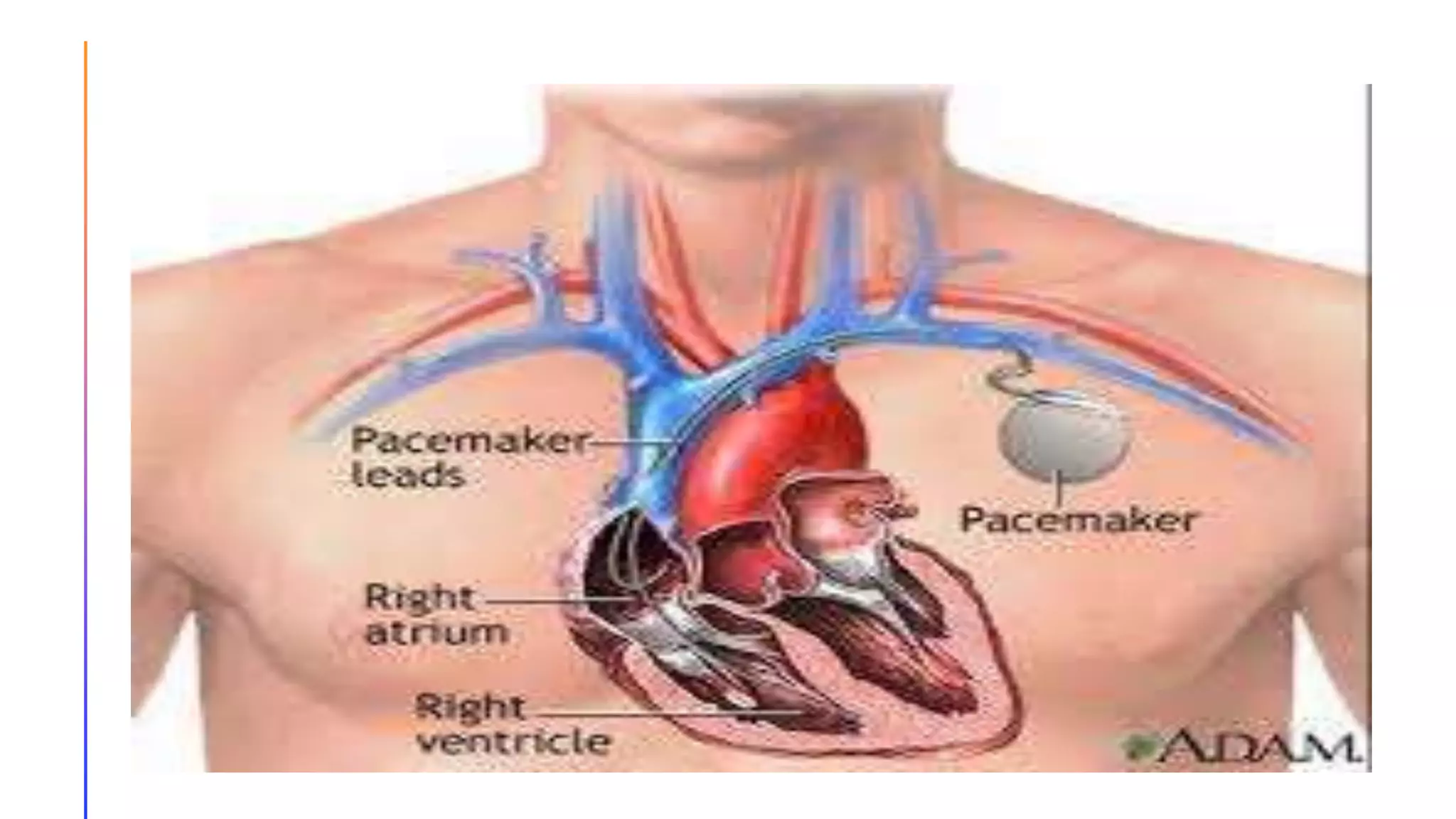
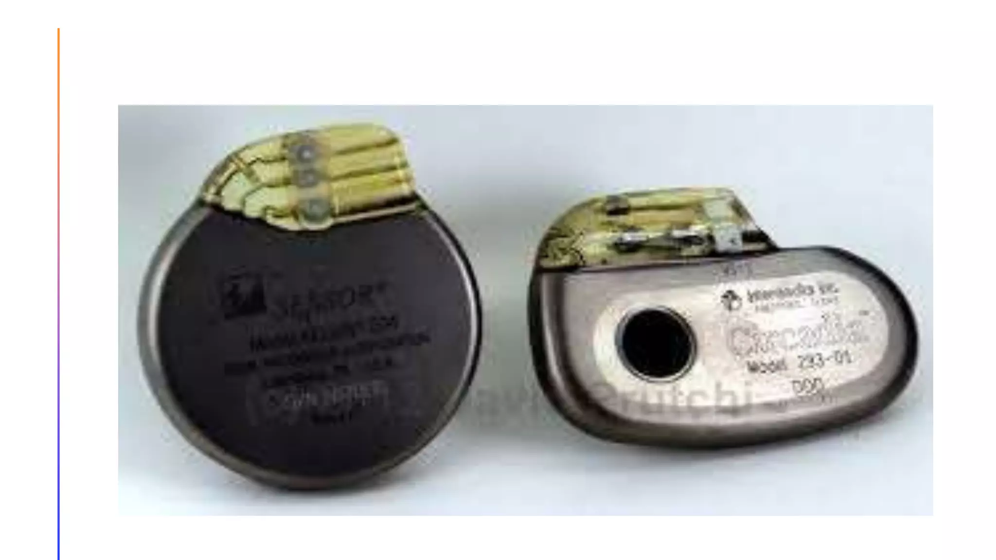
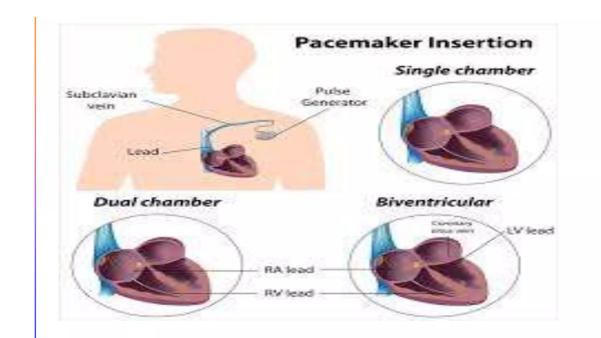
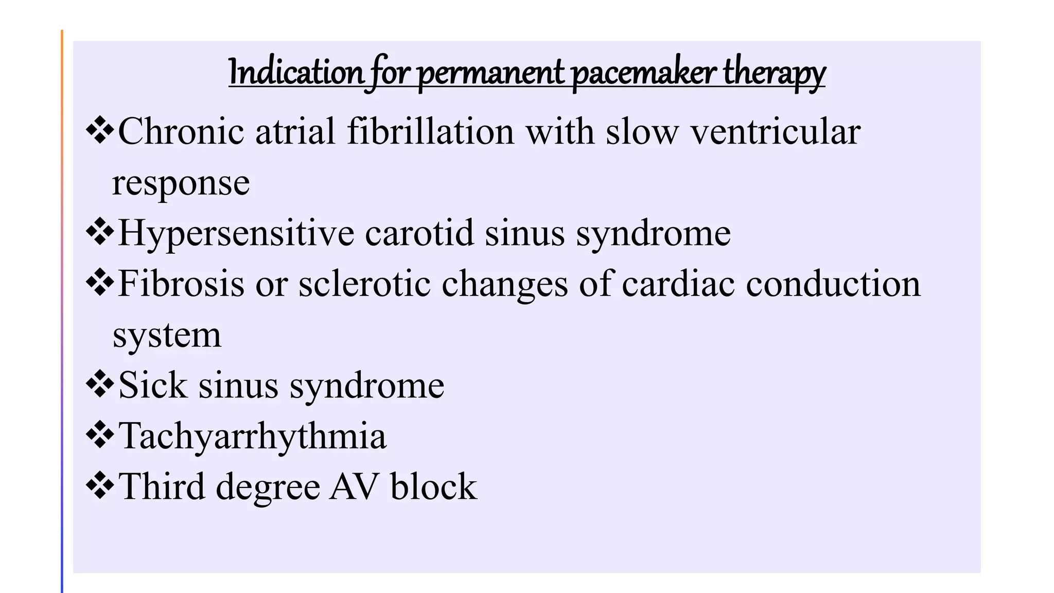
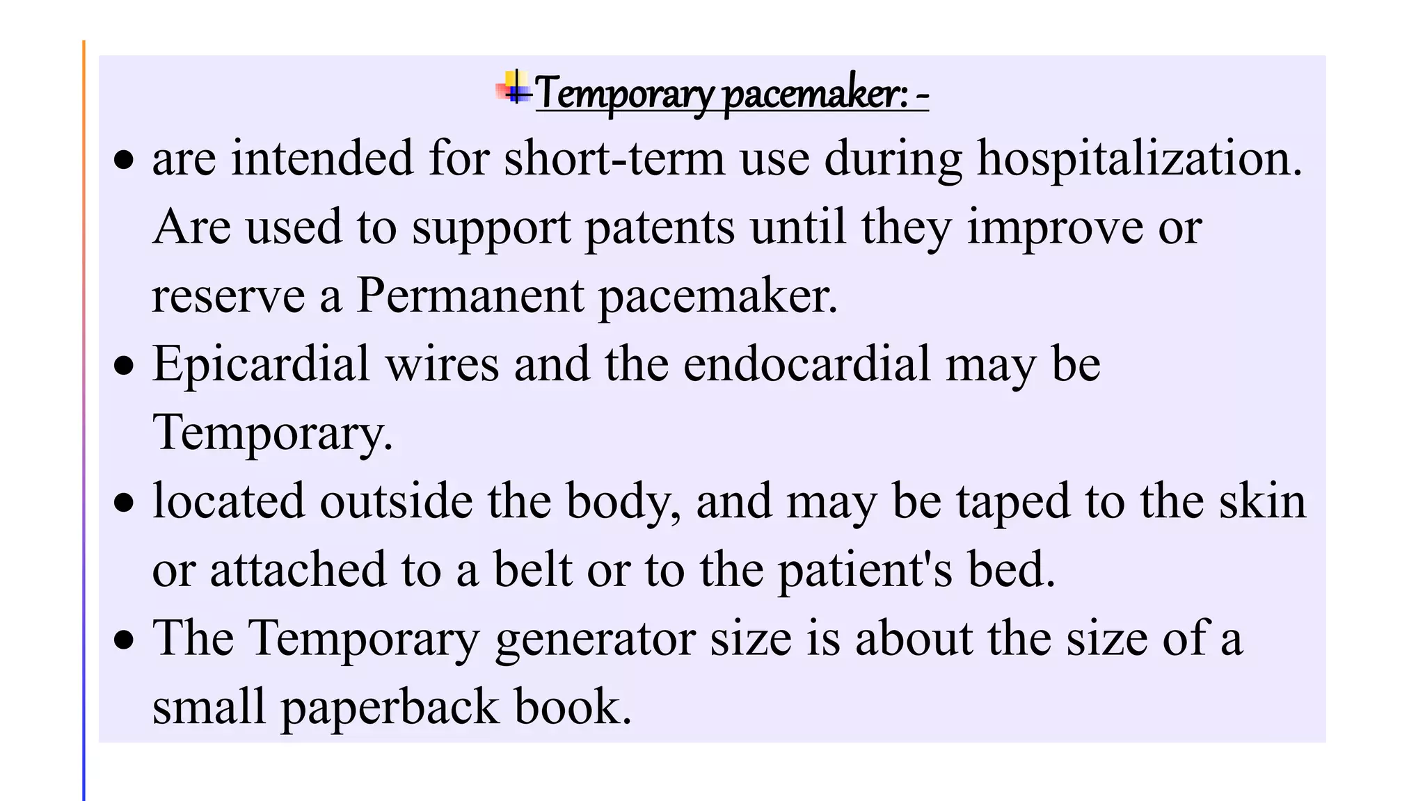
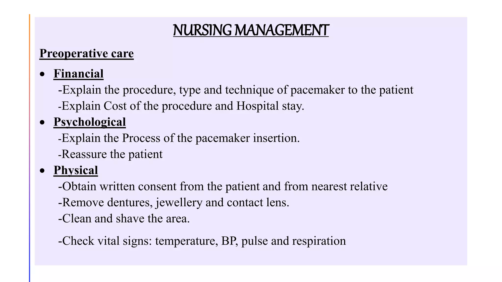
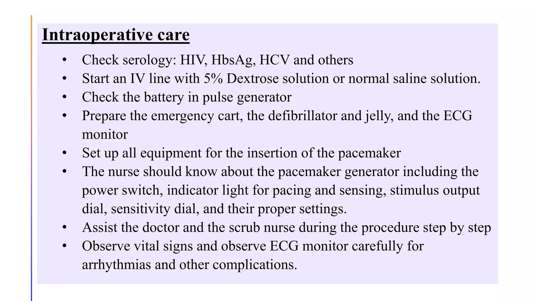
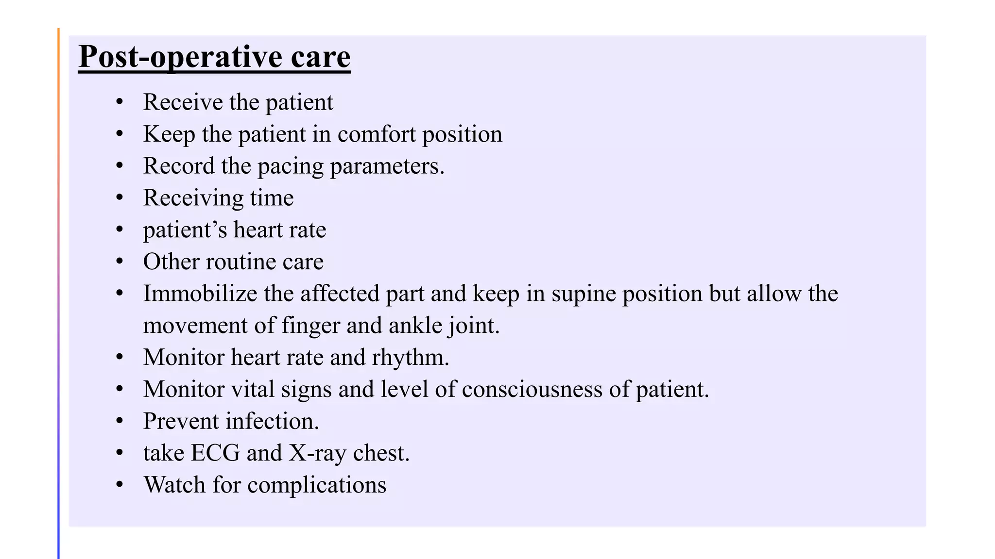
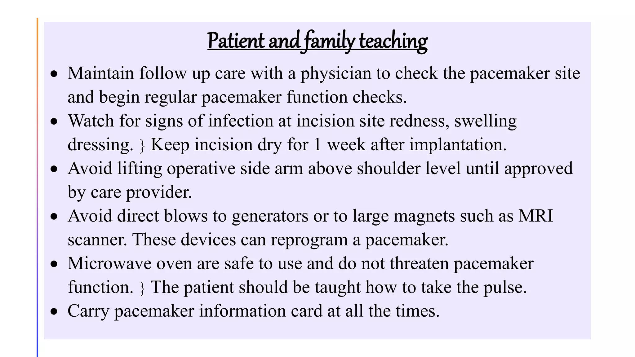
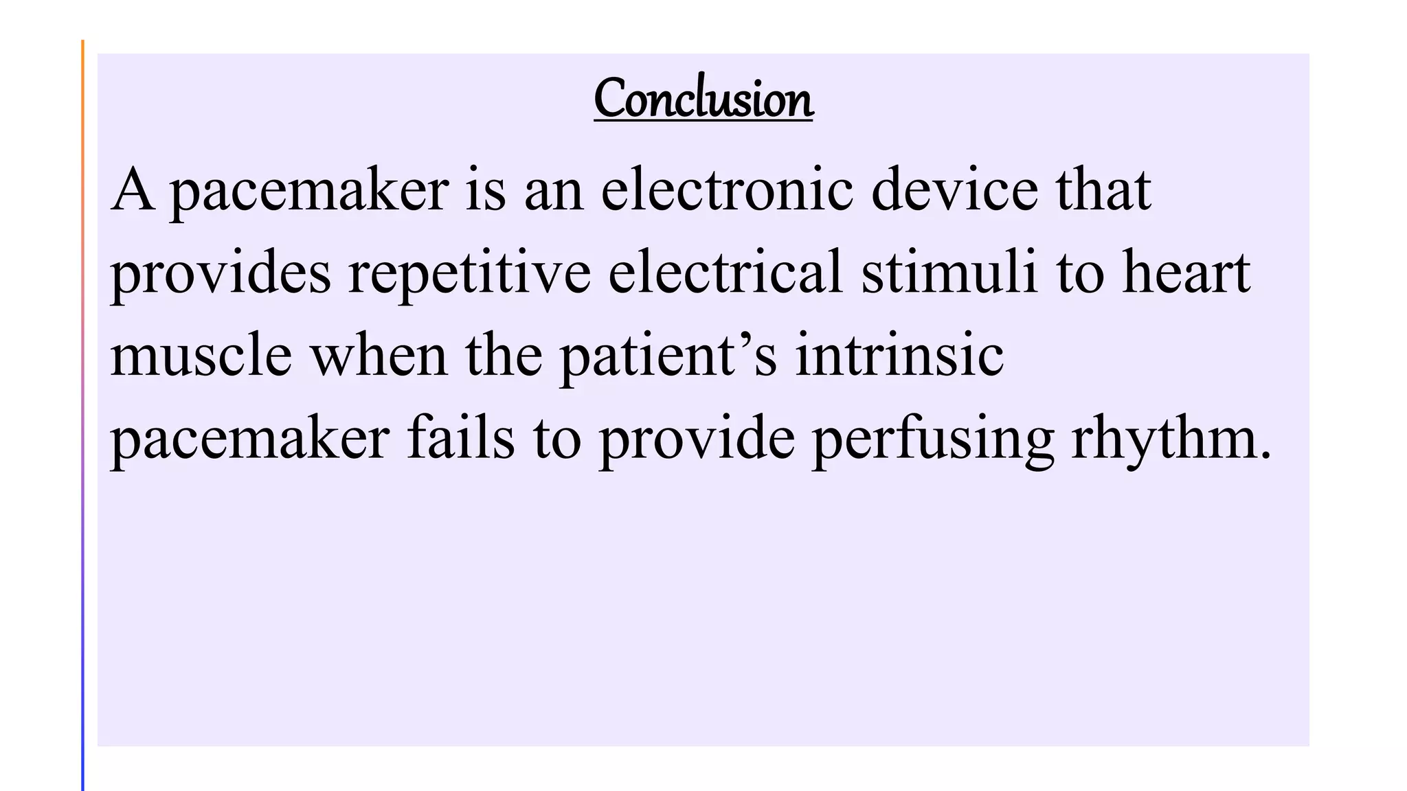
CARDIAC PACEMAKER
DEMONSTRATION
INTRODUCTION
Pacemakers are the electrode devices that can be
used to initiate the heartbeat when the hearts
intrinsic electrical system cannot effectively
generate a rate adequate to support cardiac output.
Pacemakers is an electronic device used to pace
the heart when the normal conduction pathway is
damaged or diseased.
Under normal conditions,
electrical activity is spontaneously generated by the SA node,
the cardiac pacemaker.
This electrical impulse is propagated throughout the right atrium,
and through Bachmann's bundle to the left atrium,
stimulating the myocardium of the atria to contract.
Need for pacemaker
Aging or heart disease damages your sinus node's ability to set the correct
pace for your heartbeat. Such damage can cause slower than normal
heartbeats or long pauses between heartbeats. The damage also can cause
your heart to switch between slow and fast rhythms, Fatigue, dizziness,
lightheadedness, fainting, and the inability to exercise without getting out
of breath
Surveys have shown that up to 80% of pacemakers are implanted in the
elderly and the average age of pacemaker recipients is now 75 ± 10 years.
The lifespan of the pacemaker depends on how much your heart is
depending on it, usually last four to eight years
DEFINITION
A pacemaker is a battery-operated generator that
initiate and control electrical stimulation of the
heart via an electrode that is usually in direct
contact with the myocardium.
COMPONENTS OF PACEMAKER
 Pulse generator: - contain the circuitry and batteries that determine
the rate (beat per minute) and the strength or output (milliamperes) of
the electrical stimulus delivered to the heart
 Pacemaker electrodes: - which carry the impulse created by the
generator to the heart. 1. Endocardial leads.
2. Epicardial wires.
Most pacemaker have elective replacement indicator (ERI). a signal that
indicates when the battery is approaching depletion. The pacemaker
continues to function for several months after the appearance of ERI to
ensure that there is adequate time for a battery replacement. Battery
replacement are usually performed using a local anaesthesia,
hospitalization is necessary for implantation or battery replacement.
HOWPACEMAKER WORKS
A pacemaker consists of a battery, a computerized generator and wires
with sensors at their tips (called as electrodes). The battery powers the
generator and both are surrounded by a thin metal box. The wires
connect the generator to the heart.
A pacemaker helps monitor and control the heartbeat. The electrodes
detect heart’s electrical activity and send data through the wires to the
computer in the generator. If heart rhythm is abnormal, the computer
will direct the generator to send electrical pulses to heart. The pulses
travel through the wires to reach the heart.
TYPES OF PACEMAKER
Permanent pacemaker: -
 Implanted totally in the body
 Power source is implanted subcutaneously usually
over the pectoral muscle on the patient non dominant
side.
 They last approximately 6 to 12 years.
 The permanent generator it weighs less than 1 oz and
is the size of a thick credit card.
Types of permanent pacemaker
Single-chamber pacemaker: -In this type, only one pacing
lead is placed into a chamber of the heart, either the atrium
or the ventricle
Dual-chamber pacemaker: -Wires are placed in two
chambers of the heart. One lead paces the atrium and one
paces the ventricle. Closely resembles the natural pacing of
the heart.
Rate-responsive pacemaker: - It has sensors that detect
changes in the patient's physical activity and automatically
adjust the pacing rate to fulfil the body's metabolic needs.
Indication for permanent pacemaker therapy
Chronic atrial fibrillation with slow ventricular
response
Hypersensitive carotid sinus syndrome
Fibrosis or sclerotic changes of cardiac conduction
system
Sick sinus syndrome
Tachyarrhythmia
Third degree AV block
Temporary pacemaker: -
 are intended for short-term use during hospitalization.
Are used to support patents until they improve or
reserve a Permanent pacemaker.
 Epicardial wires and the endocardial may be
Temporary.
 located outside the body, and may be taped to the skin
or attached to a belt or to the patient's bed.
 The Temporary generator size is about the size of a
small paperback book.
Types of temporary pacemaker therapy
There are 3 types of temporary pacemaker.
 Transvenous invasive pacemaker (endocardial): -It consists of
lead or leads that are threaded transvenously to the right atrium and
or right ventricle and attached to external power source.
 Transthoracic invasive pacing (Epicardial pacing): -It is achieved
by attaching an atrial and ventricle and attached to epicardium during
heart surgery. The leads are passed through the chest wall and
attached to the external power source.
 Trans cutaneous pacemaker (Non-invasive pacing): -It is used to
provide adequate heart rate and rhythm to the patient in and
emergency situation.
Trans cutaneous pacemaker (Non-invasive pacing)
Indications of temporary pacing
Maintenance of adequate heart rate and rhythm during
special circumstances such as surgery and postoperative
recovery, cardiac catheterization or coronary angioplasty.
Before implantation of a permanent pacemaker.
As prophylaxis after open heart surgery.
Acute anterior MI with second degree or third-degree AV
block or bundle branch block.
Acute inferior MI with symptomatic bradycardia and AV
block
NURSING MANAGEMENT
Preoperative care
 Financial
-Explain the procedure, type and technique of pacemaker to the patient
-Explain Cost of the procedure and Hospital stay.
 Psychological
-Explain the Process of the pacemaker insertion.
-Reassure the patient
 Physical
-Obtain written consent from the patient and from nearest relative
-Remove dentures, jewellery and contact lens.
-Clean and shave the area.
-Check vital signs: temperature, BP, pulse and respiration
Intraoperative care
• Check serology: HIV, HbsAg, HCV and others
• Start an IV line with 5% Dextrose solution or normal saline solution.
• Check the battery in pulse generator
• Prepare the emergency cart, the defibrillator and jelly, and the ECG
monitor
• Set up all equipment for the insertion of the pacemaker
• The nurse should know about the pacemaker generator including the
power switch, indicator light for pacing and sensing, stimulus output
dial, sensitivity dial, and their proper settings.
• Assist the doctor and the scrub nurse during the procedure step by step
• Observe vital signs and observe ECG monitor carefully for
arrhythmias and other complications.
Post-operative care
• Receive the patient
• Keep the patient in comfort position
• Record the pacing parameters.
• Receiving time
• patient’s heart rate
• Other routine care
• Immobilize the affected part and keep in supine position but allow the
movement of finger and ankle joint.
• Monitor heart rate and rhythm.
• Monitor vital signs and level of consciousness of patient.
• Prevent infection.
• take ECG and X-ray chest.
• Watch for complications
Patient and family teaching
 Maintain follow up care with a physician to check the pacemaker site
and begin regular pacemaker function checks.
 Watch for signs of infection at incision site redness, swelling
dressing.  Keep incision dry for 1 week after implantation.
 Avoid lifting operative side arm above shoulder level until approved
by care provider.
 Avoid direct blows to generators or to large magnets such as MRI
scanner. These devices can reprogram a pacemaker.
 Microwave oven are safe to use and do not threaten pacemaker
function.  The patient should be taught how to take the pulse.
 Carry pacemaker information card at all the times.
COMPLICATION
 Hematoma
 Pneumothorax
 Failure to sense or capture
 Perforation of atrial or ventricle septum
 Ventricular atrophy and tachycardia
 Movement or dislocation of lead
 Cardiac perforation
 Infection (endocarditis)
 A study done on “Pacemaker Endocarditis: Clinical Features and
Management of 60 Consecutive Cases “89% cases were found to be
endocarditis related to pacemaker and the causative agent was
staphyloccus .
Nursing diagnosis
Acute pain related to insertion site and prescribed post
procedure immobilization.
Disturbed self-concept related to perceived loss of health
and dependence on pacemaker.
Impaired physical mobility related to incisional site pain,
activity restrictions.  Risk for infection related to operative
site.
Risk for ineffective therapeutic regimen management related
to insufficient knowledge of activity restrictions,
precautions.
Conclusion
A pacemaker is an electronic device that
provides repetitive electrical stimuli to heart
muscle when the patient’s intrinsic
pacemaker fails to provide perfusing rhythm.

Artificial Cardiac pacemaker |medical device that generates electrical impulses

  • 1.
  • 2.
    INTRODUCTION Pacemakers are theelectrode devices that can be used to initiate the heartbeat when the hearts intrinsic electrical system cannot effectively generate a rate adequate to support cardiac output. Pacemakers is an electronic device used to pace the heart when the normal conduction pathway is damaged or diseased.
  • 3.
    Under normal conditions, electricalactivity is spontaneously generated by the SA node, the cardiac pacemaker. This electrical impulse is propagated throughout the right atrium, and through Bachmann's bundle to the left atrium, stimulating the myocardium of the atria to contract.
  • 6.
    Need for pacemaker Agingor heart disease damages your sinus node's ability to set the correct pace for your heartbeat. Such damage can cause slower than normal heartbeats or long pauses between heartbeats. The damage also can cause your heart to switch between slow and fast rhythms, Fatigue, dizziness, lightheadedness, fainting, and the inability to exercise without getting out of breath Surveys have shown that up to 80% of pacemakers are implanted in the elderly and the average age of pacemaker recipients is now 75 ± 10 years. The lifespan of the pacemaker depends on how much your heart is depending on it, usually last four to eight years
  • 7.
    DEFINITION A pacemaker isa battery-operated generator that initiate and control electrical stimulation of the heart via an electrode that is usually in direct contact with the myocardium.
  • 9.
    COMPONENTS OF PACEMAKER Pulse generator: - contain the circuitry and batteries that determine the rate (beat per minute) and the strength or output (milliamperes) of the electrical stimulus delivered to the heart  Pacemaker electrodes: - which carry the impulse created by the generator to the heart. 1. Endocardial leads. 2. Epicardial wires. Most pacemaker have elective replacement indicator (ERI). a signal that indicates when the battery is approaching depletion. The pacemaker continues to function for several months after the appearance of ERI to ensure that there is adequate time for a battery replacement. Battery replacement are usually performed using a local anaesthesia, hospitalization is necessary for implantation or battery replacement.
  • 10.
    HOWPACEMAKER WORKS A pacemakerconsists of a battery, a computerized generator and wires with sensors at their tips (called as electrodes). The battery powers the generator and both are surrounded by a thin metal box. The wires connect the generator to the heart. A pacemaker helps monitor and control the heartbeat. The electrodes detect heart’s electrical activity and send data through the wires to the computer in the generator. If heart rhythm is abnormal, the computer will direct the generator to send electrical pulses to heart. The pulses travel through the wires to reach the heart.
  • 11.
    TYPES OF PACEMAKER Permanentpacemaker: -  Implanted totally in the body  Power source is implanted subcutaneously usually over the pectoral muscle on the patient non dominant side.  They last approximately 6 to 12 years.  The permanent generator it weighs less than 1 oz and is the size of a thick credit card.
  • 13.
    Types of permanentpacemaker Single-chamber pacemaker: -In this type, only one pacing lead is placed into a chamber of the heart, either the atrium or the ventricle Dual-chamber pacemaker: -Wires are placed in two chambers of the heart. One lead paces the atrium and one paces the ventricle. Closely resembles the natural pacing of the heart. Rate-responsive pacemaker: - It has sensors that detect changes in the patient's physical activity and automatically adjust the pacing rate to fulfil the body's metabolic needs.
  • 16.
    Indication for permanentpacemaker therapy Chronic atrial fibrillation with slow ventricular response Hypersensitive carotid sinus syndrome Fibrosis or sclerotic changes of cardiac conduction system Sick sinus syndrome Tachyarrhythmia Third degree AV block
  • 17.
    Temporary pacemaker: - are intended for short-term use during hospitalization. Are used to support patents until they improve or reserve a Permanent pacemaker.  Epicardial wires and the endocardial may be Temporary.  located outside the body, and may be taped to the skin or attached to a belt or to the patient's bed.  The Temporary generator size is about the size of a small paperback book.
  • 18.
    Types of temporarypacemaker therapy There are 3 types of temporary pacemaker.  Transvenous invasive pacemaker (endocardial): -It consists of lead or leads that are threaded transvenously to the right atrium and or right ventricle and attached to external power source.  Transthoracic invasive pacing (Epicardial pacing): -It is achieved by attaching an atrial and ventricle and attached to epicardium during heart surgery. The leads are passed through the chest wall and attached to the external power source.  Trans cutaneous pacemaker (Non-invasive pacing): -It is used to provide adequate heart rate and rhythm to the patient in and emergency situation.
  • 20.
    Trans cutaneous pacemaker(Non-invasive pacing)
  • 21.
    Indications of temporarypacing Maintenance of adequate heart rate and rhythm during special circumstances such as surgery and postoperative recovery, cardiac catheterization or coronary angioplasty. Before implantation of a permanent pacemaker. As prophylaxis after open heart surgery. Acute anterior MI with second degree or third-degree AV block or bundle branch block. Acute inferior MI with symptomatic bradycardia and AV block
  • 22.
    NURSING MANAGEMENT Preoperative care Financial -Explain the procedure, type and technique of pacemaker to the patient -Explain Cost of the procedure and Hospital stay.  Psychological -Explain the Process of the pacemaker insertion. -Reassure the patient  Physical -Obtain written consent from the patient and from nearest relative -Remove dentures, jewellery and contact lens. -Clean and shave the area. -Check vital signs: temperature, BP, pulse and respiration
  • 23.
    Intraoperative care • Checkserology: HIV, HbsAg, HCV and others • Start an IV line with 5% Dextrose solution or normal saline solution. • Check the battery in pulse generator • Prepare the emergency cart, the defibrillator and jelly, and the ECG monitor • Set up all equipment for the insertion of the pacemaker • The nurse should know about the pacemaker generator including the power switch, indicator light for pacing and sensing, stimulus output dial, sensitivity dial, and their proper settings. • Assist the doctor and the scrub nurse during the procedure step by step • Observe vital signs and observe ECG monitor carefully for arrhythmias and other complications.
  • 24.
    Post-operative care • Receivethe patient • Keep the patient in comfort position • Record the pacing parameters. • Receiving time • patient’s heart rate • Other routine care • Immobilize the affected part and keep in supine position but allow the movement of finger and ankle joint. • Monitor heart rate and rhythm. • Monitor vital signs and level of consciousness of patient. • Prevent infection. • take ECG and X-ray chest. • Watch for complications
  • 25.
    Patient and familyteaching  Maintain follow up care with a physician to check the pacemaker site and begin regular pacemaker function checks.  Watch for signs of infection at incision site redness, swelling dressing.  Keep incision dry for 1 week after implantation.  Avoid lifting operative side arm above shoulder level until approved by care provider.  Avoid direct blows to generators or to large magnets such as MRI scanner. These devices can reprogram a pacemaker.  Microwave oven are safe to use and do not threaten pacemaker function.  The patient should be taught how to take the pulse.  Carry pacemaker information card at all the times.
  • 26.
    COMPLICATION  Hematoma  Pneumothorax Failure to sense or capture  Perforation of atrial or ventricle septum  Ventricular atrophy and tachycardia  Movement or dislocation of lead  Cardiac perforation  Infection (endocarditis)  A study done on “Pacemaker Endocarditis: Clinical Features and Management of 60 Consecutive Cases “89% cases were found to be endocarditis related to pacemaker and the causative agent was staphyloccus .
  • 27.
    Nursing diagnosis Acute painrelated to insertion site and prescribed post procedure immobilization. Disturbed self-concept related to perceived loss of health and dependence on pacemaker. Impaired physical mobility related to incisional site pain, activity restrictions.  Risk for infection related to operative site. Risk for ineffective therapeutic regimen management related to insufficient knowledge of activity restrictions, precautions.
  • 28.
    Conclusion A pacemaker isan electronic device that provides repetitive electrical stimuli to heart muscle when the patient’s intrinsic pacemaker fails to provide perfusing rhythm.