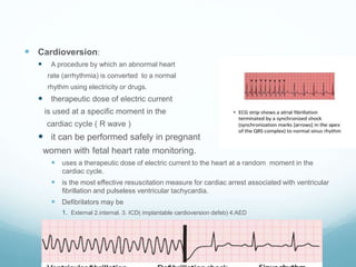
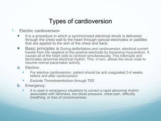
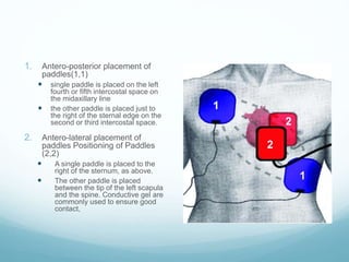
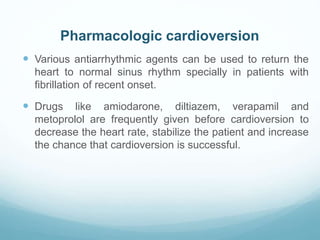
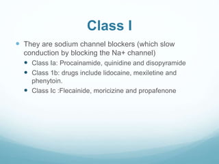
Cardioversion
Dr Kifayat khan
Resident Cardiac Surgeon
cardioversion
 Learning Objectives
 Difference between cardioversion and defebrilation
 Types of cardioversion
1. Electrical
 Elective
 Emergency
2. Chemical or pharmacological
 Cardioversion:
 A procedure by which an abnormal heart
rate (arrhythmia) is converted to a normal
rhythm using electricity or drugs.
 therapeutic dose of electric current
is used at a specific moment in the
cardiac cycle ( R wave )
 it can be performed safely in pregnant
women with fetal heart rate monitoring.
 uses a therapeutic dose of electric current to the heart at a random moment in the
cardiac cycle.
 is the most effective resuscitation measure for cardiac arrest associated with ventricular
fibrillation and pulseless ventricular tachycardia.
 Defibrilators may be
1. External 2.internal. 3. ICD( implantable cardioversion defeb) 4:AED
Cardioversion vs defebrilation
 Sync on R wave
 For peri arest tachyarhtmias
 Usually elective
 Low energy
 Escalate for next shock (
100,200,300,360)
 Done for
 A Feb
 A flutters
 V tach with pulse
 Not sync
 For arrest
 Always emergency
 High energy
 No escalate for next shock
 Done for
 Vent feb
 V tach + pluseless
Types of cardioversion
1. Electric cardioversion
 It is a procedure in which a synchronized electrical shock is delivered
through the chest wall to the heart through special electrodes or paddles
that are applied to the skin of the chest and back.
 Basic principles is During defibrillation and cardioversion, electrical current
travels from the negative to the positive electrode by traversing myocardium. It
causes all of the heart cells to contract simultaneously. This interrupts and
terminates abnormal electrical rhythm. This, in turn, allows the sinus node to
resume normal pacemaker activity.
a. Elective:
 For elective cardioversion, patient should be anti coagulated 3-4 weeks
before and after cardioversion
 Exclude Thromboembolism through TEE
b. Emergency:
 It is used in emergency situations to correct a rapid abnormal rhythm
associated with faintness, low blood pressure, chest pain, difficulty
breathing, or loss of consciousness.
Indications & Contraindications
 Indications:
 V. tachycardia with pulse (ventricular rate >150) who is unstable
(chest pain, pulmonary edema, lightheadedness, hypotension)
 Atrial fibrillation
 Atrial flutter
 Atrial tachycardia
 Contraindications •
 Presence of left atrial thrombus.
 Digitalis toxicity or hypokalemia.
 Sinus tachycardia caused by various clinical conditions and
catecholamine-induced arrhythmia.
Recommendations for Direct-current Cardioversion of Atrial
Fibrillation
1. Class I:
 When a rapid ventricular response does not respond promptly to pharmacological
measures for patients with AF with ongoing myocardial ischemia, symptomatic
hypotension, angina or hear faliure, immediate R-wave synchronized direct-current
cardioversion is recommended. (Level of Evidence: C)
 AF involving preexcitation when very rapid tachycardia or hemodynamic instability
occurs. (Level of Evidence: B).
 Cardioversion is recommended in patients without hemodynamic instability when
symptoms of AF are unacceptable to the patient. In case of early relapse of AF after
cardioversion, repeated cardioversion attempts may be made following
administration of antiarrhythmic medication. (Level of Evidence: C)
2. Class IIa :
 Direct-current cardioversion can be useful to restore sinus rhythm as part of a long-
term management strategy for patients with AF. (Level of Evidence: B).
 Patient preference is a reasonable consideration in the selection of infrequently
repeated cardioversions for the management of symptomatic or recurrent AF. (Level
of Evidence: C)
 Equipment:
 Defibrillator with a synchronising button.
 Emergency trolley with emergency drugs; ( lignocaine,
atropine, and adrenaline ).
 Oxygen mask, intubation equipment, airway .
 Monitor and continuous recording facilities (BP,ECG,
SpO2).
 Intravenous access • Suction device
1. Antero-posterior placement of
paddles(1,1)
 single paddle is placed on the left
fourth or fifth intercostal space on
the midaxillary line
 the other paddle is placed just to
the right of the sternal edge on the
second or third intercostal space.
2. Antero-lateral placement of
paddles Positioning of Paddles
(2,2)
 A single paddle is placed to the
right of the sternum, as above.
 The other paddle is placed
between the tip of the left scapula
and the spine. Conductive gel are
commonly used to ensure good
contact,
Preparing for a Cardioversion
 Do not eat or drink for at least eight hours prior to the
procedure.
 Blood thining medicines may be given with electrical
cardioversion to prevent clots
 Take your regularly scheduled medications the morning of
the procedure unless your medical practitioner has told you.
 Stop digoxin 48 hours prior to the procedure
 Do not apply any lotions or ointments to chest or back as
this may interfere with the adhesiveness of the shocking
pads.
Procedure Steps
 Place paddles so that they do not touch pts clothing or bed linens
 Ensure monitor is attached.
 Do not charge the machine untill ready to shock.
 Exert 25 pound pressure on the paddle .
 Ensure you and every body is free of the pat.
 Inspect skin for burns.
 Record the delivered energy.
 Sedate patient with a short-acting agent such as midazolam or propofol and an
opioid analgesic, such as fentanyl.
 Reversal agents, such as flumazenil and naloxone, should be available.
Complications
 uncommon but may include:
 Harmless arrhythmias, such as atrial, ventricular, and junctional
premature beats.
 Serious complications include ventricular fibrillation (VF)
 severe bradycardia or asystole
 Thromboembolization
 Bruising, burning or pain where the paddles were used.
 Myocardial necrosis can result from high-energy shocks.
 ST segment elevation can be seen immediately and usually lasts
for 1-2 minutes.
 ST segment elevation that lasts longer than 2 minutes usually
indicates myocardial injury unrelated to the shock.
 Pulmonary edema is a rare complication of cardioversion. It is
probably due to transient left atrial standstill and left ventricular
systolic dysfunction.
Pharmacologic cardioversion
 Various antiarrhythmic agents can be used to return the
heart to normal sinus rhythm specially in patients with
fibrillation of recent onset.
 Drugs like amiodarone, diltiazem, verapamil and
metoprolol are frequently given before cardioversion to
decrease the heart rate, stabilize the patient and increase
the chance that cardioversion is successful.
Class I
 They are sodium channel blockers (which slow
conduction by blocking the Na+ channel)
 Class Ia: Procainamide, quinidine and disopyramide
 Class 1b: drugs include lidocaine, mexiletine and
phenytoin.
 Class Ic :Flecainide, moricizine and propafenone
Class II
 They are beta blockers which inhibit SA and AV node
depolarization and slow heart rate.
 They also decrease cardiac oxygen demand and can
prevent cardiac remodeling.
 some are cardio selective (affecting only beta 1
receptors, metoprolol,nebivolol) while others are non-
selective (affecting beta 1 and 2 receptors).
Class III, Class IV
 agents (prolong repolarization by blocking outward K+
current).
 amiodarone and sotalol
 Class iv drugs are calcium (Ca) channel blockers. They
work by inhibiting the action potential of the SA and AV
nodes.
 Deltiazem
Cardioversion

Cardioversion

  • 1.
  • 2.
    cardioversion  Learning Objectives Difference between cardioversion and defebrilation  Types of cardioversion 1. Electrical  Elective  Emergency 2. Chemical or pharmacological
  • 3.
     Cardioversion:  Aprocedure by which an abnormal heart rate (arrhythmia) is converted to a normal rhythm using electricity or drugs.  therapeutic dose of electric current is used at a specific moment in the cardiac cycle ( R wave )  it can be performed safely in pregnant women with fetal heart rate monitoring.  uses a therapeutic dose of electric current to the heart at a random moment in the cardiac cycle.  is the most effective resuscitation measure for cardiac arrest associated with ventricular fibrillation and pulseless ventricular tachycardia.  Defibrilators may be 1. External 2.internal. 3. ICD( implantable cardioversion defeb) 4:AED
  • 4.
    Cardioversion vs defebrilation Sync on R wave  For peri arest tachyarhtmias  Usually elective  Low energy  Escalate for next shock ( 100,200,300,360)  Done for  A Feb  A flutters  V tach with pulse  Not sync  For arrest  Always emergency  High energy  No escalate for next shock  Done for  Vent feb  V tach + pluseless
  • 5.
    Types of cardioversion 1.Electric cardioversion  It is a procedure in which a synchronized electrical shock is delivered through the chest wall to the heart through special electrodes or paddles that are applied to the skin of the chest and back.  Basic principles is During defibrillation and cardioversion, electrical current travels from the negative to the positive electrode by traversing myocardium. It causes all of the heart cells to contract simultaneously. This interrupts and terminates abnormal electrical rhythm. This, in turn, allows the sinus node to resume normal pacemaker activity. a. Elective:  For elective cardioversion, patient should be anti coagulated 3-4 weeks before and after cardioversion  Exclude Thromboembolism through TEE b. Emergency:  It is used in emergency situations to correct a rapid abnormal rhythm associated with faintness, low blood pressure, chest pain, difficulty breathing, or loss of consciousness.
  • 6.
    Indications & Contraindications Indications:  V. tachycardia with pulse (ventricular rate >150) who is unstable (chest pain, pulmonary edema, lightheadedness, hypotension)  Atrial fibrillation  Atrial flutter  Atrial tachycardia  Contraindications •  Presence of left atrial thrombus.  Digitalis toxicity or hypokalemia.  Sinus tachycardia caused by various clinical conditions and catecholamine-induced arrhythmia.
  • 7.
    Recommendations for Direct-currentCardioversion of Atrial Fibrillation 1. Class I:  When a rapid ventricular response does not respond promptly to pharmacological measures for patients with AF with ongoing myocardial ischemia, symptomatic hypotension, angina or hear faliure, immediate R-wave synchronized direct-current cardioversion is recommended. (Level of Evidence: C)  AF involving preexcitation when very rapid tachycardia or hemodynamic instability occurs. (Level of Evidence: B).  Cardioversion is recommended in patients without hemodynamic instability when symptoms of AF are unacceptable to the patient. In case of early relapse of AF after cardioversion, repeated cardioversion attempts may be made following administration of antiarrhythmic medication. (Level of Evidence: C) 2. Class IIa :  Direct-current cardioversion can be useful to restore sinus rhythm as part of a long- term management strategy for patients with AF. (Level of Evidence: B).  Patient preference is a reasonable consideration in the selection of infrequently repeated cardioversions for the management of symptomatic or recurrent AF. (Level of Evidence: C)
  • 8.
     Equipment:  Defibrillatorwith a synchronising button.  Emergency trolley with emergency drugs; ( lignocaine, atropine, and adrenaline ).  Oxygen mask, intubation equipment, airway .  Monitor and continuous recording facilities (BP,ECG, SpO2).  Intravenous access • Suction device
  • 9.
    1. Antero-posterior placementof paddles(1,1)  single paddle is placed on the left fourth or fifth intercostal space on the midaxillary line  the other paddle is placed just to the right of the sternal edge on the second or third intercostal space. 2. Antero-lateral placement of paddles Positioning of Paddles (2,2)  A single paddle is placed to the right of the sternum, as above.  The other paddle is placed between the tip of the left scapula and the spine. Conductive gel are commonly used to ensure good contact,
  • 10.
    Preparing for aCardioversion  Do not eat or drink for at least eight hours prior to the procedure.  Blood thining medicines may be given with electrical cardioversion to prevent clots  Take your regularly scheduled medications the morning of the procedure unless your medical practitioner has told you.  Stop digoxin 48 hours prior to the procedure  Do not apply any lotions or ointments to chest or back as this may interfere with the adhesiveness of the shocking pads.
  • 11.
    Procedure Steps  Placepaddles so that they do not touch pts clothing or bed linens  Ensure monitor is attached.  Do not charge the machine untill ready to shock.  Exert 25 pound pressure on the paddle .  Ensure you and every body is free of the pat.  Inspect skin for burns.  Record the delivered energy.  Sedate patient with a short-acting agent such as midazolam or propofol and an opioid analgesic, such as fentanyl.  Reversal agents, such as flumazenil and naloxone, should be available.
  • 12.
    Complications  uncommon butmay include:  Harmless arrhythmias, such as atrial, ventricular, and junctional premature beats.  Serious complications include ventricular fibrillation (VF)  severe bradycardia or asystole  Thromboembolization  Bruising, burning or pain where the paddles were used.  Myocardial necrosis can result from high-energy shocks.  ST segment elevation can be seen immediately and usually lasts for 1-2 minutes.  ST segment elevation that lasts longer than 2 minutes usually indicates myocardial injury unrelated to the shock.  Pulmonary edema is a rare complication of cardioversion. It is probably due to transient left atrial standstill and left ventricular systolic dysfunction.
  • 13.
    Pharmacologic cardioversion  Variousantiarrhythmic agents can be used to return the heart to normal sinus rhythm specially in patients with fibrillation of recent onset.  Drugs like amiodarone, diltiazem, verapamil and metoprolol are frequently given before cardioversion to decrease the heart rate, stabilize the patient and increase the chance that cardioversion is successful.
  • 14.
    Class I  Theyare sodium channel blockers (which slow conduction by blocking the Na+ channel)  Class Ia: Procainamide, quinidine and disopyramide  Class 1b: drugs include lidocaine, mexiletine and phenytoin.  Class Ic :Flecainide, moricizine and propafenone
  • 15.
    Class II  Theyare beta blockers which inhibit SA and AV node depolarization and slow heart rate.  They also decrease cardiac oxygen demand and can prevent cardiac remodeling.  some are cardio selective (affecting only beta 1 receptors, metoprolol,nebivolol) while others are non- selective (affecting beta 1 and 2 receptors).
  • 16.
    Class III, ClassIV  agents (prolong repolarization by blocking outward K+ current).  amiodarone and sotalol  Class iv drugs are calcium (Ca) channel blockers. They work by inhibiting the action potential of the SA and AV nodes.  Deltiazem