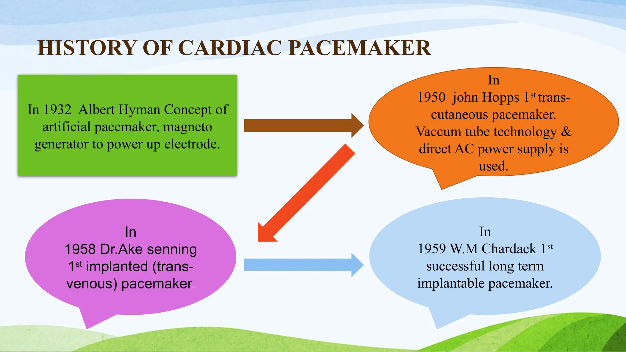
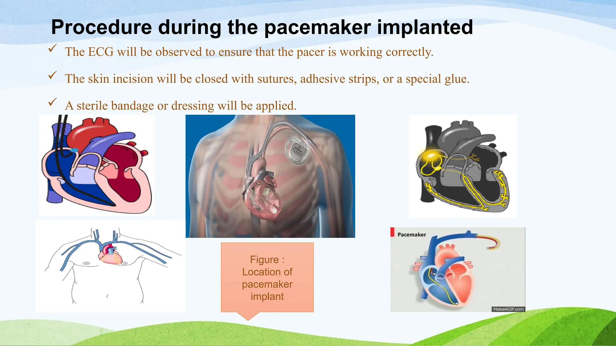
CARDIAC
PACEMAKER
Presenter
M. Sundari
Second Year, M.Sc. Nursing,
CON, MMC, Chennai.
INTRODUCTION
• A pacemaker is a small device that's placed in the chest or abdomen to
help control abnormal heart rhythms. This device uses electrical pulses
to prompt the heart to beat at a normal rate.
• Pacemakers are used to treat arrhythmias and their symptoms such as
fatigue, shortness of breath or fainting. severe arrythmias can damage the
body’s vital organ and may causes loss of consciousness or death.
• A pacemaker can relieve some arrhythmia symptoms, such as fatigue and
fainting. A pacemaker also can help a person who has abnormal heart
rhythms resume a more active lifestyle.
DEFINITION
A pacemaker is a small electronic device implanted under the skin that
sends electrical impulses to the heart muscle to maintain a suitable heart
rate and to prevent slow heart rates.
A pacemaker (or artificial pacemaker) is a medical device that uses
electrical impulses, delivered by electrodes contracting the heart muscles,
to regulate the beating of the heart.
PURPOSE
The primary purpose of a pacemaker is to initiate and maintain an adequate
heart rate, either because the heart's natural pacemaker is not fast enough,
or there is a block in the heart's electrical conduction system.
HISTORY OF CARDIAC PACEMAKER
In 1932 Albert Hyman Concept of
artificial pacemaker, magneto
generator to power up electrode.
In
1950 john Hopps 1st
trans-
cutaneous pacemaker.
Vaccum tube technology &
direct AC power supply is
used.
In
1958 Dr.Ake senning
1st
implanted (trans-
venous) pacemaker.
In
1959 W.M Chardack 1st
successful long term
implantable pacemaker.
INDICATION
Indication for temporary pacing:
 Maintenance of adequate heart rate and rhythm during special circumstances
such as surgery and post operative recovery.
 Cardiac catheterization or coronary angioplasty.
 During drug therapy that may causes bradycardia.
 Before implantation of a permanent pacemaker.
 As prophylaxis after open heart surgery.
 Acute anterior MI with heart block.
 Acute inferior MI with symptomatic bradycardia.
Indication for permanent pacing:
 Acquired Av block
 Second degree, Third degree and Bundle branch block.
 Cardiomyopathy – Dilated, Hypertrophic
 Heart Failure
 Hypersensitive carotid sinus syndrome
 SA node dysfunction
 Tachydysrhythmias
 Atrial fibrillation.
 Ablation
 Acute myocardial infarction
 Autonomic nervous system failure
 Cardiac surgery
 Drug toxicity ( antidysrhythmic)
 Electrolyte imbalance
 Myocardial ischemia
TYPES OF PACEMAKER
1. Temporary Pacemaker 2. Permanent Pacemaker
CARDIAC PACEMAKER DESIGN
They are packaged in hermetically (airtight) sealed metal packages.
 Titanium
 Stainless steel
Power supply: Currently Lithium Iodide batteries are used
 Increased life time
 Open circuit voltage of 2.8 V.
 Highly reliable
 Relatively High source resistance is a major limitation
Timing Circuit : Advanced pacemakers have timing circuits to determine
when a stimulus should be applied to the heart.
Output Circuit/Pulse Generator: Produces the actual electrical stimulus
that is applied to the heart.
• Generates an electrical stimulus pulse that has been optimized for
stimulating the myocardium through the electrode system that is being
applied with the generator.
• Constant-voltage or constant-current amplitude pulses are the two usual
types of stimuli produced by the output circuit.
Output Circuit : Constant-voltage amplitude pulses are typically in the
range of 5.0 to 5.5 V with a duration of 500 to 600 µs. Pulse rate range 70
to 90 beats per minutes.
Lead Wires : Must be mechanically strong. Must maintain good electrical
insulation to prevent the possibility of shunting important stimulating
current away from its intended point of application on the heart.
Electrodes : Can be placed on the external surface of the heart (epicardial
electrodes). Made of materials that do not dissolve during long term
implantation.
Procedure during the pacemaker implanted
 The ECG will be observed to ensure that the pacer is working correctly.
 The skin incision will be closed with sutures, adhesive strips, or a special glue.
 A sterile bandage or dressing will be applied.
Figure :
Location of
pacemaker
implant
TEMPORARY PACEMAKER
 Temporary pacemaker is one that has the power source outside the body.
 This is usually instituted to provide immediate stabilization prior to permanent
pacemaker placement or to provide pacemaker support when a bradycardia is
precipitated by what is presumed to be a transient event such as ischemia or drug
toxicity.
 Temporary pacing is usually achieved by the transvenous insertion of an
electrode catheter with the catheter positioned in the right ventricular apex and
attached to an external generator. This procedure is associated with a small risk
of cardiac perforation, infection at the insertion site, and thromboembolism; the
risk of the latter two complications increases markedly if the pacing wire is left
in place for 48 h.
TEMPORARY PACING
TRANSVENOUS
PACING
EPICARDIAL
TRANSCUTANEOUS
TRANSCUTANEOUS PACING
 Transcutaneous pacing (TCP), also called external pacing, is recommended for
the initial stabilization of hemodynamically significant bradycardias of all types.
The procedure is performed by placing two pacing pads on the patient's chest,
either in the anterior/lateral position or the anterior/posterior position.
 The rescuer selects the pacing rate, and gradually increases the pacing current
with a corresponding pulse.
 Pacing artifact on the ECG and severe muscle twitching may make this
determination difficult. External pacing should not be relied upon for an
extended period of time.
 It is an emergency procedure that acts as a bridge until transvenous pacing or
other therapies can be applied.
EPICARDIAL PACING
Temporary epicardial pacing is used during open heart surgery should the
surgical procedure create atrio-ventricular block. The electrodes are placed in
contact with the outer wall of the ventricle (epicardium) to maintain
satisfactory cardiac output until a temporary transvenous electrode has been
inserted.
TRANSVENOUS PACING
 Transvenous pacing, when used for temporary pacing, is an alternative to
transcutaneous pacing. A pacemaker wire is placed into a vein, under
sterile conditions, and then passed into either the right atrium or right
ventricle. The pacing wire is then connected to an external pacemaker
outside the body.
 Transvenous pacing is often used as a bridge to permanent pacemaker
placement. It can be kept in place until a permanent pacemaker is
implanted or until there is no longer a need for a pacemaker and then it is
removed.
PERMANENT PACING INTRODUCTION
 This mode of pacing is instituted for persistent or intermittent
symptomatic bradycardia not related to a self-limiting precipitating factor
or for documented intranodal second- or third-degree AV block.
 A permanent pacemaker is one that is implanted totally within the body.
 Permanent pacing leads are usually inserted trans venously through the
subclavian or cephalic vein with the leads positioned in the right atrial
appendage for atrial pacing and the right ventricular apex for ventricular
pacing.
 The leads are then attached to the pulse generator, which is inserted into a
subcutaneous pocket below the clavicle.
 Most pacemaker generators are powered by lithium batteries. The life
expectancy of the generator is related to
1. voltage output required for capture,
2. requirement for incessant or intermittent pacing
3. number of cardiac chambers paced.
Life expectancy of the simple ventricular demand pacemaker can exceed 10
years.
PERMANENT
PACING
PERCUSSIVE PACING
SINGLE CHAMBER
RATE RESPONSIVE
PACEMAKER
SUB-CLAVICULAR
PACING
DOUBLE CHAMBER
INTRACARDIAL
PERCUSSIVE PACING
 Percussive pacing, also known as transthoracic mechanical pacing, is the
use of the closed fist, usually on the left lower edge of the sternum over
the right ventricle in the vena cava, striking from a distance of 20 – 30 cm
to induce a ventricular beat (the British Journal of Anesthesia suggests this
must be done to raise the ventricular pressure to 10 - 15mmHg to induce
electrical activity). This is an old procedure used only as a life saving
means until an electrical pacemaker is brought to the patient.
SUB CLAVICULAR PACING
 Permanent pacing with an implantable pacemaker involves transvenous
placement of one or more pacing electrodes within a chamber, or chambers, of
the heart, while the pacemaker is implanted inside the skin under the clavicle.
 The procedure is performed by incision of a suitable vein into which the
electrode lead is inserted and passed along the vein, through the valve of the
heart, until positioned in the chamber.
 The procedure is facilitated by fluoroscopy which enables the physician to
view the passage of the electrode lead. After satisfactory lodgment of the
electrode is confirmed, the opposite end of the electrode lead is connected to
the pacemaker generator.
INTRACARDIAL
 Intracardial pacemakers are placed inside the heart. They are some 10
percent the size of a sub-claviculars. Such pacemakers do not require leads
to be connected to in the heart muscle from the distant pacemaker device.
 Intracardial are implanted into the heart using a steerable catheter fed into
the femoral vein via an incision in the inner thigh. The device cannot be
seen or felt beneath the skin.
 No leads extend to break or detach, eliminating the need for traditional
activity restrictions. The pacemaker can be retrieved in the same manner
in which it was implanted.
There are three basic types of permanent pacemakers, classified according to
the number of chambers involved and their basic operating mechanism:
Single-chamber pacemaker - In this type, only one pacing lead is placed
into a chamber of the heart, either the atrium or the ventricle.
Dual-chamber pacemaker - Here, wires are placed in two chambers of the
heart. One lead paces the atrium and one paces the ventricle. This type more
closely resembles the natural pacing of the heart by assisting the heart in
coordinating the function between the atria and ventricles.
Rate-responsive pacemaker - This pacemaker has sensors that detect
changes in the patient's physical activity and automatically adjust the pacing
rate to fulfill the body's metabolic needs
Pacemaker Modes
There are two basic types of pacemakers
1. Fixed Rate (non-demand or asynchronous)
Fixed rate pacemakers are designed to fire constantly at a preset rate without regard to
the electrical activity of the client’s heart. This mode of pacing is appropriate in the
absence of any electrical activity, but is dangerous in the presence of an intrinsic
rhythm because of the potential of the pacemaker to fire during the vulnerable period of
repolarization and initiate lethal ventricular dysrhythmias.
2. Demand pacemakers
Demand Pacemakers contains a device that senses the heart’s electrical activity and
fires at a preset rate only when the heart’s electrical activity drops below a
predetermined rate.
CLASSIFICATION SYSTEM FOR PACEMAKERS
FIRST LETTER- CHAMBER PACED
Indicates which chamber of the heart wiil be stimulated
 V = Ventricle
 A = Atrium
 D = Dual chamber (both atria and ventricles stimulated)
SECOND LETTER- CHAMBER SENSED
Indicates the chambers of the heart in which the lead is capable of
recognizing intrinsic electrical activity:
 V = Ventricle
 A = Atrium
 D = Dual chamber
 O = No sensing capability
THIRD LETTER- MODE OF RESPONSE
Indicates how the pacemaker will act based on the information it senses:
 T = Triggered (may have energy output triggered)
 I = Inhibited (pacing output inhibited by intrinsic activity)
 D = Dual chamber (may be either inhibiting or triggering of both
chambers)
FOURTH LETTER- PROGRAMMABLE FUNCTIONS
Indicates ability to change function once the pacemaker has been implanted:
 P = Programmable for one or two functions
 M = Multiprogrammable ability to change functions other than the rate or
output
FIFTH LETTER- TACHYDYSRHYTHMIC FUNCTIONS
Indicate specific methods of interrupting tachydysrhythmias.
 B = Bursts of pacing
 N = Normal rate competition
 S = Scanning
PACEMAKER FUNCTION
A simple demand pacing system works in the following manner;
The cardiac cycle normally begins with the client’s own beat. The
pacemaker’s sensor sense whether the intrinsic beat has occurred; if not, the
pacer sends out an impulse to begin myocardial depolarization through a
pulse generator. The impulse generator is said to “capture” the myocardium
and thereby maintain the heart rhythm.
For a predetermined amount of time after the pacemaker impulse, the
pacemaker cannot sense incoming signals. This feature prevents the pacer
from sensing its own generated electrical current and from acting again.
PACEMAKER FAILURE
Malfunctions can occur in the pacemaker’s sensor or pulse generator’
1. Failure to Sense
An ability of the sensor to detect the client’s intrinsic beats; as a result, the
pacemaker sends out impulses too early. A failure may be due to improper position
of the catheter, tip or lead dislodgement, battery failure, the sensitivity being set too
low, or a fractured wire in a catheter.
2. Failure to Pace
A malfunction of the pulse generator. Component failure to discharge( Pace) can be
due to battery failure, lead dislodgement, fracture of the lead wire inside the
catheter, disconnections between catheter and generator, or a sensing malfunction.
3. Failure to Capture
A disorder in the pacemaker electrodes; the impulse do not generate
depolarization. This complication can result from low voltage, battery failure,
faulty connections between the pulse generator and catheter, improper
position of the catheter, catheter wire fracture, fibrosis at the catheter tip.
Clinical manifestations associated with pacemaker malfunction include;
 Syncope
 Bradycardia/ tachycardia
 Palpitations
PATIENT EVALUATION
PREOPERATIVE
Indication for implanted pacemaker/ICD
Sustained /intermittent tachyarrhythmia or bradyarrhythmia.
Heart failure
Type of device:
 Clinical indication of the device
 Appraisal of patient’s degree of dependence on the devices(for patient
requiring pacing for bradyarrhythmia)
Assessment of device function,
 A preoperative history of vertigo, pre syncope, or syncope in a patient with
a pacemaker could reflect pacemaker dysfunction.
 A 10% decrease in heart rate from the initial heart rate setting may reflect
battery depletion.
 An irregular heart rate could indicate competition of the pulse generator
with the patient's intrinsic heart rate or failure of the pulse generator to
sense R waves.
Continue antiarrhythmic drug and other cardiac drugs as mandated
Consider Electromagnetic and Mechanical Interference (EMI)
MONITORING
Manual pulse palpation
Pulse oximetry
Continuous ECG monitoring
Auscultation of heart sounds
Intra-arterial blood pressure
INVESTIGATION
 Routine investigation along with s. electrolytes/acid –base analysis.
 Chest x-ray:
 Location and external condition of pacemaker electrodes.
 If bi-ventricular pace maker(position of coronary sinus lead when insertion of
central line or PA catheter planned ).
Management of a Patient - Intra Operatively
Application of magnet over pulse generator of pace maker…no longer an
acceptable practice.
 Results in asynchronous fixed rate.(chance of R on T phenomenon)
But Difficult to assess the effect of magnet on cardioverter- defibrillator.
Transcutaneous pacing is always kept ready.
Rate responsive pacemakers should have rate responsive mode disabled
before surgery.
Central venous catheterization: chance of pacing leads dislodgement.
Factors affecting the pacing threshold
Increase threshold Decrease threshold
1-4 wks after implantation Increases catecholamines
MI Stress, anxiety
Hypothermia/Hypothyroidism Sympathomimetic drugs
Hyperkaliemia/acidosis/alkalosis Anticholinergics
Antiarrhythmics(class 1a,1b,1c) Glucocorticoids
Severe hypoxia/hyperglycemia Hyperthyroidism
Inhalational –local anesthesia Hypermetabolic status
POST OPERATIVE MANAGEMENT
Interrogating the device & restoring baseline settings( like anti tachycardia
therapy).
Cardiac rate ,rhythm monitoring continuously, Hypothermia prevention.
Reprogramming.
NURSING MANAGEMENT OF PATIENTS WITH
PACEMAKERS
ASSESSMENT
Assess the client for subjective clinical manifestations of dysrhythmias and
alterations in cardiac output;
 Palpitations
 Syncope
 Fatigue
 Shortness of breath
 Chest pain
 Skipped beats felt in the chest
 The client may also feel anxiety, nervousness, fear, sleeplessness, or
uncertainty or hopelessness.
Objective clinical manifestations may include;
Diaphoresis, pallor or cyanosis, variations in radial and apical pulse, rhythm
changes, hypotension, crackles, and decreased mental acuity.
Explain the purpose of the pacemaker and the experience of having a
pacemaker inserted to the client and family. A preoperative ECG is obtained,
and a patent IV line is maintained. Prophylactic antibiotics may be given.
After insertion, monitor vital signs and pacemaker function. Initially instruct
the client to avoid excessive extension or abduction of the arm on the
operative side. Perform passive range of motion exercises on the arm.
Obtain paced and non-paced ECGs. The location of pacemaker electrodes is
determined by X ray. The model and serial number of the pulse generator
and leads, along with the date of implantation and programmed functions of
the initial implant are recorded.
PATIENT EDUCATION
WOUND CARE
1. Assess your wound daily and keep the incision clean and dry until it
heals.
2. Report any fever, redness, drainage, warmth and discoloration, or
swelling to the physician.
3. Avoid constrictive clothing which puts excessive pressure on the wound
and the pulse generator.
4. Avoid extensive “toying” with the pulse generator because this may
cause pacemaker malfunction and local skin inflammation.
PACEMAKER MANAGEMENT
1. Measure pulse rate as instructed by the physician in the wrist and neck.
2. Notify the physician if pulse rate is slower than the set rate, also report
sensations of feeling heart “racing”, beating irregularly, fatigue or
dizziness.
3. Avoid being near the areas with high voltage, magnetic force fields or
radiation that can cause pacemaker problems.
4. Avoid being near large running motors and standing near high tension
wires, power plants, radio transmitters, large industrial magnets and arc
welding machines. Riding in a car is safe, but do not bring the pacemaker
to within 6-12 inches of the distributor coil of a running engine.
5. Client can operate televisions microwave oven, radio.
6. At all times carry a pacemaker identity card and wear a medical alert
bracelet.
7. Avoid activity that might damage the pulse generator, such as playing
football.
8. If radiation therapy has been prescribed to the area in which the pulse
generator was implanted, it must be relocated.
9. Do not lift more than 2-4 kg for the first 6 weeks after surgery.
10. Do not move your arms and shoulders vigorously for the first 6 weeks.
Normal activities can be resumed in 6 weeks.
11. Follow up should be done periodically.
COMPLICATIONS
Complications of permanent pacing
Causes of pacing system malfunction include undersensing, oversensing,
loss of capture, loss of output, inappropriate rate, inappropriate lead
position, inappropriate mode, extracardiac stimulation, true pulse generator
failure, pacemaker-mediated tachycardia (in dual-chamber pacemakers
with DDD, VDD, and DDDR modes), pacemaker syndrome and
inappropriate fiddling of the pulse generator by the patient. Most pacing
system malfunctions are benign, but some can be life-threatening.
 Complications related to venous access include pneumothorax,
hemothorax, and air embolism.
Lead-related complications include perforation, dislodgment,
diaphragmatic stimulation, and malposition (including passage into the
left side of the heart via a septal defect).Cardiac tamponade, usually due
to chamber perforation, should be suspected whenever hypotension
occurs.
Local pocket-related complications include hematoma, wound pain,
pocket erosion, and infection.
Pacemaker infection ranges from mild local pain and erythema to life-
threaten ingsepticaemia. The most common pathogens are coagulase-
negative staphylococci, Staphylococcus aureus Gram-negative enteric
bacilli and mixed infections.
Delayed complications of permanent pacing leads include venous
thrombosis, exit block, insulation failure, and conductor fracture. Late
lead damage may be reduced by use of axillary or cephalic venous
access.
Most modern pulse generators have an expected longevity of 5-9 years
and unexpected pulse generator (electrical) failure is rare.
Lead-related problems (increased thresholds, decreased impedance)
resulting in increased current drain are the most common causes of
premature battery depletion.
Lithium-iodine batteries used in current pulse generators are not
rechargeable and surgical replacement of the entire generator is required.
Complications of temporary pacing
• Immediate complications include:
• Ventricular tachycardia or fibrillation
• Arterial puncture
• Pneumothorax
• Brachial plexus injury
• Late complications include:
• Ventricular arrhythmias
• Septicemia (especially staphylococcal infection)
• Wrong position requiring repositioning
PACEMAKER SYNDROME
Pacemaker syndrome refers to the occurrence of symptoms relating to the loss of
atrioventricular (AV) synchrony in patients with a pacemaker.
 Ventricular pacing has been shown to sacrifice the atrial contribution to ventricular
output.
 In some cases, atrial contraction occurs against closed AV valves, producing reverse
blood flow.
 In response to decreased cardiac output, total peripheral resistance is usually increased
in order to maintain blood pressure but does not increase in some patients, resulting in
decreased blood pressure.
 This combination of decreased cardiac output with a loss of the usual compensatory
increase in total peripheral resistance contributes to the development of pacemaker
syndrome.
Pacemaker Circuit
• A pacemaker has three components :
 This is kept in small metal case and the circuit is connected to a battery. The
circuit generates the impulse at a specific time.
 These insulated wires carry the impulse to the heart.
 This is kept in the hospital or clinic to program the pacemaker and to
adjust the setting of impulse amplitudes and frequency. The programmer
is a specialized computer with a hardware and software interface.
1.Pacemaker electronic circuit
2. Pacing leds
3. Programmer
cardiac pacemaker internal cardiac device

cardiac pacemaker internal cardiac device

  • 1.
    CARDIAC PACEMAKER Presenter M. Sundari Second Year,M.Sc. Nursing, CON, MMC, Chennai.
  • 2.
    INTRODUCTION • A pacemakeris a small device that's placed in the chest or abdomen to help control abnormal heart rhythms. This device uses electrical pulses to prompt the heart to beat at a normal rate. • Pacemakers are used to treat arrhythmias and their symptoms such as fatigue, shortness of breath or fainting. severe arrythmias can damage the body’s vital organ and may causes loss of consciousness or death. • A pacemaker can relieve some arrhythmia symptoms, such as fatigue and fainting. A pacemaker also can help a person who has abnormal heart rhythms resume a more active lifestyle.
  • 3.
    DEFINITION A pacemaker isa small electronic device implanted under the skin that sends electrical impulses to the heart muscle to maintain a suitable heart rate and to prevent slow heart rates. A pacemaker (or artificial pacemaker) is a medical device that uses electrical impulses, delivered by electrodes contracting the heart muscles, to regulate the beating of the heart.
  • 4.
    PURPOSE The primary purposeof a pacemaker is to initiate and maintain an adequate heart rate, either because the heart's natural pacemaker is not fast enough, or there is a block in the heart's electrical conduction system.
  • 5.
    HISTORY OF CARDIACPACEMAKER In 1932 Albert Hyman Concept of artificial pacemaker, magneto generator to power up electrode. In 1950 john Hopps 1st trans- cutaneous pacemaker. Vaccum tube technology & direct AC power supply is used. In 1958 Dr.Ake senning 1st implanted (trans- venous) pacemaker. In 1959 W.M Chardack 1st successful long term implantable pacemaker.
  • 6.
    INDICATION Indication for temporarypacing:  Maintenance of adequate heart rate and rhythm during special circumstances such as surgery and post operative recovery.  Cardiac catheterization or coronary angioplasty.  During drug therapy that may causes bradycardia.  Before implantation of a permanent pacemaker.  As prophylaxis after open heart surgery.  Acute anterior MI with heart block.  Acute inferior MI with symptomatic bradycardia.
  • 7.
    Indication for permanentpacing:  Acquired Av block  Second degree, Third degree and Bundle branch block.  Cardiomyopathy – Dilated, Hypertrophic  Heart Failure  Hypersensitive carotid sinus syndrome  SA node dysfunction  Tachydysrhythmias  Atrial fibrillation.
  • 8.
     Ablation  Acutemyocardial infarction  Autonomic nervous system failure  Cardiac surgery  Drug toxicity ( antidysrhythmic)  Electrolyte imbalance  Myocardial ischemia
  • 9.
    TYPES OF PACEMAKER 1.Temporary Pacemaker 2. Permanent Pacemaker
  • 10.
    CARDIAC PACEMAKER DESIGN Theyare packaged in hermetically (airtight) sealed metal packages.  Titanium  Stainless steel Power supply: Currently Lithium Iodide batteries are used  Increased life time  Open circuit voltage of 2.8 V.  Highly reliable  Relatively High source resistance is a major limitation
  • 11.
    Timing Circuit :Advanced pacemakers have timing circuits to determine when a stimulus should be applied to the heart. Output Circuit/Pulse Generator: Produces the actual electrical stimulus that is applied to the heart. • Generates an electrical stimulus pulse that has been optimized for stimulating the myocardium through the electrode system that is being applied with the generator. • Constant-voltage or constant-current amplitude pulses are the two usual types of stimuli produced by the output circuit.
  • 12.
    Output Circuit :Constant-voltage amplitude pulses are typically in the range of 5.0 to 5.5 V with a duration of 500 to 600 µs. Pulse rate range 70 to 90 beats per minutes. Lead Wires : Must be mechanically strong. Must maintain good electrical insulation to prevent the possibility of shunting important stimulating current away from its intended point of application on the heart. Electrodes : Can be placed on the external surface of the heart (epicardial electrodes). Made of materials that do not dissolve during long term implantation.
  • 13.
    Procedure during thepacemaker implanted  The ECG will be observed to ensure that the pacer is working correctly.  The skin incision will be closed with sutures, adhesive strips, or a special glue.  A sterile bandage or dressing will be applied. Figure : Location of pacemaker implant
  • 14.
    TEMPORARY PACEMAKER  Temporarypacemaker is one that has the power source outside the body.  This is usually instituted to provide immediate stabilization prior to permanent pacemaker placement or to provide pacemaker support when a bradycardia is precipitated by what is presumed to be a transient event such as ischemia or drug toxicity.  Temporary pacing is usually achieved by the transvenous insertion of an electrode catheter with the catheter positioned in the right ventricular apex and attached to an external generator. This procedure is associated with a small risk of cardiac perforation, infection at the insertion site, and thromboembolism; the risk of the latter two complications increases markedly if the pacing wire is left in place for 48 h.
  • 15.
  • 16.
    TRANSCUTANEOUS PACING  Transcutaneouspacing (TCP), also called external pacing, is recommended for the initial stabilization of hemodynamically significant bradycardias of all types. The procedure is performed by placing two pacing pads on the patient's chest, either in the anterior/lateral position or the anterior/posterior position.  The rescuer selects the pacing rate, and gradually increases the pacing current with a corresponding pulse.  Pacing artifact on the ECG and severe muscle twitching may make this determination difficult. External pacing should not be relied upon for an extended period of time.  It is an emergency procedure that acts as a bridge until transvenous pacing or other therapies can be applied.
  • 17.
    EPICARDIAL PACING Temporary epicardialpacing is used during open heart surgery should the surgical procedure create atrio-ventricular block. The electrodes are placed in contact with the outer wall of the ventricle (epicardium) to maintain satisfactory cardiac output until a temporary transvenous electrode has been inserted.
  • 18.
    TRANSVENOUS PACING  Transvenouspacing, when used for temporary pacing, is an alternative to transcutaneous pacing. A pacemaker wire is placed into a vein, under sterile conditions, and then passed into either the right atrium or right ventricle. The pacing wire is then connected to an external pacemaker outside the body.  Transvenous pacing is often used as a bridge to permanent pacemaker placement. It can be kept in place until a permanent pacemaker is implanted or until there is no longer a need for a pacemaker and then it is removed.
  • 19.
    PERMANENT PACING INTRODUCTION This mode of pacing is instituted for persistent or intermittent symptomatic bradycardia not related to a self-limiting precipitating factor or for documented intranodal second- or third-degree AV block.  A permanent pacemaker is one that is implanted totally within the body.  Permanent pacing leads are usually inserted trans venously through the subclavian or cephalic vein with the leads positioned in the right atrial appendage for atrial pacing and the right ventricular apex for ventricular pacing.
  • 20.
     The leadsare then attached to the pulse generator, which is inserted into a subcutaneous pocket below the clavicle.  Most pacemaker generators are powered by lithium batteries. The life expectancy of the generator is related to 1. voltage output required for capture, 2. requirement for incessant or intermittent pacing 3. number of cardiac chambers paced. Life expectancy of the simple ventricular demand pacemaker can exceed 10 years.
  • 21.
    PERMANENT PACING PERCUSSIVE PACING SINGLE CHAMBER RATERESPONSIVE PACEMAKER SUB-CLAVICULAR PACING DOUBLE CHAMBER INTRACARDIAL
  • 22.
    PERCUSSIVE PACING  Percussivepacing, also known as transthoracic mechanical pacing, is the use of the closed fist, usually on the left lower edge of the sternum over the right ventricle in the vena cava, striking from a distance of 20 – 30 cm to induce a ventricular beat (the British Journal of Anesthesia suggests this must be done to raise the ventricular pressure to 10 - 15mmHg to induce electrical activity). This is an old procedure used only as a life saving means until an electrical pacemaker is brought to the patient.
  • 23.
    SUB CLAVICULAR PACING Permanent pacing with an implantable pacemaker involves transvenous placement of one or more pacing electrodes within a chamber, or chambers, of the heart, while the pacemaker is implanted inside the skin under the clavicle.  The procedure is performed by incision of a suitable vein into which the electrode lead is inserted and passed along the vein, through the valve of the heart, until positioned in the chamber.  The procedure is facilitated by fluoroscopy which enables the physician to view the passage of the electrode lead. After satisfactory lodgment of the electrode is confirmed, the opposite end of the electrode lead is connected to the pacemaker generator.
  • 24.
    INTRACARDIAL  Intracardial pacemakersare placed inside the heart. They are some 10 percent the size of a sub-claviculars. Such pacemakers do not require leads to be connected to in the heart muscle from the distant pacemaker device.  Intracardial are implanted into the heart using a steerable catheter fed into the femoral vein via an incision in the inner thigh. The device cannot be seen or felt beneath the skin.  No leads extend to break or detach, eliminating the need for traditional activity restrictions. The pacemaker can be retrieved in the same manner in which it was implanted.
  • 25.
    There are threebasic types of permanent pacemakers, classified according to the number of chambers involved and their basic operating mechanism: Single-chamber pacemaker - In this type, only one pacing lead is placed into a chamber of the heart, either the atrium or the ventricle. Dual-chamber pacemaker - Here, wires are placed in two chambers of the heart. One lead paces the atrium and one paces the ventricle. This type more closely resembles the natural pacing of the heart by assisting the heart in coordinating the function between the atria and ventricles. Rate-responsive pacemaker - This pacemaker has sensors that detect changes in the patient's physical activity and automatically adjust the pacing rate to fulfill the body's metabolic needs
  • 26.
    Pacemaker Modes There aretwo basic types of pacemakers 1. Fixed Rate (non-demand or asynchronous) Fixed rate pacemakers are designed to fire constantly at a preset rate without regard to the electrical activity of the client’s heart. This mode of pacing is appropriate in the absence of any electrical activity, but is dangerous in the presence of an intrinsic rhythm because of the potential of the pacemaker to fire during the vulnerable period of repolarization and initiate lethal ventricular dysrhythmias. 2. Demand pacemakers Demand Pacemakers contains a device that senses the heart’s electrical activity and fires at a preset rate only when the heart’s electrical activity drops below a predetermined rate.
  • 27.
    CLASSIFICATION SYSTEM FORPACEMAKERS FIRST LETTER- CHAMBER PACED Indicates which chamber of the heart wiil be stimulated  V = Ventricle  A = Atrium  D = Dual chamber (both atria and ventricles stimulated)
  • 28.
    SECOND LETTER- CHAMBERSENSED Indicates the chambers of the heart in which the lead is capable of recognizing intrinsic electrical activity:  V = Ventricle  A = Atrium  D = Dual chamber  O = No sensing capability
  • 29.
    THIRD LETTER- MODEOF RESPONSE Indicates how the pacemaker will act based on the information it senses:  T = Triggered (may have energy output triggered)  I = Inhibited (pacing output inhibited by intrinsic activity)  D = Dual chamber (may be either inhibiting or triggering of both chambers)
  • 30.
    FOURTH LETTER- PROGRAMMABLEFUNCTIONS Indicates ability to change function once the pacemaker has been implanted:  P = Programmable for one or two functions  M = Multiprogrammable ability to change functions other than the rate or output FIFTH LETTER- TACHYDYSRHYTHMIC FUNCTIONS Indicate specific methods of interrupting tachydysrhythmias.  B = Bursts of pacing  N = Normal rate competition  S = Scanning
  • 31.
    PACEMAKER FUNCTION A simpledemand pacing system works in the following manner; The cardiac cycle normally begins with the client’s own beat. The pacemaker’s sensor sense whether the intrinsic beat has occurred; if not, the pacer sends out an impulse to begin myocardial depolarization through a pulse generator. The impulse generator is said to “capture” the myocardium and thereby maintain the heart rhythm. For a predetermined amount of time after the pacemaker impulse, the pacemaker cannot sense incoming signals. This feature prevents the pacer from sensing its own generated electrical current and from acting again.
  • 32.
    PACEMAKER FAILURE Malfunctions canoccur in the pacemaker’s sensor or pulse generator’ 1. Failure to Sense An ability of the sensor to detect the client’s intrinsic beats; as a result, the pacemaker sends out impulses too early. A failure may be due to improper position of the catheter, tip or lead dislodgement, battery failure, the sensitivity being set too low, or a fractured wire in a catheter. 2. Failure to Pace A malfunction of the pulse generator. Component failure to discharge( Pace) can be due to battery failure, lead dislodgement, fracture of the lead wire inside the catheter, disconnections between catheter and generator, or a sensing malfunction.
  • 33.
    3. Failure toCapture A disorder in the pacemaker electrodes; the impulse do not generate depolarization. This complication can result from low voltage, battery failure, faulty connections between the pulse generator and catheter, improper position of the catheter, catheter wire fracture, fibrosis at the catheter tip. Clinical manifestations associated with pacemaker malfunction include;  Syncope  Bradycardia/ tachycardia  Palpitations
  • 34.
    PATIENT EVALUATION PREOPERATIVE Indication forimplanted pacemaker/ICD Sustained /intermittent tachyarrhythmia or bradyarrhythmia. Heart failure Type of device:  Clinical indication of the device  Appraisal of patient’s degree of dependence on the devices(for patient requiring pacing for bradyarrhythmia)
  • 35.
    Assessment of devicefunction,  A preoperative history of vertigo, pre syncope, or syncope in a patient with a pacemaker could reflect pacemaker dysfunction.  A 10% decrease in heart rate from the initial heart rate setting may reflect battery depletion.  An irregular heart rate could indicate competition of the pulse generator with the patient's intrinsic heart rate or failure of the pulse generator to sense R waves. Continue antiarrhythmic drug and other cardiac drugs as mandated Consider Electromagnetic and Mechanical Interference (EMI)
  • 36.
    MONITORING Manual pulse palpation Pulseoximetry Continuous ECG monitoring Auscultation of heart sounds Intra-arterial blood pressure
  • 37.
    INVESTIGATION  Routine investigationalong with s. electrolytes/acid –base analysis.  Chest x-ray:  Location and external condition of pacemaker electrodes.  If bi-ventricular pace maker(position of coronary sinus lead when insertion of central line or PA catheter planned ).
  • 38.
    Management of aPatient - Intra Operatively Application of magnet over pulse generator of pace maker…no longer an acceptable practice.  Results in asynchronous fixed rate.(chance of R on T phenomenon) But Difficult to assess the effect of magnet on cardioverter- defibrillator. Transcutaneous pacing is always kept ready. Rate responsive pacemakers should have rate responsive mode disabled before surgery. Central venous catheterization: chance of pacing leads dislodgement.
  • 39.
    Factors affecting thepacing threshold Increase threshold Decrease threshold 1-4 wks after implantation Increases catecholamines MI Stress, anxiety Hypothermia/Hypothyroidism Sympathomimetic drugs Hyperkaliemia/acidosis/alkalosis Anticholinergics Antiarrhythmics(class 1a,1b,1c) Glucocorticoids Severe hypoxia/hyperglycemia Hyperthyroidism Inhalational –local anesthesia Hypermetabolic status
  • 40.
    POST OPERATIVE MANAGEMENT Interrogatingthe device & restoring baseline settings( like anti tachycardia therapy). Cardiac rate ,rhythm monitoring continuously, Hypothermia prevention. Reprogramming.
  • 41.
    NURSING MANAGEMENT OFPATIENTS WITH PACEMAKERS ASSESSMENT Assess the client for subjective clinical manifestations of dysrhythmias and alterations in cardiac output;  Palpitations  Syncope  Fatigue  Shortness of breath  Chest pain  Skipped beats felt in the chest
  • 42.
     The clientmay also feel anxiety, nervousness, fear, sleeplessness, or uncertainty or hopelessness. Objective clinical manifestations may include; Diaphoresis, pallor or cyanosis, variations in radial and apical pulse, rhythm changes, hypotension, crackles, and decreased mental acuity. Explain the purpose of the pacemaker and the experience of having a pacemaker inserted to the client and family. A preoperative ECG is obtained, and a patent IV line is maintained. Prophylactic antibiotics may be given.
  • 43.
    After insertion, monitorvital signs and pacemaker function. Initially instruct the client to avoid excessive extension or abduction of the arm on the operative side. Perform passive range of motion exercises on the arm. Obtain paced and non-paced ECGs. The location of pacemaker electrodes is determined by X ray. The model and serial number of the pulse generator and leads, along with the date of implantation and programmed functions of the initial implant are recorded.
  • 44.
    PATIENT EDUCATION WOUND CARE 1.Assess your wound daily and keep the incision clean and dry until it heals. 2. Report any fever, redness, drainage, warmth and discoloration, or swelling to the physician. 3. Avoid constrictive clothing which puts excessive pressure on the wound and the pulse generator. 4. Avoid extensive “toying” with the pulse generator because this may cause pacemaker malfunction and local skin inflammation.
  • 45.
    PACEMAKER MANAGEMENT 1. Measurepulse rate as instructed by the physician in the wrist and neck. 2. Notify the physician if pulse rate is slower than the set rate, also report sensations of feeling heart “racing”, beating irregularly, fatigue or dizziness. 3. Avoid being near the areas with high voltage, magnetic force fields or radiation that can cause pacemaker problems. 4. Avoid being near large running motors and standing near high tension wires, power plants, radio transmitters, large industrial magnets and arc welding machines. Riding in a car is safe, but do not bring the pacemaker to within 6-12 inches of the distributor coil of a running engine.
  • 46.
    5. Client canoperate televisions microwave oven, radio. 6. At all times carry a pacemaker identity card and wear a medical alert bracelet. 7. Avoid activity that might damage the pulse generator, such as playing football. 8. If radiation therapy has been prescribed to the area in which the pulse generator was implanted, it must be relocated. 9. Do not lift more than 2-4 kg for the first 6 weeks after surgery. 10. Do not move your arms and shoulders vigorously for the first 6 weeks. Normal activities can be resumed in 6 weeks. 11. Follow up should be done periodically.
  • 47.
    COMPLICATIONS Complications of permanentpacing Causes of pacing system malfunction include undersensing, oversensing, loss of capture, loss of output, inappropriate rate, inappropriate lead position, inappropriate mode, extracardiac stimulation, true pulse generator failure, pacemaker-mediated tachycardia (in dual-chamber pacemakers with DDD, VDD, and DDDR modes), pacemaker syndrome and inappropriate fiddling of the pulse generator by the patient. Most pacing system malfunctions are benign, but some can be life-threatening.  Complications related to venous access include pneumothorax, hemothorax, and air embolism.
  • 48.
    Lead-related complications includeperforation, dislodgment, diaphragmatic stimulation, and malposition (including passage into the left side of the heart via a septal defect).Cardiac tamponade, usually due to chamber perforation, should be suspected whenever hypotension occurs. Local pocket-related complications include hematoma, wound pain, pocket erosion, and infection. Pacemaker infection ranges from mild local pain and erythema to life- threaten ingsepticaemia. The most common pathogens are coagulase- negative staphylococci, Staphylococcus aureus Gram-negative enteric bacilli and mixed infections.
  • 49.
    Delayed complications ofpermanent pacing leads include venous thrombosis, exit block, insulation failure, and conductor fracture. Late lead damage may be reduced by use of axillary or cephalic venous access. Most modern pulse generators have an expected longevity of 5-9 years and unexpected pulse generator (electrical) failure is rare. Lead-related problems (increased thresholds, decreased impedance) resulting in increased current drain are the most common causes of premature battery depletion. Lithium-iodine batteries used in current pulse generators are not rechargeable and surgical replacement of the entire generator is required.
  • 50.
    Complications of temporarypacing • Immediate complications include: • Ventricular tachycardia or fibrillation • Arterial puncture • Pneumothorax • Brachial plexus injury • Late complications include: • Ventricular arrhythmias • Septicemia (especially staphylococcal infection) • Wrong position requiring repositioning
  • 51.
    PACEMAKER SYNDROME Pacemaker syndromerefers to the occurrence of symptoms relating to the loss of atrioventricular (AV) synchrony in patients with a pacemaker.  Ventricular pacing has been shown to sacrifice the atrial contribution to ventricular output.  In some cases, atrial contraction occurs against closed AV valves, producing reverse blood flow.  In response to decreased cardiac output, total peripheral resistance is usually increased in order to maintain blood pressure but does not increase in some patients, resulting in decreased blood pressure.  This combination of decreased cardiac output with a loss of the usual compensatory increase in total peripheral resistance contributes to the development of pacemaker syndrome.
  • 52.
    Pacemaker Circuit • Apacemaker has three components :  This is kept in small metal case and the circuit is connected to a battery. The circuit generates the impulse at a specific time.  These insulated wires carry the impulse to the heart.  This is kept in the hospital or clinic to program the pacemaker and to adjust the setting of impulse amplitudes and frequency. The programmer is a specialized computer with a hardware and software interface. 1.Pacemaker electronic circuit 2. Pacing leds 3. Programmer