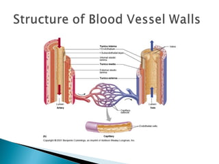
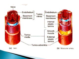
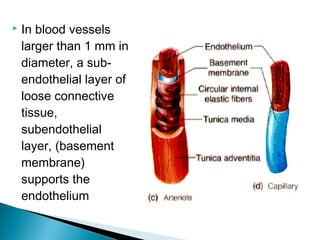
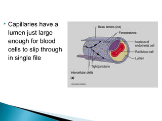
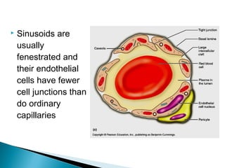
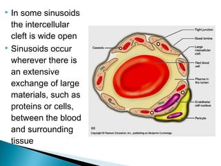
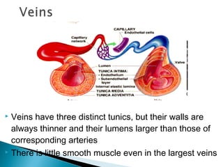
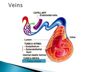
The document summarizes the structure and function of the cardiovascular system. It describes the three main types of blood vessels - arteries, capillaries, and veins - and their roles in circulating blood throughout the body. Arteries carry oxygenated blood away from the heart, branching into smaller vessels. Capillaries allow for exchange of oxygen, nutrients, waste at the cellular level. Veins then collect deoxygenated blood and return it to the heart. The document provides detailed information on the layers, tissue composition, and regulatory mechanisms of different sections of the cardiovascular system.
The blood vessels are the components of the circulatory system that transport blood throughout the human body. These vessels transport blood cells, nutrients, and oxygen to the tissues of the body. They also take waste and carbon dioxide away from the tissues.
BONE – AN INTRODUCTION
A bone is a rigid organ that constitutes part of the vertebrate skeleton.
There are around 270 to 300+ bones in Infants which gets reduced to 206 bones in adults.
Bones are dynamic structures that are undergoing constant change and remodelling in
response to the ever-changing environment.
Bones support and protect the various organs of the body, produce red and white blood cells,
store minerals, provide structure and support for the body, and enable mobility.
It has a honeycomb-like matrix internally, which helps to give the bone rigidity.
The largest bone in the body is the femur or thigh-bone, and the smallest is the stapes in
the middle ear.
Blood vessels: Arteries, Veins and CapillariesAmir Rifaat
It is one of the circulatory systems. This explains the roles of arteries, veins and capillaries. It also differentiate between the arteries, veins and capillaries. This slide also explained the pulmonary circuit and systemic curcuit. This is an interesting notes and easy to be understand.
Blood is carried through the body via blood vessels. An artery is a blood vessel that carries blood away from the heart, where it branches into ever-smaller vessels.
Artery and veins, capillaries, arteriole and venules, systemic circulation an...Dr Shahid Alam
Artery and veins, capillaries, arteriole and venules, systemic circulation and pulmonary circulation, blood vessels, heart, chambers of heart, dr shahid alam, shahid alam, doctor shahid alam, shahid, alam
Amazing WTF Facts about animals that will blow your mindUsama Nasir
Amazing WTF Facts about animals that will blow your mind away. These WTF Facts are collected from credible resources and are brought to you by http://www.wtf-facts.net
For more amazing WTF Facts, visit www.wtf-facts.net
Till then enjoy these amazing facts!
Elite Gamespeed | The concept of a game of Rugby is simple: two teams try and gain the most points in 80 minutes by trying to score points in the other team's half.
Blood Vessels
By: Saiyed Falakaara
Assistant Professor
Department of Pharmacy
Sumandeep Vidyapeeth
Introduction
Blood vessels form a closed system of tubes that carry blood away from the heart, transport it to the tissues of the body, and then return it to the heart.
The blood vessels of human body carry blood to every tissue and organ.
Vessels decrease in size as they move away from the heart (arteries and arterioles), ending in the capillaries, and then increase in size as they move towards the heart (venules and veins)
The largest artery in the blood is Aorta.
Types of blood vessels
1. Arteries
2. Arterioles
3. Capillaries
4. Venules
5. veins
Learning Objectives:
Compare and contrast the structure and function
of
Arteries
Veins
Capillaries
ulatory
system
Arteries
Arterioles
Capillaries
Venules
Veins
3 tunics
Lume
The Vessels
Functions:
Distribution of blood
Exchange of materials with tissues
Return of blood to the heart
Structure:
Most have the same basic structure:
– 3 layers surrounding a hollow lumen
The circulatory system transports fluids throughout the body;
it consists of the cardiovascular and lymphatic systems.
The heart and blood vessels make up the blood transportation network, the cardiovascular system.
Through this system, the heart pumps blood through the body’s vast system of blood vessels.
The blood carries nutrients, oxygen, and waste products to and from the cells.
VASCULAR CIRCUITS
The heart consists of two muscular pumps dividing the circulation into two components:
pulmonary circulations
systemic circulations or circuit
Pulmonary Circulation
Rt ventricle propels low O2 blood into the lungs via the pulmonary arteries.
CO2 is exchanged for O2 in the capillaries of the lungs.
Then the O2 -rich blood is returned via the pulmonary veins to the Lft atrium.
This circuit, from the right ventricle through the lungs to the left atrium, is the pulmonary circulation.
Systemic Circulation
Left ventricle propels the O2 -rich blood through systemic arteries (the aorta and its branches),
exchanging O2 and nutrients for CO2 in the remainder of the body’s capillaries.
Low- O2 blood returns to right atrium via systemic veins (tributaries of the superior and inferior vena cava).
This circuit, from left ventricle to right atrium, is the systemic circulation.
The first topic in the practical histology coarse for pharmacy students
In this lecture the student will be able to recognize the histological layers of the circulatory system parts such as veins and arteries and the similarities and differences between each layer
There are three kinds of blood vessels: arteries, veins, and capillaries.
Each of these plays a very specific role in the circulation process/blood circulation.
Arteries transport blood away from the heart.
Veins return blood back toward the heart.
Capillaries surround body cells and tissues to deliver and absorb oxygen, nutrients, and other substances.
The capillaries also connect the branches of arteries and to the branches of veins.
The walls of most blood vessels have three distinct layers: the tunica externa, the tunica media, and the tunica intima.
The French Revolution, which began in 1789, was a period of radical social and political upheaval in France. It marked the decline of absolute monarchies, the rise of secular and democratic republics, and the eventual rise of Napoleon Bonaparte. This revolutionary period is crucial in understanding the transition from feudalism to modernity in Europe.
For more information, visit-www.vavaclasses.com
The Roman Empire A Historical Colossus.pdfkaushalkr1407
The Roman Empire, a vast and enduring power, stands as one of history's most remarkable civilizations, leaving an indelible imprint on the world. It emerged from the Roman Republic, transitioning into an imperial powerhouse under the leadership of Augustus Caesar in 27 BCE. This transformation marked the beginning of an era defined by unprecedented territorial expansion, architectural marvels, and profound cultural influence.
The empire's roots lie in the city of Rome, founded, according to legend, by Romulus in 753 BCE. Over centuries, Rome evolved from a small settlement to a formidable republic, characterized by a complex political system with elected officials and checks on power. However, internal strife, class conflicts, and military ambitions paved the way for the end of the Republic. Julius Caesar’s dictatorship and subsequent assassination in 44 BCE created a power vacuum, leading to a civil war. Octavian, later Augustus, emerged victorious, heralding the Roman Empire’s birth.
Under Augustus, the empire experienced the Pax Romana, a 200-year period of relative peace and stability. Augustus reformed the military, established efficient administrative systems, and initiated grand construction projects. The empire's borders expanded, encompassing territories from Britain to Egypt and from Spain to the Euphrates. Roman legions, renowned for their discipline and engineering prowess, secured and maintained these vast territories, building roads, fortifications, and cities that facilitated control and integration.
The Roman Empire’s society was hierarchical, with a rigid class system. At the top were the patricians, wealthy elites who held significant political power. Below them were the plebeians, free citizens with limited political influence, and the vast numbers of slaves who formed the backbone of the economy. The family unit was central, governed by the paterfamilias, the male head who held absolute authority.
Culturally, the Romans were eclectic, absorbing and adapting elements from the civilizations they encountered, particularly the Greeks. Roman art, literature, and philosophy reflected this synthesis, creating a rich cultural tapestry. Latin, the Roman language, became the lingua franca of the Western world, influencing numerous modern languages.
Roman architecture and engineering achievements were monumental. They perfected the arch, vault, and dome, constructing enduring structures like the Colosseum, Pantheon, and aqueducts. These engineering marvels not only showcased Roman ingenuity but also served practical purposes, from public entertainment to water supply.
The Indian economy is classified into different sectors to simplify the analysis and understanding of economic activities. For Class 10, it's essential to grasp the sectors of the Indian economy, understand their characteristics, and recognize their importance. This guide will provide detailed notes on the Sectors of the Indian Economy Class 10, using specific long-tail keywords to enhance comprehension.
For more information, visit-www.vavaclasses.com
The Art Pastor's Guide to Sabbath | Steve ThomasonSteve Thomason
What is the purpose of the Sabbath Law in the Torah. It is interesting to compare how the context of the law shifts from Exodus to Deuteronomy. Who gets to rest, and why?
How to Create Map Views in the Odoo 17 ERPCeline George
The map views are useful for providing a geographical representation of data. They allow users to visualize and analyze the data in a more intuitive manner.
Synthetic Fiber Construction in lab .pptxPavel ( NSTU)
Synthetic fiber production is a fascinating and complex field that blends chemistry, engineering, and environmental science. By understanding these aspects, students can gain a comprehensive view of synthetic fiber production, its impact on society and the environment, and the potential for future innovations. Synthetic fibers play a crucial role in modern society, impacting various aspects of daily life, industry, and the environment. ynthetic fibers are integral to modern life, offering a range of benefits from cost-effectiveness and versatility to innovative applications and performance characteristics. While they pose environmental challenges, ongoing research and development aim to create more sustainable and eco-friendly alternatives. Understanding the importance of synthetic fibers helps in appreciating their role in the economy, industry, and daily life, while also emphasizing the need for sustainable practices and innovation.
Operation “Blue Star” is the only event in the history of Independent India where the state went into war with its own people. Even after about 40 years it is not clear if it was culmination of states anger over people of the region, a political game of power or start of dictatorial chapter in the democratic setup.
The people of Punjab felt alienated from main stream due to denial of their just demands during a long democratic struggle since independence. As it happen all over the word, it led to militant struggle with great loss of lives of military, police and civilian personnel. Killing of Indira Gandhi and massacre of innocent Sikhs in Delhi and other India cities was also associated with this movement.
We all have good and bad thoughts from time to time and situation to situation. We are bombarded daily with spiraling thoughts(both negative and positive) creating all-consuming feel , making us difficult to manage with associated suffering. Good thoughts are like our Mob Signal (Positive thought) amidst noise(negative thought) in the atmosphere. Negative thoughts like noise outweigh positive thoughts. These thoughts often create unwanted confusion, trouble, stress and frustration in our mind as well as chaos in our physical world. Negative thoughts are also known as “distorted thinking”.
Palestine last event orientationfvgnh .pptxRaedMohamed3
An EFL lesson about the current events in Palestine. It is intended to be for intermediate students who wish to increase their listening skills through a short lesson in power point.
2. The blood vessels of the body form a closed
delivery system that begins and ends at the heart
Often compared to a plumbing system, it is a far
more dynamic system of structures that pulse,
constrict and relax and even proliferate to meet
changing body needs
3. The
major types of blood vessels are:
◦ Arteries
The large distributing vessels that bring blood to the body
◦ Capillaries
The tiny vessels that distribute blood to the cells
◦ Veins
The large collecting vessels that bring blood back to the
heart
Intermediate vessels connect
◦ Arterioles bring blood to the capillaries
◦ Venules drain blood from the capillaries
4. The pattern of distribution starts with arteries to
arterioles to capillaries to venules to veins
The blood vessels in the adult human body
carry blood in a distribution network that is
approximately 60,000 miles in length
Only capillaries come into intimate contact with
tissue cells and serve cellular needs
5.
6.
7. • The walls of blood vessels are composed of
three distinct layers or tunics
1.Tunica Intima
2.Tunica Media
3.Tunica Adventitia
The tunics surround a
central opening called a lumen
8. The innermost tunic is the tunica intima
This tunic contains the endothelium, the simple
squamous endothelium that lines all vessels
Its flat cells fit closely together, forming a slick
surface that minimizes friction as blood moves
through the vessel lumen
9.
10.
In blood vessels
larger than 1 mm in
diameter, a subendothelial layer of
loose connective
tissue,
subendothelial
layer, (basement
membrane)
supports the
endothelium
11. The middle tunic, the tunica media, is mostly
circularly arranged smooth muscle cells and
sheets of elastin
The activity of the smooth muscle is regulated by
vasomotor nerve fibers of the sympathetic
division of the autonomic nervous system
13. Depending on the needs of the body, the
vasomotor fibers can cause vaso-constriction or
vasodilation
The activities of the tunica media are critical in
regulating circulatory dynamics
Generally, the tunica media is the bulkiest layer
in arteries, which bear the chief responsibility for
maintaining blood pressure and continuous
blood circulation
14. The outermost layer of a blood vessel is the tunica
externa or tunica adventitia
This tunic is composed largely of loosely woven
collagen fibers that protect blood vessels and
anchor it to surrounding structures
16. The tunica externa is infiltrated with nerve fibers
and lymphatic vessels and, in larger vessels, a
system of tiny blood vessels
These vessels, the vasa vasorum nourish the
external tissues of the blood vessel wall
17.
18. Arteries are vessels that carry blood away from
the heart
All arteries carry oxygen rich blood with the
exception of those in the pulmonary circuit
Blood proceeds to the tissues through these
arteries, which are classified into three types:
◦ Elastic arteries
◦ Muscular arteries
◦ Arterioles
19. Elastic arteries are thick walled arteries near the
heart - the aorta and its major branches
These arteries are the largest in diameter and the
most elastic
A large lumen allows them to serve as low
resistance pathways that conduct blood from the
heart to medium-sized arteries and thus are called
conducting arteries
20. The elastic arteries contain more elastin than
any other type of vessel
While present in all three layers, the tunica
media contains the most
The abundant elastin enables these arteries to
withstand and smooth out large pressure
fluctuations by expanding when the heart forces
blood into them and then recoiling to propel
blood onward into the circulation when the heart
relaxes
21. Elastic arteries also contain substantial amounts
of smooth muscle, but they are relatively inactive
in vasoconstriction
Because elastic arteries expand and recoil
passively to accommodate changes in blood
volume, the blood is kept under pressure
Thus, blood flows continuously rather than starting
and stopping with each heart beat
22. The muscular distributing arteries deliver blood
to specific body organs and account for most of
the named arteries
Proportionately, they have the thickest media
of all vessels
Their tunica media contains relatively more
smooth muscle and less elastic tissue than that
of elastic arteries
They are more active in vasoconstriction and
are less distensible
23.
As in all vessels, concentric sheets of elastin
occur within the tunica media of muscular arteries
although these sheets are not as thick or
abundant as those of elastic arteries
24.
A feature unique
to muscular
arteries,
especially thick
sheets of elastin
lie on each side
of the tunica
media
An external
elastic lamina lies
between the
tunica media and
tunica externa
25.
The elastin in muscular arteries, like that in elastic
arteries, helps dampen the pulsatile pressure
produced by the heartbeat
26.
Arterioles have a lumen diameter from 0.3 mm to
10 µm, and are the smallest of the arteries
Larger arterioles exhibit all three tunics, but their
tunica media is chiefly smooth muscle with a few
scattered muscle fibers
The smaller arterioles that lead into capillary beds,
are little more than a single layer of smooth
muscle cells spiraling around the endothelial lining
27.
The diameter of each arteriole is regulated in two
ways:
◦ Local factors in the tissues signal the smooth
musculature to contract or relax, thus regulating the
amount of blood sent downstream to each capillary bed
◦ Sympathetic nervous system adjusts the diameter of
arterioles throughout the body to regulate systemic
blood pressure
28.
29.
The microscopic
capillaries are the
smallest blood
vessels
In some cases, one
endothelial cell forms
the entire
circumference of the
capillary wall
The average length of
a capillary is 1 mm
and the average
diameter is 8-10 µm
31. Capillaries are the body’s most important blood
vessels because they renew and refresh the
surrounding tissue fluid (interstitial fluid) with
which all cells in the body are in contract
Capillaries deliver to interstitial fluid the oxygen
and nutrients that cells need while removing
carbon dioxide and nitrogenous wastes that cells
deposit in the fluid
32. Given their location and the thinness of their
walls capillaries are ideally suited for their role
of providing access to nearly every cell
Along with the universal functions just
described some capillaries also perform sitespecific functions
◦
◦
◦
◦
Lungs: gas exchanges
Endocrine glands: pick up hormones
Small intestine: nutrients
Kidneys: removal of nitrogenous wastes
33. A capillary bed is a network of the body’s smallest
vessels that run throughout almost all tissues,
especially the loose connective tissue
This flow is also called a “microcirculation”
34. The
capillary bed consist of two
types of vessels: Shunt vessels(met-
arterioles) and true capillaries
The terminal arteriole leads into a
metarteriole which is directly continuous with
the thorough- fare channel
35. The thoroughfare channel joins the post- capillary
venule that drains the capillary bed
The true capillaries number 10 to 100 per
capillary bed, depending on the organ served
Branch from metarteriole to thoroughfare channel
36. A cuff of smooth muscle fibers, called a precapillary sphincter surrounds the root of each
capillary at the metarteriole and acts as a valve to
regulate the flow of blood into the capillary
When the pre-capillary sphincters are relaxed,
blood flows through the true capillaries and takes
part in exchanges with tissue cells
When the pre-capillary sphincters are contracted,
blood flows through the shunts and bypasses the
tissue cells
37.
Most tissues have a rich supply, but there are a
few exceptions
◦
◦
◦
◦
Tendons and ligaments / poorly vascularized
Cartilage / from adjacent connective tissue
Epithelia / from adjacent connective tissue
Cornea / nourished by aqueous humor
38. The relative amount of blood entering a capillary
bed is regulated by vasomotor nerve fibers and
local chemical conditions
A capillary bed may be flooded with blood or
almost completely bypassed, depending on
conditions in the body or in that specific organ
Example of shunting blood from digestive organs
to skeletal muscles
39. The structure of capillaries is well suited for their
function in the exchange of nutrients and wastes
between the blood and the tissues through the
tissue fluid
A capillary is a tube consisting of thin endothelial
cells surrounded by a basal lamina
The endothelial cells are held together by tight
junctions and occasional desmosomes
40. Tight junctions block the passage of small
molecules, but such junctions do not surround the
whole perimeter of the endothelial cells
Instead, gaps of unjoined membrane called
intercellular clefts occur through which small
molecules exit and enter the capillary
42.
The pericytes are
spider shaped
cells whose thin
processes form a
network that is
widely spaced so
as to not to
interfere with
capillary
permeability
44. Continuous
capillaries are
abundant in the
CNS, skin and
muscles and are
the most common
They are
continuous in the
sense that their
endothelial cells
provide an
uninterrupted
lining
45. Adjacent cells are joined laterally by tight
junctions
However, these are usually incomplete and leave
gaps of unjoined membrane called intracellular
clefts that are just large enough to allow limited
passage of fluids
46. Fenestrated capillaries have fenestrations (pores)
spanning the endothelial cells
Fenestrated capillaries occur only where there are
exceptionally high rates of exchange of small
molecules between blood and the surrounding
tissue
47.
48. The fenestrations are usually covered by a thin
diaphragm but this variety has much greater
permeability to fluids and small solutes
Fenestrated capillaries are found where active
capillary absorption or filtrate formation occurs
49.
Fenestrated
capillaries are
found in the small
intestine to receive
digested nutrients
Intercellular
clefts
These capillaries are also found in the synovial
membranes of joints to allow water molecules to
exit the blood to form synovial fluid
50.
Molecules pass into and out of capillaries via four
routes
◦
◦
◦
◦
Direct diffusion through endothelial cell membranes
Through the intercellular clefts
Through cytoplasmic vesicles or caveolae
Through fenestrations in fenestrated capillaries
51. Most exchange of small molecules is thought to
occur through intercellular clefts
Caveolae apparently transport a few larger
molecules, such as small proteins
Carbon dioxide and oxygen seem to be the only
important molecules that diffuse directly through
endothelial cells because these uncharged
molecules easily diffuse through lipid containing
membranes of cells
52. The blood-brain barrier prevents all but the most
vital molecules(even leukocytes) from leaving the
blood and entering brain tissue
The blood-brain barrier derives its structure from
the capillaries of the brain
Brain capillaries have complete tight junctions, so
intercellular clefts are absent
53. Brain capillaries are continuous, not fenestrated
and they also lack caveolae
Vital capillaries that must cross brain capillaries
are “ushered through” by highly selective transport
mechanisms in the plasma membranes of the
endothelial cells
54. Some organs
contain wide,
leaky capillaries
called sinusoids
Each sinusoid
follows a twisted
path and has both
expanded and
narrowed regions
56. In some sinusoids
the intercellular
cleft is wide open
Sinusoids occur
wherever there is
an extensive
exchange of large
materials, such as
proteins or cells,
between the blood
and surrounding
tissue
57. Sinusoids are found in only in bone marrow and
spleen, where many blood cells move through
their walls
The large diameter and twisted course of
sinusoids ensure that blood slows when flowing
through these vessels, allowing time for the many
exchanges that occur across their walls
58.
59. Veins are the blood vessels that conduct blood
from the capillaries back to the heart
Because blood pressure declines substantially
while passing through the high-resistance
arterioles and capillary beds, blood pressure in
the venous part of the circulation is much lower
than in the arterial part
60. Because they need not withstand as much
pressure, the walls of veins are thinner than those
of comparable arteries
The venous vessels increase in diameter, and
their walls gradually thicken as they progress from
venules to the larger and larger veins leading to
the heart
61. Venules, ranging from 8 to 100 µm in diameter are
formed when capillaries unite
The smallest venules, the postcapillary venules,
consist of endothelium on which lie pericytes
62. Venules join to form veins
With their large lumens and thin walls, veins can
accommodate a fairly large blood volume
Up to 65%of the body’s total blood supply is found
in the veins at any one time although the veins are
normally only partially filled with blood
63. Veins have three distinct tunics, but their walls are
always thinner and their lumens larger than those of
corresponding arteries
There is little smooth muscle even in the largest veins
64. The tunica externa is the heaviest wall layer
and is often several times thicker than the
tunica media
In the venae cavae, the largest veins, which
return blood directly to the heart the tunica
externa is further thickened by longitudinal
bands of smooth muscle
66. Veins have less elastin in their walls than do
arteries, because veins do not dampen any
pulsations (these have been smoothed out by
the arteries)
Because blood pressure within veins is low, they
can be much thinner walled than arterioles
without danger of bursting
67. Low-pressure conditions demand some special
adaptations to help return blood to the heart at
the same rate as it was pumped into circulation
One structural feature that prevents the
backflow of blood away from the heart is the
presence of valves within veins
68. Venous valves are formed
from folds of the tunica
intima and they resemble
the semilunar valves of the
heart in structure and
function
Venous valves are most
abundant in the veins of
the limbs, where the
upward flow of blood is
opposed by gravity
69. A few valves occur in the veins of the head and
neck, but none are located in veins of the thoracic
and abdominal cavities
A functional mechanism that aids the return of
venous blood to the heart is the normal movement
of our body and limbs
70. Another mechanism of
venous return is called
the skeletal muscular
pump
Here contracting
muscles press against
the thin-walled veins
forcing valves proximal
to the contraction to
open and propelling the
blood toward the heart
71.
72. Where vessels unite or interconnect, they form
vascular anastomoses
Most organ receive blood from more than one
arterial branch and arteries supplying the same
area often merge, forming arterial anastomoses
Arterial anastomoses provide alternative pathways
called collateral channels for blood to reach a
given body region
73. If one arterial branch is blocked arterial
anastomoses provide the region with an adequate
blood supply
Arterial anastomoses are abundant in abdominal
organs and around joints, where active movement
may hinder blood flow through one channel
74. Anastomoses are also prevalent in the abdominal
organs, brain, and heart
Because of the many anastomoses among the
smaller branches of the coronary artery in the
heart wall, a coronary artery can be 90% occluded
by atherosclerosis (plaque) before a myocardial
infarction (heart attack) occurs
75. Arteries that do not anastomose, or which have
a poorly developed collateral circulation (retina,
kidneys, spleen) may be vulnerable if their
blood flow is interrupted
Veins anastomoses much more freely than
arteries and because of abundant collateral
circulation occlusion of a vein rarely blocks
blood flow leading to tissue death
76. The wall of the blood vessels contain living cells and
therefore require a blood supply of their own
For this reason the larger arteries and veins have
tiny arteries, capillaries and veins in their tunica
externa
These tiny vessels the vasa vasorum nourish the
outer half of the wall of a large vessel with the inner
half being nourished by the blood in the lumen