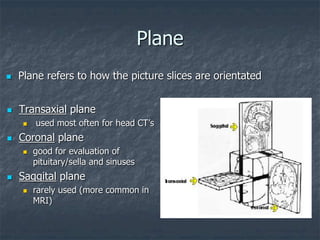
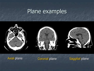
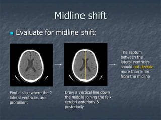
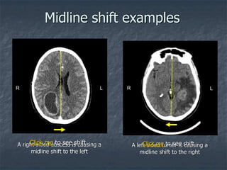
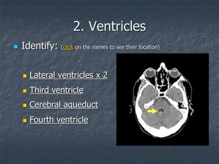
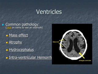
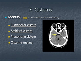
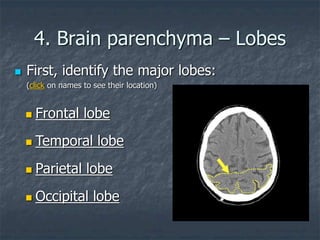
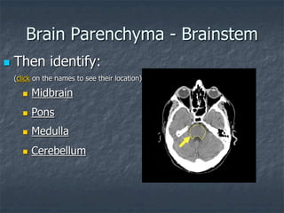
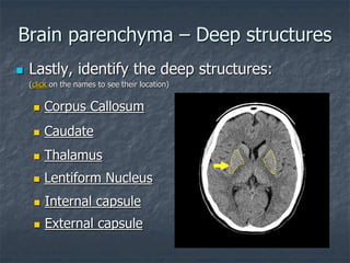
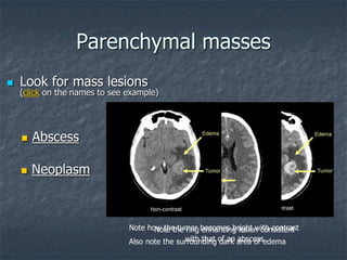
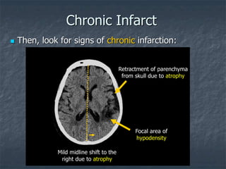
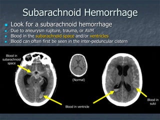
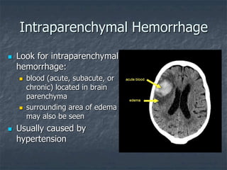
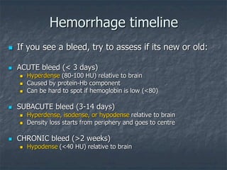
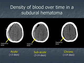
Approach to Head CT
Wesley Chan
CC3 Queen’s University
with
Dr. Danny Mandell
Dr. Nasir Jaffer
Department of Medical Imaging
University of Toronto
About this presentation
 This presentation will give you a
systematic approach to head CT
 By the end you should be familiar with
normal anatomy and be able to identify
classic abnormalities on CT
 You can test your knowledge with the
short cases at the end
Types of head CT’s
 Non-contrast
 Contrast
 IV contrast is given to better evaluate:
 Vascular structures
 Tumors
 Sites of infection
 Relative contraindications:
 Allergy, renal failure
Common Indications for Head CT
 Cranial-facial trauma
 Acute stroke
 Suspected subarachnoid or intracranial
hemorrhage
 Evaluation of headache
 Evaluation of sensory or motor function loss
 Evaluation of sinus cavities
CT basics
 Before we begin, there are key concepts
you should be familiar with:
 Hounsfield units
 Windowing & leveling
 Planes
What’s a Hounsfield Unit?
 Named after the inventor of CT
 CT scanners record the attenuation
(brightness) of each pixel in Hounsfield Units
(HU)
 This number represents the relative density
of the scanned substance
 Ranges from -1000 to +1000
Hounsfield Unit (HU)
 Different substances have different relative densities and
thus, different Hounsfield units
 Air: -1000 HU
 Fat: -50 HU
 Water: 0 HU
 Soft tissue: +40 HU
 Blood: +40-80 HU
 Stones: +100 to +400 HU
 Bone: +1000 HU
 Therefore, if you’re not sure what you’re looking at,
measure its Hounsfield Unit!
How to measure HU
 In EFILM, you can
measure the HU using
the oval ROI tool:
 On the right, you can
see sample
measurements of
different structures
 Note how bone, CSF,
brain tissue, and air all
have different mean HUs
Windowing
 The human eye can only perceive ~ 16 shades
of gray
 The CT scanner records levels of gray far
beyond what the eye can see
 Therefore, to interpret images, we have to limit
the number of Hounsfield units shown
(windowing)
 The computer then converts this set range of
HU into shades of gray we can see
Windows & levels
 Window width:
 The range of HU of all tissues of interest
 Tissues in this range will be displayed in
various shades of gray
 Tissues with HU outside the range are
displayed as black or white
 Window level:
 The central HU of all the numbers in the
window width
Windowing
+400
+300
+200
+100
0
-100
-200
-300
-400
Wide
Window
Narrow
Window
Hounsfield Units
Window examples
BRAIN window
W:80 L:40
BONE window
W:2500 L:480
SUBDURAL window
W:350 L:90
 In head CT, 3 windows are commonly used
Hounsfield Unit (HU)
 Different substances have different relative densities and
thus, different Hounsfield units
 Air: -1000 HU
 Fat: -50 HU
 Water: 0 HU
 Soft tissue: +40 HU
 Blood: +40-80 HU
 Stones: +100 to +400 HU
 Bone: +1000 HU
 Therefore, if you’re not sure what you’re looking at,
measure its Hounsfield Unit!
Plane
 Transaxial plane
 used most often for head CT’s
 Coronal plane
 good for evaluation of
pituitary/sella and sinuses
 Saggital plane
 rarely used (more common in
MRI)
 Plane refers to how the picture slices are orientated
Plane examples
Axial plane Coronal plane Saggital plane
Identification
 Now we can begin our basic approach to
the head CT
 Start with the easy stuff:
 PATIENT NAME (make sure you have the right patient !!)
 MEDICAL RECORD # (MRN)
 AGE
 DATE OF EXAM
Previous studies
 Always check for any previous scans for
comparison
 Findings can be very subtle
 A good way to spot them is to look for changes
between the current and previous scans
 Even old chest and abdominal films can give
you clues to possible brain pathology
 ie. Brain mets from lung cancer
Study parameters
 Make note of the study technique:
 Anatomic region of scan: head, neck, spine
 Slice thickness (mm)
 Window level & width
 Plane: Transaxial, coronal, saggital
 Use of contrast?
 Look for the Circle of Willis. It will be enhanced on
studies using contrast
Image analysis
 Now that you have noted all the basic
information about the scan, it’s time to
look at the scan itself
 Use a systematic order & approach to
what you look at
 Use the same approach for all scans to
ensure that you don’t miss anything
Regions to inspect
 We will start from the inside and move
outwards:
1. Midline structures &
symmetry
2. Ventricles
3. Cisterns
4. Brain parenchyma
5. Sulci
6. Sinuses
7. Bones
8. Skin/soft tissue
1. Midline structures
 Identify: (click on the names to see their location)
 Falx Cerebri
 Pineal gland
(usually calcified)
 Great vein of Galen
 Fornix
Midline shift
 Evaluate for midline shift:
Find a slice where the 2
lateral ventricles are
prominent
Draw a vertical line down
the middle joining the falx
cerebri anteriorly &
posteriorly
The septum
between the
lateral ventricles
should not deviate
more than 5mm
from the midline
Midline shift examples
A right-sided abscess is causing a
midline shift to the left
A left-sided tumor is causing a
midline shift to the right
L L
R R
Click me to see shift Click me to see shift
2. Ventricles
 Third ventricle
 Fourth ventricle
 Identify: (click on the names to see their location)
 Lateral ventricles x 2
 Cerebral aqueduct
Ventricles
 Evaluate for any changes in
 Symmetry
 Size
 Shape
 Density
 A displaced ventricle is often the product
of mass effect or atrophy
 Common pathology:
(click on name to see an example)
 Hydrocephalus
 Intra-ventricular Hemorrhage
 Mass effect
 Atrophy
Ventricles
3. Cisterns
 Identify: (click on the names to see their location)
 Supracellar cistern
 Ambient cistern
 Prepontine cistern
 Cisterna magna
Cisterns
 Evaluate for any changes in
 Symmetry
 Size
 Density
 Cisterns often contain blood with
subarachnoid hemorrhage
 Cisterns can fill with pus in the setting of
meningitis
4. Brain parenchyma – Lobes
 First, identify the major lobes:
(click on names to see their location)
 Temporal lobe
 Occipital lobe
 Frontal lobe
 Parietal lobe
Brain Parenchyma - Brainstem
 Then identify:
(click on the names to see their location)
 Pons
 Cerebellum
 Midbrain
 Medulla
Brain parenchyma – Deep structures
 Lastly, identify the deep structures:
(click on the names to see their location)
 Thalamus
 Lentiform Nucleus
 Caudate
 Corpus Callosum
 Internal capsule
 External capsule
 Look for mass lesions
(click on the names to see example)
 Abscess
 Neoplasm
Parenchymal masses
Note how the tumor becomes bright with contrast
Also note the surrounding dark area of edema
Note the ring enhancing lesion consistent
with that of an abscess
Acute Infarct
The middle cerebral artery (MCA)
becomes hyperdense due to occlusion
The usual border between grey and white
matter is lost due to vasogenic edema
 Hyperdense MCA sign
 Look for signs of acute infarction
 Loss of gray-white
differentiation
Click me to see Click me to see
Chronic Infarct
 Then, look for signs of chronic infarction:
Mild midline shift to the
right due to atrophy
Retractment of parenchyma
from skull due to atrophy
Focal area of
hypodensity
Infarction locations
Microangiopathic change
 You may encounter the term
ā€œmicroangiopathic changeā€ in reports
and wonder what it is
 Microangiopathic change refers to
age-related white matter ischemia
due to microvessel disease
 Very commonly seen in the elderly
 Its clinical significance is still not
known
Microangiopathic change
Normal
Types of Hematoma
 Look for evidence of a bleed:
 Subdural Hematoma
 Due to tear of bridging veins
 Look for crescentic shape along brain surface
 Crosses suture lines
 Epidural Hematoma
 Due to rupture of middle meningeal artery
 Associated with skull fractures
 Look for biconvex, lenticular shape
 Does not cross suture lines
Subdural vs. Epidural
SUBDURAL EPIDURAL
Note the cresentic shape Note the lenticular shape
Click me to see shape Click me to see shape
Subarachnoid Hemorrhage
Blood in
subarachnoid
space
Blood in
sulci
Blood in ventricle
 Look for a subarachnoid hemorrhage
 Due to aneurysm rupture, trauma, or AVM
 Blood in the subarachnoid space and/or ventricles
 Blood can often first be seen in the inter-peduncular cistern
(Normal)
Intraparenchymal Hemorrhage
 Look for intraparenchymal
hemorrhage:
 blood (acute, subacute, or
chronic) located in brain
parenchyma
 surrounding area of edema
may also be seen
 Usually caused by
hypertension
Hemorrhage timeline
 If you see a bleed, try to assess if its new or old:
 ACUTE bleed (< 3 days)
 Hyperdense (80-100 HU) relative to brain
 Caused by protein-Hb component
 Can be hard to spot if hemoglobin is low (<80)
 SUBACUTE bleed (3-14 days)
 Hyperdense, isodense, or hypodense relative to brain
 Density loss starts from periphery and goes to centre
 CHRONIC bleed (>2 weeks)
 Hypodense (<40 HU) relative to brain
Density of blood over time in a
subdural hematoma
Acute
(<3 days)
Sub-acute
(3-14 days)
Chronic
(>14 days)
Hyperdense
blood
Isodense
blood
Hypodense
blood
5. Sulci
 Central sulcus
 Precentral sulcus
 Sulci
 Sylvian fissures
 Postcentral sulcus
 Identify:
(click on the names to see their location)
Sulci
 Remember that sulci will become deeper and more prominent
with age
 Look for blood in the sulci & Sylvian Fissure which are
indications of a sub-arachnoid bleed
Acute blood in
Sylvian fissure
Acute blood in
sulci
6. Sinuses
 Switch to Bone Window to better evaluate the sinuses
 Identify: (click on the names to see their location)
 Ethmoid Sinus
 Sphenoid Sinus
 Superior Saggital Sinus
 Frontal Sinus
 Maxillary Sinus
Sinuses
 Evaluate for any sinusitis:
fluid in sinuses
(notice the air/fluid level)
normal
sinusitis
Sinuses
 Also look for any:
 Mucosal thickening
 Blood in sinuses (especially with history of trauma)
 Polyps or mucous retention cysts
7. Bone
 Stay on the Bone Window and look at the bones now
 Identify:
(click on the names to see their location)
 Skull
 Sutures
 Mastoid air cells
Bone
 Evaluate for any:
 Fractures
 Surgical changes
(ie. craniotomies)
8. Skin & Soft tissue
 Evaluate for any:
 Surgical changes
 Sub-galeal hematoma
 Foreign body
Congratulations!
 You now know an easy, systematic
approach to head CT!
 You have also learned how to identify
normal anatomy and how to spot classic
abnormalities on CT
 What follows is a brief summary of what
you have learned followed by some short
cases
Recap
 Begin with the basic identification
 Remember to check for previous scans
 Check the technique
 Look at each region of the brain
systematically
 We started from the middle and worked out:
1. Midline structures
2. Ventricles
3. Cisterns
4. Brain parenchyma
5. Sulci
6. Sinuses
7. Bones
8. Skin/soft tissue
Recap
 In each area, identify the major anatomy
 Then look for findings
 Below is a list of important things not to miss:
 Midline: midline shift
 Ventricles: blood and mass effect
 Cisterns: blood and pus
 Parenchyma: signs of ischemia and/or bleeding
 Sulci: for blood
 Sinuses: signs of sinusitis
 Bones: fractures
 Soft tissue: hematoma
Recap
 Remember to use the same approach
every time so that you don’t miss
anything!
 Try out the cases in the next slides to test
your knowledge
CASES
Case #1
 Ms. EW is an 80 y/o female presenting
with:
 Expressive aphasia/apraxia
 Mild right facial droop
 Atrial fibrillation
 A non-contrast CT scan of her brain is
performed
Your analysis
 What are your findings?
 What is your impression?
 What would be your top diagnosis?
Normal
Case #1 - Answer
 Mrs. EW had an infarction
of her Left Parietal Lobe
 The location is consistent
with MCA infarction
 The cause was emboli
related to her atrial
fibrillation
Case #2
 Mr. GH is a 56 y/o male presenting with:
 A sudden onset 10/10 headache while running
 Photophobia, nausea & vomiting
 No history of trauma or LOC
 Otherwise well
 A non-contrast CT scan of his brain is
performed
Your analysis
 What are your findings?
 What is your impression?
 What would be your top diagnosis?
 Is this pathology acute, subacute, or
chronic
Case #2 - Answer
 Mr. GH had a large
subarachnoid hemorrhage
 The bleed was acute
 This was caused by rupture of
an ACA aneurysm
 He was admitted to ICU where
his condition deteriorated
rapidly
 He passed away shortly after
admission
Case #3
 Ms. JH is a 66 y/o female who slipped
down the stairs yesterday and hit the back
of her head.
 She presents with
 Generalized left sided weakness
 Light headache
 A non-contrast CT scan of her brain is
performed
R L
Your analysis
 What are your findings?
 What is your impression?
 What would be your top diagnosis?
 Is this pathology acute, subacute, or
chronic
Case #3 - Answer
 Mrs. JH had a large right-
sided subdural hematoma
 The hematoma is acute
 This was caused by
rupture of bridging veins
when she hit her head
 A craniotomy was
performed and the bleed
was drained
Bonus case
 Mr. LR is a 80 y/o male presenting with:
 3 month history of delirium
 Recent fall from bed
 Large scalp laceration
 No focal neurological findings
 An non-contrast CT scan of his brain is
performed
Subdural
Look closely at the midline structures
Hint?
Analysis
 Can you spot the abnormalities?
 What is your impression?
 What would be your top diagnosis?
Bonus case - Answer
 Mr. LR had a tiny right-sided
subdural hematoma
 Blood is seen along the left
subdural space as well as in the
falx cerebri anteriorly (arrows)
 The hematoma is acute
 Because of its small size, no
immediate treatment was
required
 Follow-up CT scans showed
resolution of the subdural
hematoma
Normal scan for comparison

Approach to Head CT.ppt

  • 1.
    Approach to HeadCT Wesley Chan CC3 Queen’s University with Dr. Danny Mandell Dr. Nasir Jaffer Department of Medical Imaging University of Toronto
  • 2.
    About this presentation This presentation will give you a systematic approach to head CT  By the end you should be familiar with normal anatomy and be able to identify classic abnormalities on CT  You can test your knowledge with the short cases at the end
  • 3.
    Types of headCT’s  Non-contrast  Contrast  IV contrast is given to better evaluate:  Vascular structures  Tumors  Sites of infection  Relative contraindications:  Allergy, renal failure
  • 4.
    Common Indications forHead CT  Cranial-facial trauma  Acute stroke  Suspected subarachnoid or intracranial hemorrhage  Evaluation of headache  Evaluation of sensory or motor function loss  Evaluation of sinus cavities
  • 5.
    CT basics  Beforewe begin, there are key concepts you should be familiar with:  Hounsfield units  Windowing & leveling  Planes
  • 6.
    What’s a HounsfieldUnit?  Named after the inventor of CT  CT scanners record the attenuation (brightness) of each pixel in Hounsfield Units (HU)  This number represents the relative density of the scanned substance  Ranges from -1000 to +1000
  • 7.
    Hounsfield Unit (HU) Different substances have different relative densities and thus, different Hounsfield units  Air: -1000 HU  Fat: -50 HU  Water: 0 HU  Soft tissue: +40 HU  Blood: +40-80 HU  Stones: +100 to +400 HU  Bone: +1000 HU  Therefore, if you’re not sure what you’re looking at, measure its Hounsfield Unit!
  • 8.
    How to measureHU  In EFILM, you can measure the HU using the oval ROI tool:  On the right, you can see sample measurements of different structures  Note how bone, CSF, brain tissue, and air all have different mean HUs
  • 9.
    Windowing  The humaneye can only perceive ~ 16 shades of gray  The CT scanner records levels of gray far beyond what the eye can see  Therefore, to interpret images, we have to limit the number of Hounsfield units shown (windowing)  The computer then converts this set range of HU into shades of gray we can see
  • 10.
    Windows & levels Window width:  The range of HU of all tissues of interest  Tissues in this range will be displayed in various shades of gray  Tissues with HU outside the range are displayed as black or white  Window level:  The central HU of all the numbers in the window width
  • 11.
  • 12.
    Window examples BRAIN window W:80L:40 BONE window W:2500 L:480 SUBDURAL window W:350 L:90  In head CT, 3 windows are commonly used
  • 13.
    Hounsfield Unit (HU) Different substances have different relative densities and thus, different Hounsfield units  Air: -1000 HU  Fat: -50 HU  Water: 0 HU  Soft tissue: +40 HU  Blood: +40-80 HU  Stones: +100 to +400 HU  Bone: +1000 HU  Therefore, if you’re not sure what you’re looking at, measure its Hounsfield Unit!
  • 14.
    Plane  Transaxial plane used most often for head CT’s  Coronal plane  good for evaluation of pituitary/sella and sinuses  Saggital plane  rarely used (more common in MRI)  Plane refers to how the picture slices are orientated
  • 15.
    Plane examples Axial planeCoronal plane Saggital plane
  • 16.
    Identification  Now wecan begin our basic approach to the head CT  Start with the easy stuff:  PATIENT NAME (make sure you have the right patient !!)  MEDICAL RECORD # (MRN)  AGE  DATE OF EXAM
  • 17.
    Previous studies  Alwayscheck for any previous scans for comparison  Findings can be very subtle  A good way to spot them is to look for changes between the current and previous scans  Even old chest and abdominal films can give you clues to possible brain pathology  ie. Brain mets from lung cancer
  • 18.
    Study parameters  Makenote of the study technique:  Anatomic region of scan: head, neck, spine  Slice thickness (mm)  Window level & width  Plane: Transaxial, coronal, saggital  Use of contrast?  Look for the Circle of Willis. It will be enhanced on studies using contrast
  • 19.
    Image analysis  Nowthat you have noted all the basic information about the scan, it’s time to look at the scan itself  Use a systematic order & approach to what you look at  Use the same approach for all scans to ensure that you don’t miss anything
  • 20.
    Regions to inspect We will start from the inside and move outwards: 1. Midline structures & symmetry 2. Ventricles 3. Cisterns 4. Brain parenchyma 5. Sulci 6. Sinuses 7. Bones 8. Skin/soft tissue
  • 21.
    1. Midline structures Identify: (click on the names to see their location)  Falx Cerebri  Pineal gland (usually calcified)  Great vein of Galen  Fornix
  • 22.
    Midline shift  Evaluatefor midline shift: Find a slice where the 2 lateral ventricles are prominent Draw a vertical line down the middle joining the falx cerebri anteriorly & posteriorly The septum between the lateral ventricles should not deviate more than 5mm from the midline
  • 23.
    Midline shift examples Aright-sided abscess is causing a midline shift to the left A left-sided tumor is causing a midline shift to the right L L R R Click me to see shift Click me to see shift
  • 24.
    2. Ventricles  Thirdventricle  Fourth ventricle  Identify: (click on the names to see their location)  Lateral ventricles x 2  Cerebral aqueduct
  • 25.
    Ventricles  Evaluate forany changes in  Symmetry  Size  Shape  Density  A displaced ventricle is often the product of mass effect or atrophy
  • 26.
     Common pathology: (clickon name to see an example)  Hydrocephalus  Intra-ventricular Hemorrhage  Mass effect  Atrophy Ventricles
  • 27.
    3. Cisterns  Identify:(click on the names to see their location)  Supracellar cistern  Ambient cistern  Prepontine cistern  Cisterna magna
  • 28.
    Cisterns  Evaluate forany changes in  Symmetry  Size  Density  Cisterns often contain blood with subarachnoid hemorrhage  Cisterns can fill with pus in the setting of meningitis
  • 29.
    4. Brain parenchyma– Lobes  First, identify the major lobes: (click on names to see their location)  Temporal lobe  Occipital lobe  Frontal lobe  Parietal lobe
  • 30.
    Brain Parenchyma -Brainstem  Then identify: (click on the names to see their location)  Pons  Cerebellum  Midbrain  Medulla
  • 31.
    Brain parenchyma –Deep structures  Lastly, identify the deep structures: (click on the names to see their location)  Thalamus  Lentiform Nucleus  Caudate  Corpus Callosum  Internal capsule  External capsule
  • 32.
     Look formass lesions (click on the names to see example)  Abscess  Neoplasm Parenchymal masses Note how the tumor becomes bright with contrast Also note the surrounding dark area of edema Note the ring enhancing lesion consistent with that of an abscess
  • 33.
    Acute Infarct The middlecerebral artery (MCA) becomes hyperdense due to occlusion The usual border between grey and white matter is lost due to vasogenic edema  Hyperdense MCA sign  Look for signs of acute infarction  Loss of gray-white differentiation Click me to see Click me to see
  • 34.
    Chronic Infarct  Then,look for signs of chronic infarction: Mild midline shift to the right due to atrophy Retractment of parenchyma from skull due to atrophy Focal area of hypodensity
  • 35.
  • 36.
    Microangiopathic change  Youmay encounter the term ā€œmicroangiopathic changeā€ in reports and wonder what it is  Microangiopathic change refers to age-related white matter ischemia due to microvessel disease  Very commonly seen in the elderly  Its clinical significance is still not known Microangiopathic change Normal
  • 37.
    Types of Hematoma Look for evidence of a bleed:  Subdural Hematoma  Due to tear of bridging veins  Look for crescentic shape along brain surface  Crosses suture lines  Epidural Hematoma  Due to rupture of middle meningeal artery  Associated with skull fractures  Look for biconvex, lenticular shape  Does not cross suture lines
  • 38.
    Subdural vs. Epidural SUBDURALEPIDURAL Note the cresentic shape Note the lenticular shape Click me to see shape Click me to see shape
  • 39.
    Subarachnoid Hemorrhage Blood in subarachnoid space Bloodin sulci Blood in ventricle  Look for a subarachnoid hemorrhage  Due to aneurysm rupture, trauma, or AVM  Blood in the subarachnoid space and/or ventricles  Blood can often first be seen in the inter-peduncular cistern (Normal)
  • 40.
    Intraparenchymal Hemorrhage  Lookfor intraparenchymal hemorrhage:  blood (acute, subacute, or chronic) located in brain parenchyma  surrounding area of edema may also be seen  Usually caused by hypertension
  • 41.
    Hemorrhage timeline  Ifyou see a bleed, try to assess if its new or old:  ACUTE bleed (< 3 days)  Hyperdense (80-100 HU) relative to brain  Caused by protein-Hb component  Can be hard to spot if hemoglobin is low (<80)  SUBACUTE bleed (3-14 days)  Hyperdense, isodense, or hypodense relative to brain  Density loss starts from periphery and goes to centre  CHRONIC bleed (>2 weeks)  Hypodense (<40 HU) relative to brain
  • 42.
    Density of bloodover time in a subdural hematoma Acute (<3 days) Sub-acute (3-14 days) Chronic (>14 days) Hyperdense blood Isodense blood Hypodense blood
  • 43.
    5. Sulci  Centralsulcus  Precentral sulcus  Sulci  Sylvian fissures  Postcentral sulcus  Identify: (click on the names to see their location)
  • 44.
    Sulci  Remember thatsulci will become deeper and more prominent with age  Look for blood in the sulci & Sylvian Fissure which are indications of a sub-arachnoid bleed Acute blood in Sylvian fissure Acute blood in sulci
  • 45.
    6. Sinuses  Switchto Bone Window to better evaluate the sinuses  Identify: (click on the names to see their location)  Ethmoid Sinus  Sphenoid Sinus  Superior Saggital Sinus  Frontal Sinus  Maxillary Sinus
  • 46.
    Sinuses  Evaluate forany sinusitis: fluid in sinuses (notice the air/fluid level) normal sinusitis
  • 47.
    Sinuses  Also lookfor any:  Mucosal thickening  Blood in sinuses (especially with history of trauma)  Polyps or mucous retention cysts
  • 48.
    7. Bone  Stayon the Bone Window and look at the bones now  Identify: (click on the names to see their location)  Skull  Sutures  Mastoid air cells
  • 49.
    Bone  Evaluate forany:  Fractures  Surgical changes (ie. craniotomies)
  • 50.
    8. Skin &Soft tissue  Evaluate for any:  Surgical changes  Sub-galeal hematoma  Foreign body
  • 51.
    Congratulations!  You nowknow an easy, systematic approach to head CT!  You have also learned how to identify normal anatomy and how to spot classic abnormalities on CT  What follows is a brief summary of what you have learned followed by some short cases
  • 52.
    Recap  Begin withthe basic identification  Remember to check for previous scans  Check the technique  Look at each region of the brain systematically  We started from the middle and worked out: 1. Midline structures 2. Ventricles 3. Cisterns 4. Brain parenchyma 5. Sulci 6. Sinuses 7. Bones 8. Skin/soft tissue
  • 53.
    Recap  In eacharea, identify the major anatomy  Then look for findings  Below is a list of important things not to miss:  Midline: midline shift  Ventricles: blood and mass effect  Cisterns: blood and pus  Parenchyma: signs of ischemia and/or bleeding  Sulci: for blood  Sinuses: signs of sinusitis  Bones: fractures  Soft tissue: hematoma
  • 54.
    Recap  Remember touse the same approach every time so that you don’t miss anything!  Try out the cases in the next slides to test your knowledge
  • 55.
  • 56.
    Case #1  Ms.EW is an 80 y/o female presenting with:  Expressive aphasia/apraxia  Mild right facial droop  Atrial fibrillation  A non-contrast CT scan of her brain is performed
  • 58.
    Your analysis  Whatare your findings?  What is your impression?  What would be your top diagnosis?
  • 59.
  • 60.
    Case #1 -Answer  Mrs. EW had an infarction of her Left Parietal Lobe  The location is consistent with MCA infarction  The cause was emboli related to her atrial fibrillation
  • 61.
    Case #2  Mr.GH is a 56 y/o male presenting with:  A sudden onset 10/10 headache while running  Photophobia, nausea & vomiting  No history of trauma or LOC  Otherwise well  A non-contrast CT scan of his brain is performed
  • 63.
    Your analysis  Whatare your findings?  What is your impression?  What would be your top diagnosis?  Is this pathology acute, subacute, or chronic
  • 64.
    Case #2 -Answer  Mr. GH had a large subarachnoid hemorrhage  The bleed was acute  This was caused by rupture of an ACA aneurysm  He was admitted to ICU where his condition deteriorated rapidly  He passed away shortly after admission
  • 65.
    Case #3  Ms.JH is a 66 y/o female who slipped down the stairs yesterday and hit the back of her head.  She presents with  Generalized left sided weakness  Light headache  A non-contrast CT scan of her brain is performed
  • 66.
  • 67.
    Your analysis  Whatare your findings?  What is your impression?  What would be your top diagnosis?  Is this pathology acute, subacute, or chronic
  • 68.
    Case #3 -Answer  Mrs. JH had a large right- sided subdural hematoma  The hematoma is acute  This was caused by rupture of bridging veins when she hit her head  A craniotomy was performed and the bleed was drained
  • 69.
    Bonus case  Mr.LR is a 80 y/o male presenting with:  3 month history of delirium  Recent fall from bed  Large scalp laceration  No focal neurological findings  An non-contrast CT scan of his brain is performed
  • 70.
    Subdural Look closely atthe midline structures Hint?
  • 71.
    Analysis  Can youspot the abnormalities?  What is your impression?  What would be your top diagnosis?
  • 72.
    Bonus case -Answer  Mr. LR had a tiny right-sided subdural hematoma  Blood is seen along the left subdural space as well as in the falx cerebri anteriorly (arrows)  The hematoma is acute  Because of its small size, no immediate treatment was required  Follow-up CT scans showed resolution of the subdural hematoma Normal scan for comparison