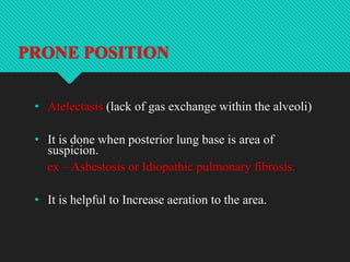
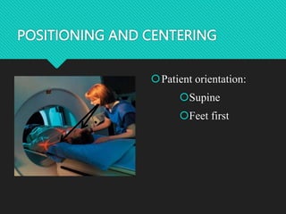
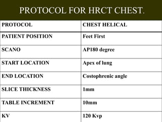
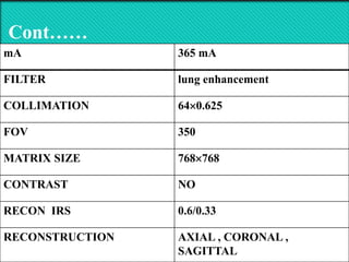
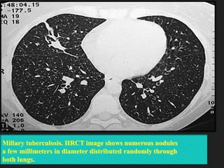
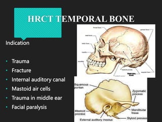
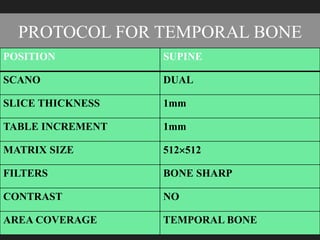
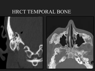
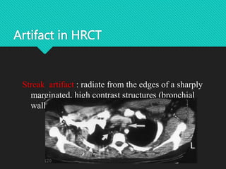
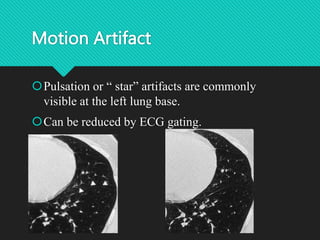
HRCT TECHNICAL ASPECTS HRCT
PROTOCOL
Mr.SAMEERAHMADGANAIE
ASSISTANTPROFESSOR
COPMSADESHUNIVERSITY,BATHINDAPUNJAB151001
Contents
 History
 Objective
 Purpose
 Introduction
 Indications
 Patientpreparations
 Technicalaspects
 HRCTchestprotocol
 HRCTtemporalboneprotocol
 Artifactsin HRCT
 Clinicalapplicationsof HRCT
History
• 1982– The term HRCT was first used by TODO et. Al
• 1985 – Nakata et.al and Naidich et.al published first
report on HRCT.
Objective
The main objective of HRCT is to detect,
characterize, and determine the extent of
diseases that involve the lung parenchyma
and airways.
Purpose
 The main purpose of High-resolution computed
tomography (HRCT) is to diagnose and monitoring
of disease of lung tissue ,airways and the temporal
bone.
Introduction
 By eliminating superimposition of structures, computed tomography (CT)
allows for a better assessment of the type, distribution, and severity of
Parenchymal abnormalities.
 HRCT is a technique which allows us for better evaluate these conditions.
 It is usually done for chest and temporal bone.
• Use of thin section CT images (0.625 to 2 mm slice thickness) often with a
high-spatial-frequency
Indication
Chronic lung disease
Diffuse infiltrative lung disease
Pulmonary tuberculosis
Sarcoidosis
Hypersensitivity pneumonitis
Langerhans cell histiocytosis
Lymphangitic carcinomatosis
Silicosis
Idiopathic pulmonary fibrosis
Usual interstitial pneumonitis (UIP)
PATIENT PREPARATION
• Fasting is not required.
• Patient is should remove radio opaque materials (chain
and other neck ornaments ) from the region of interest.
• Pacemaker is not contraindicated.
• Patient is asked to change to hospital gown.
•Proper instruction to patient leads to successful
HRCT scan.
•Breath holding techniques
•Routine HRCT during suspended full
inspiration.
•It also reduces motion Artifact
Technical aspects
HRCT CHEST
It can be done in three position
1.Antero posterior (AP) i.e. Supine
2.Postero anterior (PA) i.e. Prone
3.Lateral decubitus
SUPINE HRCT
 Bronchiectasis
 Diffuse infiltrative lung disease
 Scan direction from down to up
PRONE POSITION
• Atelectasis (lack of gas exchange within the alveoli)
• It is done when posterior lung base is area of
suspicion.
ex – Asbestosis or Idiopathic pulmonary fibrosis.
• It is helpful to Increase aeration to the area.
LATERAL DECUBITUS
• It is helpful to distinguish between complex
pleural and pulmonary pathological conditions.
POSITIONING AND CENTERING
Patient orientation:
Supine
Feet first
PROTOCOL FOR HRCT CHEST.
PROTOCOL CHEST HELICAL
PATIENT POSITION Feet First
SCANO AP180 degree
START LOCATION Apex of lung
END LOCATION Costophrenic angle
SLICE THICKNESS 1mm
TABLE INCREMENT 10mm
KV 120 Kvp
Cont……
mA 365 mA
FILTER lung enhancement
COLLIMATION 640.625
FOV 350
MATRIX SIZE 768768
CONTRAST NO
RECON IRS 0.6/0.33
RECONSTRUCTION AXIAL , CORONAL ,
SAGITTAL
Miliary tuberculosis. HRCT image shows numerous nodules
a few millimeters in diameter distributed randomly through
both lungs.
FINDINGS……
HRCT PLAIN
HRCT TEMPORAL BONE
Indication
• Trauma
• Fracture
• Internal auditory canal
• Mastoid air cells
• Trauma in middle ear
• Facial paralysis
POSITIONING AND CENTERING
Patient orientation:
Supine
Head first
Anatomical landmark:
glabella
PLANNING…
PROTOCOL FOR TEMPORAL BONE
POSITION SUPINE
SCANO DUAL
SLICE THICKNESS 1mm
TABLE INCREMENT 1mm
MATRIX SIZE 512512
FILTERS BONE SHARP
CONTRAST NO
AREA COVERAGE TEMPORAL BONE
HRCT TEMPORAL BONE
Artifact in HRCT
Streak artifact : radiate from the edges of a sharply
marginated, high contrast structures (bronchial
walls, ribs or vertebral bodies)
Motion Artifact
Pulsation or “ star” artifacts are commonly
visible at the left lung base.
Can be reduced by ECG gating.
ADVANTAGES
• Diagnosis of very small lesions. Eg:
small metastatic lesions, ILD.
DISADVANTAGES
• Increased image noise.
• Radiation dose is more.
Clinical Application of HRCT
To detect diffuse lung disease
To investigate patient with unexplained
severe obstructive airway
To investigate patient with hemoptysis
To guide the type and style of lung biopsy
Thank you

HRCT CHEST/TEMPORAL BONE PROTOCOL.pptx

  • 1.
    HRCT TECHNICAL ASPECTSHRCT PROTOCOL Mr.SAMEERAHMADGANAIE ASSISTANTPROFESSOR COPMSADESHUNIVERSITY,BATHINDAPUNJAB151001
  • 2.
    Contents  History  Objective Purpose  Introduction  Indications  Patientpreparations  Technicalaspects  HRCTchestprotocol  HRCTtemporalboneprotocol  Artifactsin HRCT  Clinicalapplicationsof HRCT
  • 3.
    History • 1982– Theterm HRCT was first used by TODO et. Al • 1985 – Nakata et.al and Naidich et.al published first report on HRCT.
  • 4.
    Objective The main objectiveof HRCT is to detect, characterize, and determine the extent of diseases that involve the lung parenchyma and airways.
  • 5.
    Purpose  The mainpurpose of High-resolution computed tomography (HRCT) is to diagnose and monitoring of disease of lung tissue ,airways and the temporal bone.
  • 6.
    Introduction  By eliminatingsuperimposition of structures, computed tomography (CT) allows for a better assessment of the type, distribution, and severity of Parenchymal abnormalities.  HRCT is a technique which allows us for better evaluate these conditions.  It is usually done for chest and temporal bone. • Use of thin section CT images (0.625 to 2 mm slice thickness) often with a high-spatial-frequency
  • 7.
    Indication Chronic lung disease Diffuseinfiltrative lung disease Pulmonary tuberculosis Sarcoidosis Hypersensitivity pneumonitis Langerhans cell histiocytosis Lymphangitic carcinomatosis Silicosis Idiopathic pulmonary fibrosis Usual interstitial pneumonitis (UIP)
  • 8.
    PATIENT PREPARATION • Fastingis not required. • Patient is should remove radio opaque materials (chain and other neck ornaments ) from the region of interest. • Pacemaker is not contraindicated. • Patient is asked to change to hospital gown.
  • 9.
    •Proper instruction topatient leads to successful HRCT scan. •Breath holding techniques •Routine HRCT during suspended full inspiration. •It also reduces motion Artifact Technical aspects
  • 10.
    HRCT CHEST It canbe done in three position 1.Antero posterior (AP) i.e. Supine 2.Postero anterior (PA) i.e. Prone 3.Lateral decubitus
  • 11.
    SUPINE HRCT  Bronchiectasis Diffuse infiltrative lung disease  Scan direction from down to up
  • 12.
    PRONE POSITION • Atelectasis(lack of gas exchange within the alveoli) • It is done when posterior lung base is area of suspicion. ex – Asbestosis or Idiopathic pulmonary fibrosis. • It is helpful to Increase aeration to the area.
  • 13.
    LATERAL DECUBITUS • Itis helpful to distinguish between complex pleural and pulmonary pathological conditions.
  • 14.
    POSITIONING AND CENTERING Patientorientation: Supine Feet first
  • 15.
    PROTOCOL FOR HRCTCHEST. PROTOCOL CHEST HELICAL PATIENT POSITION Feet First SCANO AP180 degree START LOCATION Apex of lung END LOCATION Costophrenic angle SLICE THICKNESS 1mm TABLE INCREMENT 10mm KV 120 Kvp
  • 16.
    Cont…… mA 365 mA FILTERlung enhancement COLLIMATION 640.625 FOV 350 MATRIX SIZE 768768 CONTRAST NO RECON IRS 0.6/0.33 RECONSTRUCTION AXIAL , CORONAL , SAGITTAL
  • 17.
    Miliary tuberculosis. HRCTimage shows numerous nodules a few millimeters in diameter distributed randomly through both lungs.
  • 18.
  • 19.
    HRCT TEMPORAL BONE Indication •Trauma • Fracture • Internal auditory canal • Mastoid air cells • Trauma in middle ear • Facial paralysis
  • 20.
    POSITIONING AND CENTERING Patientorientation: Supine Head first Anatomical landmark: glabella
  • 21.
  • 22.
    PROTOCOL FOR TEMPORALBONE POSITION SUPINE SCANO DUAL SLICE THICKNESS 1mm TABLE INCREMENT 1mm MATRIX SIZE 512512 FILTERS BONE SHARP CONTRAST NO AREA COVERAGE TEMPORAL BONE
  • 23.
  • 24.
    Artifact in HRCT Streakartifact : radiate from the edges of a sharply marginated, high contrast structures (bronchial walls, ribs or vertebral bodies)
  • 25.
    Motion Artifact Pulsation or“ star” artifacts are commonly visible at the left lung base. Can be reduced by ECG gating.
  • 26.
    ADVANTAGES • Diagnosis ofvery small lesions. Eg: small metastatic lesions, ILD. DISADVANTAGES • Increased image noise. • Radiation dose is more.
  • 27.
    Clinical Application ofHRCT To detect diffuse lung disease To investigate patient with unexplained severe obstructive airway To investigate patient with hemoptysis To guide the type and style of lung biopsy
  • 28.

Editor's Notes

  • #7 It is often used for anything and everything to do with “high resolution”. Resolution : Means ability to resolve small object that are close together ,as separate form. Actual meaning A scan performed using high- spatial frequency algorithm to accentuate the contrast between tissue of widely differing densities, eg., - air & vessels (lung) - air & bone (temporal & paranasal sinus)