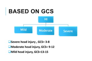
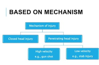
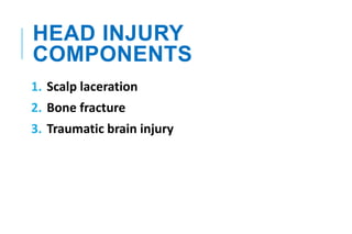
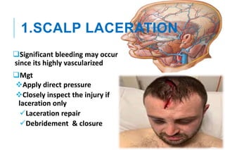
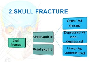
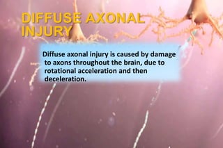
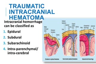
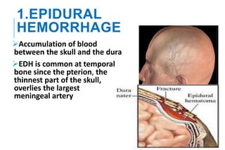
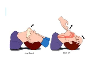
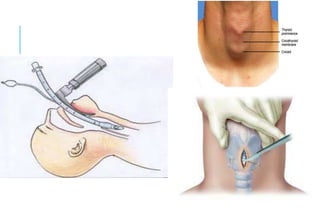
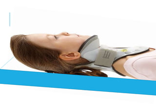
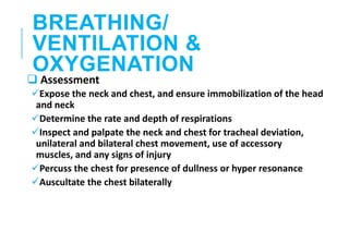
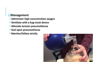
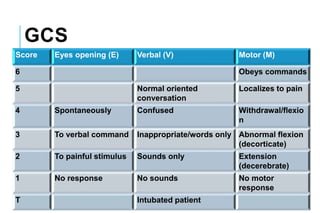
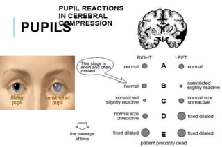
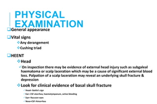
HEAD
INJURY
DR.DANIEL FENTANEH(MD)
WDU,CHS
OUTLINE
Introduction
Pathophysiology
Classification of HI
Head injury components
Scalp laceration
Skull #
TBI
Approach to HI patients
INTRODUCTION
PATHOPHYSIOLOGY OF
HI
Monro-kellie doctrine states
that the total volume of the
intracranial contents must
remain constant. Because the
cranium is a rigid & non-
expansible container.
Total volume=Brain tissue(80%) + CSF
fluid(10%) + intravascular blood(10%)
Venous blood & CSF fluid may be displaced out of the
container to provide pressure buffering (keeping the total
volume constant). So early after injury the pt may have
normal ICP, but once the limit of displacement reached
ICP rapidly increases
CPP(cerebral perfusion pressure- The pressure gradient
required to perfuse the cerebral tissue)= Mean arterial
pressure (MAP) – ICP
MAP = 1/3 (SBP) + 2/3 (DBP)
Normal CPP range = 50 – 70 mmHg
Normal ICP in adults< 10-15mmHg
1. Subfalcine hernation
2. Uncal hernation
3. Transtentorial hernation
4. Tonsillar hernation
What are symptoms & signs of raised ICP?
Brain herination
CLASSIFICATIO
N OF HI
BASED ON GCS
Severe head injury , GCS= 3-8
Moderate head injury, GCS= 9-12
Mild head injury, GCS=13-15
HI
Mild Moderate Severe
BASED ON MECHANISM
Mechanism of injury
Closed head injury Penetrating head injury
High velocity
e.g., gun shot
Low velocity
e.g., stab injury
HEAD INJURY
COMPONENTS
1. Scalp laceration
2. Bone fracture
3. Traumatic brain injury
1.SCALP LACERATION
Significant bleeding may occur
since its highly vascularized
Mgt
Apply direct pressure
Closely inspect the injury if
laceration only
Laceration repair
Debridement & closure
2.SKULL FRACTURE
SKULL VAULT
A closed fracture is covered by intact skin. An open
or compound fracture is associated with disrupted
overlying skin
Simple or linear fractures do not need any Rx. But
patient should be observed for 24 hrs for possible
intracranial bleeding
Depressed skull fracture can cause brain or dural laceration
and brain compression
Depressed skull # is managed by elevation of bigger
fragments and removal of smaller fragments
Indication for elevation
 Neurologic deficit
 Loss of consciousness
 Seizure
 Cosmetic areas
BASAL SKULL #
Otorrhea
Presence of CSF drainage from the ears /Copious clear drainage
from the ears/ will make the Dx obvious
Rhinorrhea
Presence of CSF drainage from the nostrils /Copious clear drainage
from the nostrils/ will make the Dx obvious
Battle sign
Post-auricular/mastoid ecchymoses
Raccoons eye
Periorbital ecchymoses
CN palsy
Anosmia
MANAGEMENT
CSF leak
Elevation of head off the bed for several days may heal
it. In addition lumbar drain can augment this method. Lumbar
drain allows the defect to heal by eliminating normal
hydrostatic pressure.
Traumatic cranial neuropathy
Facial nerve palsy—steroids. If no response after 48-72hrs
surgical decompression of the petrous portion of CN-VII may
be considered
3.TRAUMATIC BRAIN
INJURY
TBI
Primary Secondary
Primary traumatic brain injury occurs at the time of
impact and includes injuries such as brainstem and
hemispheric contusions, diffuse axonal injury & cortical
lacerations
Secondary brain injury occurs at some time after the
moment of impact and is often preventable. The
principle causes of secondary brain injury can be
classified as extra cranial & intracranial causes
Extra cranial
 Hypoxia
 Hypotension
Intracranial
Hematoma
Brain swelling(edema)
Infection
CPP=MAP – ICP
CLOSED HEAD INJURY
Types
Concussion
Contusion
Diffuse axonal injury
CONCUSSION
Defined as temporary neuronal dysfunction following
non-penetrating head trauma.
The head CT is normal, and deficits resolve over
minutes to hours.
CONTUSION
A bruise of the brain
The contused areas appear bright on CT scan
Frontal, occipital, temporal are common sites for
contusion
Coup--contusion at the point of
impact
Counter coup--Contusions also may
occur in brain tissue opposite the site
of impact. This is known as a contre-
coup injury. These contusions result
from deceleration of the brain against
the skull.
Contusion also occurs as the brain
slides forwards and backwards over
the ridged cranial fossa floor
DIFFUSE AXONAL
INJURY
Diffuse axonal injury is caused by damage
to axons throughout the brain, due to
rotational acceleration and then
deceleration.
TRAUMATIC
INTRACRANIAL
HEMATOMA
Intracranial hemorrhage
can be classified as
1. Epidural
2. Subdural
3. Subarachinoid
4. Intra-parenchymal/
intra-cerebral
1.EPIDURAL
HEMORRHAGE
Accumulation of blood
between the skull and the dura
EDH is common at temporal
bone since the pterion, the
thinnest part of the skull,
overlies the largest
meningeal artery
CLINICAL PRESENTATION
Classic presentation/20%
1. Loss of consiousness –due to concussive effect of head
trauma
2. Awakens & “lucid interval”—subclinical hematoma
expansion
3. Deterioration—as a result of brain compression &
herniation
 Contralateral hemiparesis
 Reduced consciousness level and
 Ipsilateral pupillary dilatation
CT-SCAN
EDH
Bright/hyperdense
Biconvex in shape (lentiform), and has a well-
defined border that usually respects cranial suture
lines
MANAGEMENT
Craniotomy--for evacuation of the congealed clot
and hemostasis
SUBDURAL
HEMORRHAGE
SDH is the accumulation of blood between the
arachnoid membrane and the dura. It can be
classified as;
Acute SDH
Chronic SDH
ACUTE SDH
Usually results from venous bleeding, typically from
tearing of a bridging vein running from the cerebral
cortex to the dural sinuses
Elderly and alcoholic patients are at higher risk for
acute SDH formation after head trauma due to brain
atrophy.
CT-SCAN
Acute SDH
The clot is bright or mixed-density
Crescent-shaped (lunate), may have a less distinct
border, and does not cross the midline due to the
presence of the falx
MANAGEMENT
Open craniotomy for evacuation of acute SDH is
indicated for any of the following: thickness >1 cm,
midline shift >5 mm, or GCS drop by two or more
points from the time of injury to hospitalization
CHRONIC SDH
Chronic SDH is a collection of blood breakdown products
that is at least 2 to 3 weeks old
Alcoholics, the elderly, and patients on anticoagulation
are at higher risk for developing chronic SDH
Patients may present with headache, seizure, confusion,
contralateral hemiparesis, or coma
Chronic SDHs often occur in patients without a clear
history of head trauma, as they may arise from minor
head injury
CT-SCAN
Chronic SDH
Hypodense
MANAGEMENT
A chronic SDH >1 cm or any symptomatic SDH
should be surgically drained
Burr hole
INTRA-PARENCHYMAL
HEMORRHAGE
Isolated hematomas within the
brain parenchyma are most often
associated with hypertensive
hemorrhage or arteriovenous
malformations (AVMs).
Traumatic--Bleeding may occur in
a contused area of brain
Delayed traumatic intracerebral hemorrhage is most
likely to occur within the first 24 hours
Patients with contusion on the initial head CT scan
should be reimaged 24 hours
Indication for craniotomy
Any clot volume >50cm3
Clot volume >20cm3 with neurologic deterioration (GCS=6-8)
& midline shift >5mm or basal cistern compression
Primary survey
Airway
Breathing
Circulation
Disability
Exposure
Secondary survey
History
Physical examination
Investigation
Definitive management
PRIMARY
SURVEY
AIRWAY ASSESSMENT
WITH SPINE
PROTECTION
 Assessment
i. Ascertain patency
ii. Rapidly assess for airway obstruction
Management
 Establish a patent airway
i. Perform a chin-lift or jaw-thrust maneuver
ii. Clear the airway of foreign bodies
iii. Insert an oro-pharyngeal airway
 Establish a definitive airway
1. Intubation
2. Surgical cricothyroidotomy
 Maintain the cervical spine in a neutral position with
manual immobilization as necessary when establishing
an airway
 Reinstate immobilization of the cervical spine with
appropriate devices after establishing an airway
BREATHING/
VENTILATION &
OXYGENATION
 Assessment
Expose the neck and chest, and ensure immobilization of the head
and neck
Determine the rate and depth of respirations
Inspect and palpate the neck and chest for tracheal deviation,
unilateral and bilateral chest movement, use of accessory
muscles, and any signs of injury
Percuss the chest for presence of dullness or hyper resonance
Auscultate the chest bilaterally
Management
Administer high-concentration oxygen
Ventilate with a bag-mask device
Alleviate tension pneumothorax
Seal open pneumothorax
Monitor/follow strictly
CIRCULATION &
HEMORRHAGE
CONTROL
 Assessment
Identify source of external, exsanguinating hemorrhage
Identify potential source(s) of internal hemorrhage
Assess pulse: Quality, rate, regularity, and paradox
Evaluate skin color
Measure blood pressure, if time permits
Management
Apply direct pressure to external bleeding site(s)
Insert two large-caliber IV catheters
Simultaneously obtain blood for hematologic and chemical
analyses; BG & RH, cross-match…
Initiate IV fluid therapy with warmed crystalloid solution and
blood replacement
Prevent hypothermia
Consider presence of internal hemorrhage and potential need for
operative intervention, and obtain surgical consult
DISABILITY /BRIEF
NEUROLOGY EXAM
Determine the level of consciousness using the GCS
Check pupils for size and reaction
GCS
Score Eyes opening (E) Verbal (V) Motor (M)
6 Obeys commands
5 Normal oriented
conversation
Localizes to pain
4 Spontaneously Confused Withdrawal/flexio
n
3 To verbal command Inappropriate/words only Abnormal flexion
(decorticate)
2 To painful stimulus Sounds only Extension
(decerebrate)
1 No response No sounds No motor
response
T Intubated patient
PUPILS
EXPOSURE
Completely undress the patient, but prevent
hypothermia
SECONDARY
SURVEY
HISTORY
Mechanism of injury
History of loss of consciousness, abnormal body
movement…
Medication history
Alcohol /illicit drug use
Psychiatric problem
PHYSICAL
EXAMINATION
General appearance
Vital signs
Any derangement
Cushing triad
HEENT
Head
On inspection there may be evidence of external head injury such as subgaleal
haematoma or scalp laceration which may be a cause of significant external blood
loss. Palpation of a scalp laceration may reveal an underlying skull fracture &
depression
Look for clinical evidence of basal skull fracture
Head= Battle’s sign
Ear= CSF otorrhea, haemotympanum, active bleeding
Eye= Raccoon eyes
Nose=CSF rhinorrhea
Chest examination
CVS examination
Abdominal examination
GUS examination
IGS examination
MSS examination
Nervous system examination
GCS
MMSE
Cranial nerve examination
Motor examination
INVESTIGATION
Skull x-ray (AP & lateral)
Is there # or not?
If fractured. Is it depressed or not?
If depressed. Is it significant?
Linear Vs comminuted?
CT-SCAN
Epidural hematoma Acute SDH Chronic SDH
hyperdense (bright)
lesion
Hyperdense (acute
blood)
hypodense
lentiform (lens-
shaped or
biconvex)
crescent shaped
(lunate)
crescent shaped
(lunate)
well defined border
(between the skull
and brain)
may have less
distinct border
may have less
distinct border
may or may not
cross the midline
doesn’t cross
midline
doesn’t cross
midline
LABORATORY
Hct
Blood group & Rh
X-match
DEFINITIVE
MANAGEMENT
Observation, Craniotomy or Burr hole based on the
underlying disease condition & indication
MILD HEAD INJURY MGT
Observation for 24 hrs
Criteria for discharge
Pt must have GCS=15/15 with no focal neurological deficit
verbal and written head injury advice must be given to the pt and pt’s
attendant
advice to return if the pt experience persistent or worsening of
headache despite analgesia, persistent vomiting, drowsiness, visual
disturbance such as double or blurred vision, and development of
weakness or numbness in the limbs.
MODERATE TO SEVERE
HEAD INJURY
Aim --To prevent secondary brain injury
The cervical spine must be immobilized
Cerebral contusion
admit for observation--because these lesions tend to mature
and expand 48–72 hours following injury
rarely requires emergent evacuation
some pts may require delayed evacuation to reduce mass
effect
Extradural (epidural) hematoma
Immediate surgical evacuation via craniotomy & hemostasis
Subdural hematoma
Acute SDH
Evacuation via craniotomy based on indications
Chronic SDH--Evacuation via burr hole(s) based on indications
Intraparenchymal hemorrhage
Reimage after 24hrs & craniotomy if indication met
MEDICAL MGT
Head elevation 300
Seizure control
Normothermia
Sedation +/– muscle relaxant
Normocapnia
Avoid obstruction of venous drainage from head--check the
cervical collar
Mannitol
Serum electrolyte balance
GI prophylaxis
THANK YOU!
REFERENCES
Schwartz's principle of surgery 10th edition
Sabiston 18th edition
Uptodate 21.6
Bailey & love 25th edition
ATLS 9th edition (student course manual)

Head injury

  • 1.
  • 2.
    OUTLINE Introduction Pathophysiology Classification of HI Headinjury components Scalp laceration Skull # TBI Approach to HI patients
  • 3.
  • 7.
    PATHOPHYSIOLOGY OF HI Monro-kellie doctrinestates that the total volume of the intracranial contents must remain constant. Because the cranium is a rigid & non- expansible container. Total volume=Brain tissue(80%) + CSF fluid(10%) + intravascular blood(10%)
  • 8.
    Venous blood &CSF fluid may be displaced out of the container to provide pressure buffering (keeping the total volume constant). So early after injury the pt may have normal ICP, but once the limit of displacement reached ICP rapidly increases CPP(cerebral perfusion pressure- The pressure gradient required to perfuse the cerebral tissue)= Mean arterial pressure (MAP) – ICP MAP = 1/3 (SBP) + 2/3 (DBP) Normal CPP range = 50 – 70 mmHg Normal ICP in adults< 10-15mmHg
  • 9.
    1. Subfalcine hernation 2.Uncal hernation 3. Transtentorial hernation 4. Tonsillar hernation What are symptoms & signs of raised ICP? Brain herination
  • 10.
  • 11.
    BASED ON GCS Severehead injury , GCS= 3-8 Moderate head injury, GCS= 9-12 Mild head injury, GCS=13-15 HI Mild Moderate Severe
  • 12.
    BASED ON MECHANISM Mechanismof injury Closed head injury Penetrating head injury High velocity e.g., gun shot Low velocity e.g., stab injury
  • 13.
    HEAD INJURY COMPONENTS 1. Scalplaceration 2. Bone fracture 3. Traumatic brain injury
  • 14.
    1.SCALP LACERATION Significant bleedingmay occur since its highly vascularized Mgt Apply direct pressure Closely inspect the injury if laceration only Laceration repair Debridement & closure
  • 15.
  • 17.
    SKULL VAULT A closedfracture is covered by intact skin. An open or compound fracture is associated with disrupted overlying skin Simple or linear fractures do not need any Rx. But patient should be observed for 24 hrs for possible intracranial bleeding
  • 18.
    Depressed skull fracturecan cause brain or dural laceration and brain compression Depressed skull # is managed by elevation of bigger fragments and removal of smaller fragments Indication for elevation  Neurologic deficit  Loss of consciousness  Seizure  Cosmetic areas
  • 20.
    BASAL SKULL # Otorrhea Presenceof CSF drainage from the ears /Copious clear drainage from the ears/ will make the Dx obvious Rhinorrhea Presence of CSF drainage from the nostrils /Copious clear drainage from the nostrils/ will make the Dx obvious
  • 22.
  • 23.
  • 24.
  • 25.
    MANAGEMENT CSF leak Elevation ofhead off the bed for several days may heal it. In addition lumbar drain can augment this method. Lumbar drain allows the defect to heal by eliminating normal hydrostatic pressure. Traumatic cranial neuropathy Facial nerve palsy—steroids. If no response after 48-72hrs surgical decompression of the petrous portion of CN-VII may be considered
  • 26.
    3.TRAUMATIC BRAIN INJURY TBI Primary Secondary Primarytraumatic brain injury occurs at the time of impact and includes injuries such as brainstem and hemispheric contusions, diffuse axonal injury & cortical lacerations
  • 27.
    Secondary brain injuryoccurs at some time after the moment of impact and is often preventable. The principle causes of secondary brain injury can be classified as extra cranial & intracranial causes Extra cranial  Hypoxia  Hypotension Intracranial Hematoma Brain swelling(edema) Infection CPP=MAP – ICP
  • 28.
  • 29.
    CONCUSSION Defined as temporaryneuronal dysfunction following non-penetrating head trauma. The head CT is normal, and deficits resolve over minutes to hours.
  • 30.
    CONTUSION A bruise ofthe brain The contused areas appear bright on CT scan Frontal, occipital, temporal are common sites for contusion
  • 31.
    Coup--contusion at thepoint of impact Counter coup--Contusions also may occur in brain tissue opposite the site of impact. This is known as a contre- coup injury. These contusions result from deceleration of the brain against the skull. Contusion also occurs as the brain slides forwards and backwards over the ridged cranial fossa floor
  • 32.
    DIFFUSE AXONAL INJURY Diffuse axonalinjury is caused by damage to axons throughout the brain, due to rotational acceleration and then deceleration.
  • 33.
    TRAUMATIC INTRACRANIAL HEMATOMA Intracranial hemorrhage can beclassified as 1. Epidural 2. Subdural 3. Subarachinoid 4. Intra-parenchymal/ intra-cerebral
  • 34.
    1.EPIDURAL HEMORRHAGE Accumulation of blood betweenthe skull and the dura EDH is common at temporal bone since the pterion, the thinnest part of the skull, overlies the largest meningeal artery
  • 35.
    CLINICAL PRESENTATION Classic presentation/20% 1.Loss of consiousness –due to concussive effect of head trauma 2. Awakens & “lucid interval”—subclinical hematoma expansion 3. Deterioration—as a result of brain compression & herniation  Contralateral hemiparesis  Reduced consciousness level and  Ipsilateral pupillary dilatation
  • 36.
    CT-SCAN EDH Bright/hyperdense Biconvex in shape(lentiform), and has a well- defined border that usually respects cranial suture lines
  • 38.
    MANAGEMENT Craniotomy--for evacuation ofthe congealed clot and hemostasis
  • 39.
    SUBDURAL HEMORRHAGE SDH is theaccumulation of blood between the arachnoid membrane and the dura. It can be classified as; Acute SDH Chronic SDH
  • 40.
    ACUTE SDH Usually resultsfrom venous bleeding, typically from tearing of a bridging vein running from the cerebral cortex to the dural sinuses Elderly and alcoholic patients are at higher risk for acute SDH formation after head trauma due to brain atrophy.
  • 41.
    CT-SCAN Acute SDH The clotis bright or mixed-density Crescent-shaped (lunate), may have a less distinct border, and does not cross the midline due to the presence of the falx
  • 43.
    MANAGEMENT Open craniotomy forevacuation of acute SDH is indicated for any of the following: thickness >1 cm, midline shift >5 mm, or GCS drop by two or more points from the time of injury to hospitalization
  • 44.
    CHRONIC SDH Chronic SDHis a collection of blood breakdown products that is at least 2 to 3 weeks old Alcoholics, the elderly, and patients on anticoagulation are at higher risk for developing chronic SDH Patients may present with headache, seizure, confusion, contralateral hemiparesis, or coma Chronic SDHs often occur in patients without a clear history of head trauma, as they may arise from minor head injury
  • 45.
  • 46.
    MANAGEMENT A chronic SDH>1 cm or any symptomatic SDH should be surgically drained Burr hole
  • 47.
    INTRA-PARENCHYMAL HEMORRHAGE Isolated hematomas withinthe brain parenchyma are most often associated with hypertensive hemorrhage or arteriovenous malformations (AVMs). Traumatic--Bleeding may occur in a contused area of brain
  • 48.
    Delayed traumatic intracerebralhemorrhage is most likely to occur within the first 24 hours Patients with contusion on the initial head CT scan should be reimaged 24 hours Indication for craniotomy Any clot volume >50cm3 Clot volume >20cm3 with neurologic deterioration (GCS=6-8) & midline shift >5mm or basal cistern compression
  • 49.
  • 50.
  • 51.
    AIRWAY ASSESSMENT WITH SPINE PROTECTION Assessment i. Ascertain patency ii. Rapidly assess for airway obstruction Management  Establish a patent airway i. Perform a chin-lift or jaw-thrust maneuver ii. Clear the airway of foreign bodies iii. Insert an oro-pharyngeal airway
  • 52.
     Establish adefinitive airway 1. Intubation 2. Surgical cricothyroidotomy  Maintain the cervical spine in a neutral position with manual immobilization as necessary when establishing an airway  Reinstate immobilization of the cervical spine with appropriate devices after establishing an airway
  • 57.
    BREATHING/ VENTILATION & OXYGENATION  Assessment Exposethe neck and chest, and ensure immobilization of the head and neck Determine the rate and depth of respirations Inspect and palpate the neck and chest for tracheal deviation, unilateral and bilateral chest movement, use of accessory muscles, and any signs of injury Percuss the chest for presence of dullness or hyper resonance Auscultate the chest bilaterally
  • 58.
    Management Administer high-concentration oxygen Ventilatewith a bag-mask device Alleviate tension pneumothorax Seal open pneumothorax Monitor/follow strictly
  • 59.
    CIRCULATION & HEMORRHAGE CONTROL  Assessment Identifysource of external, exsanguinating hemorrhage Identify potential source(s) of internal hemorrhage Assess pulse: Quality, rate, regularity, and paradox Evaluate skin color Measure blood pressure, if time permits
  • 60.
    Management Apply direct pressureto external bleeding site(s) Insert two large-caliber IV catheters Simultaneously obtain blood for hematologic and chemical analyses; BG & RH, cross-match… Initiate IV fluid therapy with warmed crystalloid solution and blood replacement Prevent hypothermia Consider presence of internal hemorrhage and potential need for operative intervention, and obtain surgical consult
  • 61.
    DISABILITY /BRIEF NEUROLOGY EXAM Determinethe level of consciousness using the GCS Check pupils for size and reaction
  • 62.
    GCS Score Eyes opening(E) Verbal (V) Motor (M) 6 Obeys commands 5 Normal oriented conversation Localizes to pain 4 Spontaneously Confused Withdrawal/flexio n 3 To verbal command Inappropriate/words only Abnormal flexion (decorticate) 2 To painful stimulus Sounds only Extension (decerebrate) 1 No response No sounds No motor response T Intubated patient
  • 63.
  • 64.
    EXPOSURE Completely undress thepatient, but prevent hypothermia
  • 65.
  • 66.
    HISTORY Mechanism of injury Historyof loss of consciousness, abnormal body movement… Medication history Alcohol /illicit drug use Psychiatric problem
  • 67.
    PHYSICAL EXAMINATION General appearance Vital signs Anyderangement Cushing triad HEENT Head On inspection there may be evidence of external head injury such as subgaleal haematoma or scalp laceration which may be a cause of significant external blood loss. Palpation of a scalp laceration may reveal an underlying skull fracture & depression Look for clinical evidence of basal skull fracture Head= Battle’s sign Ear= CSF otorrhea, haemotympanum, active bleeding Eye= Raccoon eyes Nose=CSF rhinorrhea
  • 68.
    Chest examination CVS examination Abdominalexamination GUS examination IGS examination MSS examination
  • 69.
    Nervous system examination GCS MMSE Cranialnerve examination Motor examination
  • 70.
    INVESTIGATION Skull x-ray (AP& lateral) Is there # or not? If fractured. Is it depressed or not? If depressed. Is it significant? Linear Vs comminuted?
  • 73.
    CT-SCAN Epidural hematoma AcuteSDH Chronic SDH hyperdense (bright) lesion Hyperdense (acute blood) hypodense lentiform (lens- shaped or biconvex) crescent shaped (lunate) crescent shaped (lunate) well defined border (between the skull and brain) may have less distinct border may have less distinct border may or may not cross the midline doesn’t cross midline doesn’t cross midline
  • 74.
  • 75.
    DEFINITIVE MANAGEMENT Observation, Craniotomy orBurr hole based on the underlying disease condition & indication
  • 76.
    MILD HEAD INJURYMGT Observation for 24 hrs Criteria for discharge Pt must have GCS=15/15 with no focal neurological deficit verbal and written head injury advice must be given to the pt and pt’s attendant advice to return if the pt experience persistent or worsening of headache despite analgesia, persistent vomiting, drowsiness, visual disturbance such as double or blurred vision, and development of weakness or numbness in the limbs.
  • 77.
    MODERATE TO SEVERE HEADINJURY Aim --To prevent secondary brain injury The cervical spine must be immobilized Cerebral contusion admit for observation--because these lesions tend to mature and expand 48–72 hours following injury rarely requires emergent evacuation some pts may require delayed evacuation to reduce mass effect
  • 78.
    Extradural (epidural) hematoma Immediatesurgical evacuation via craniotomy & hemostasis Subdural hematoma Acute SDH Evacuation via craniotomy based on indications Chronic SDH--Evacuation via burr hole(s) based on indications
  • 79.
    Intraparenchymal hemorrhage Reimage after24hrs & craniotomy if indication met
  • 80.
    MEDICAL MGT Head elevation300 Seizure control Normothermia Sedation +/– muscle relaxant Normocapnia Avoid obstruction of venous drainage from head--check the cervical collar Mannitol Serum electrolyte balance GI prophylaxis
  • 81.
    THANK YOU! REFERENCES Schwartz's principleof surgery 10th edition Sabiston 18th edition Uptodate 21.6 Bailey & love 25th edition ATLS 9th edition (student course manual)