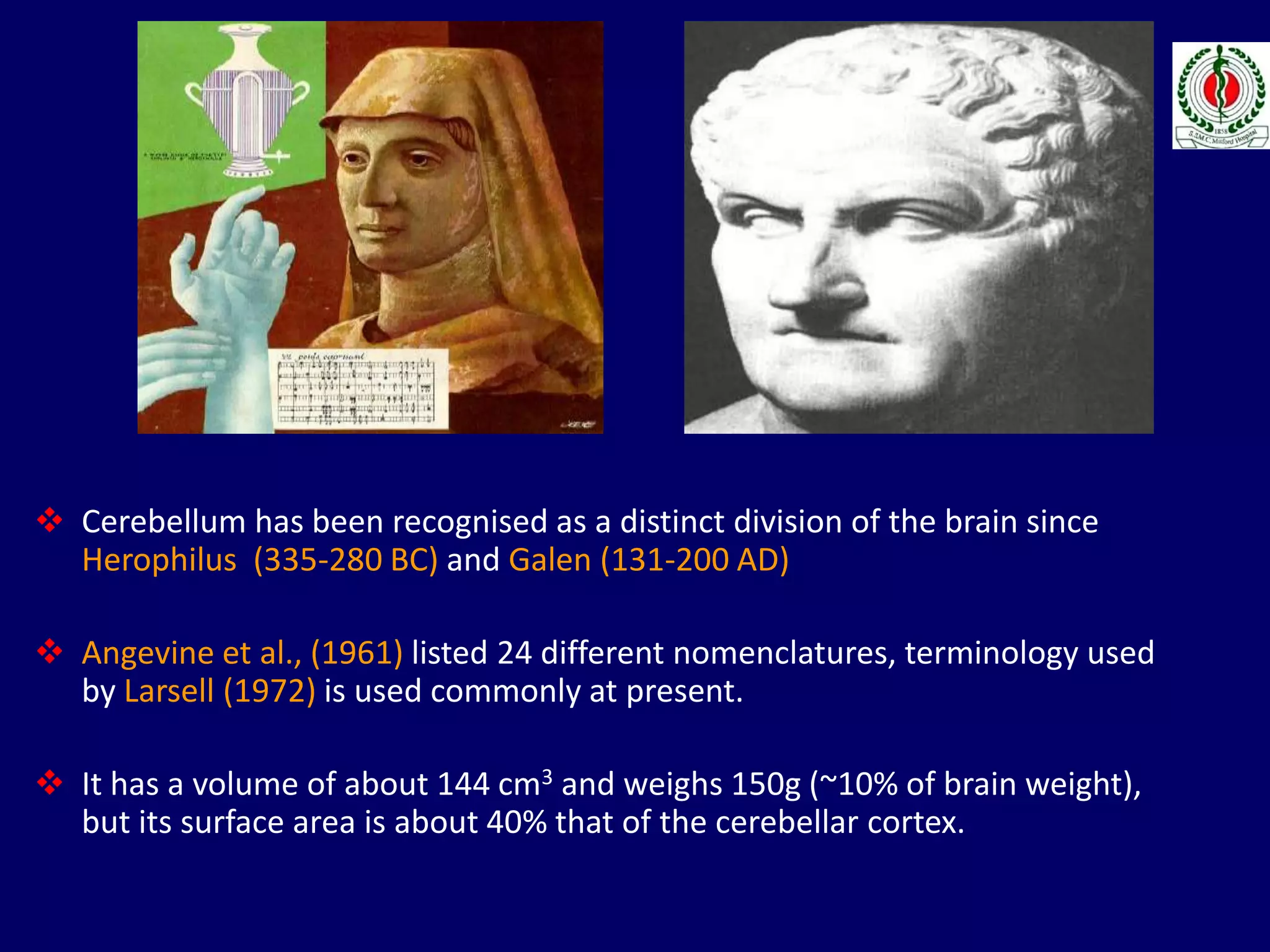
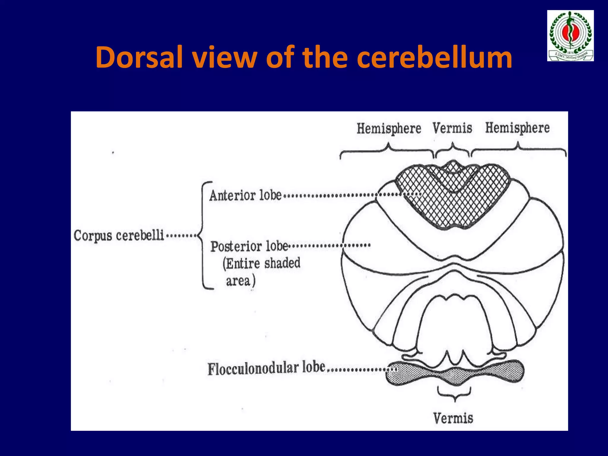
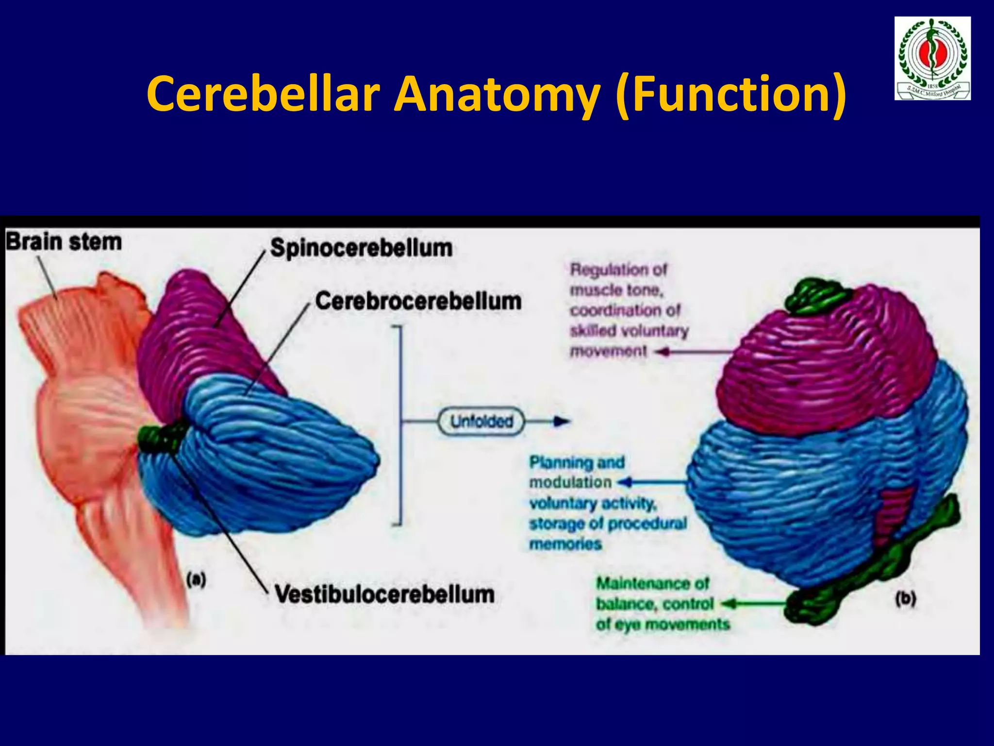
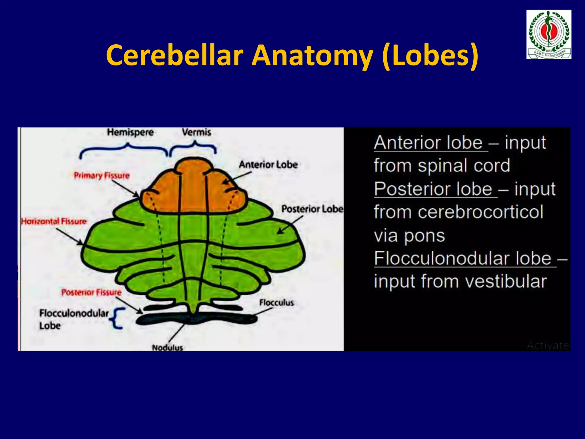
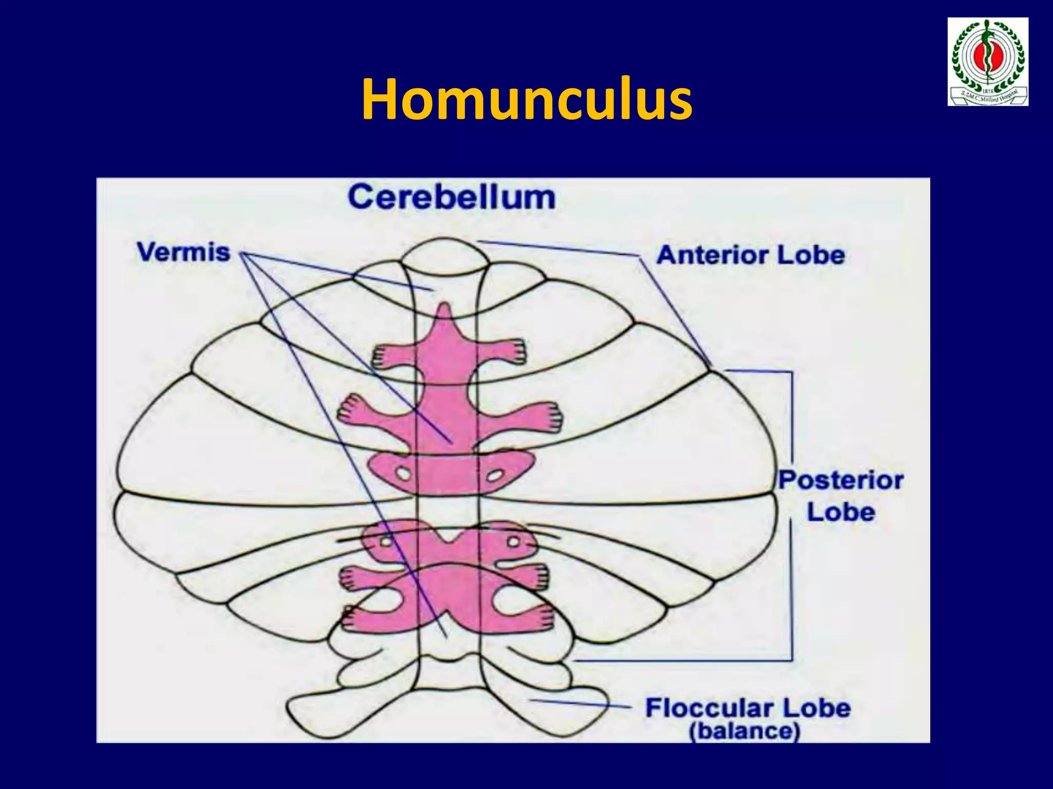
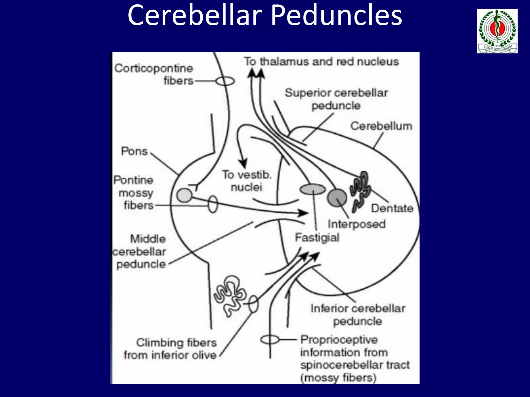
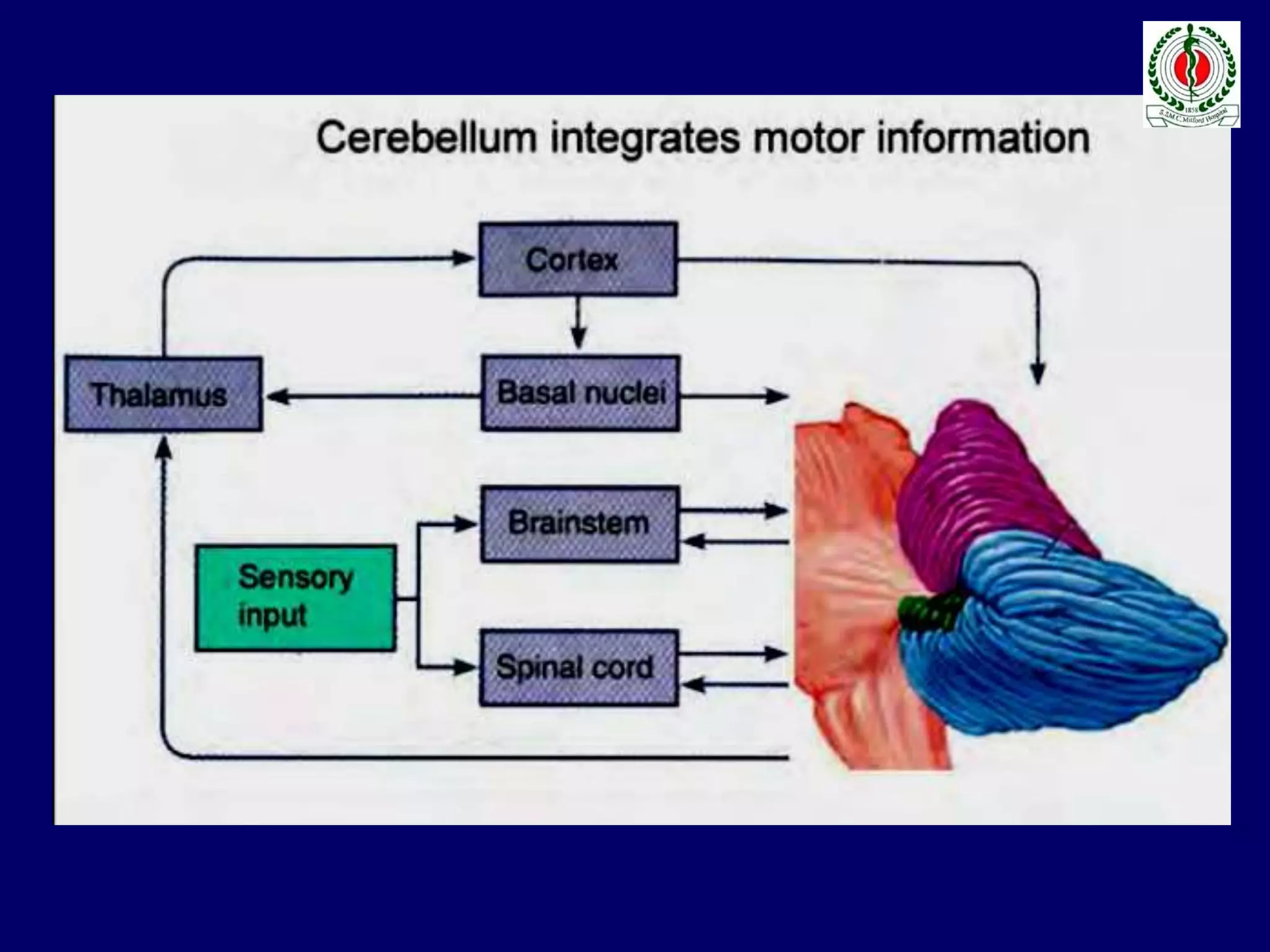
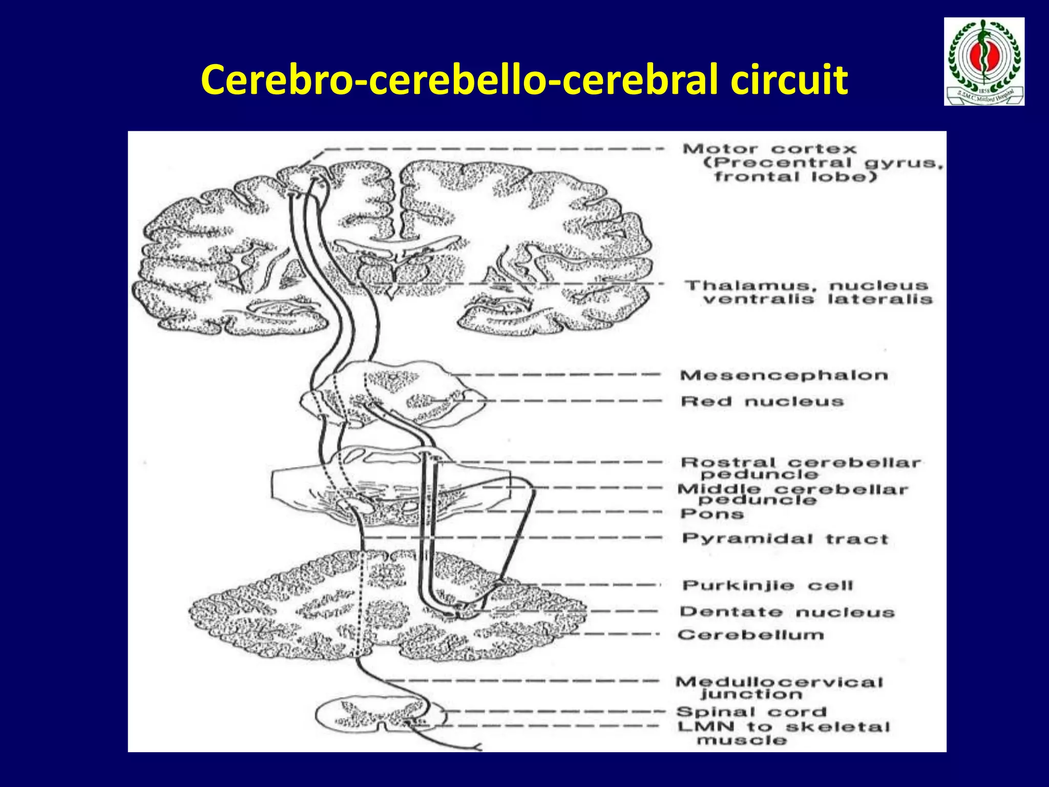
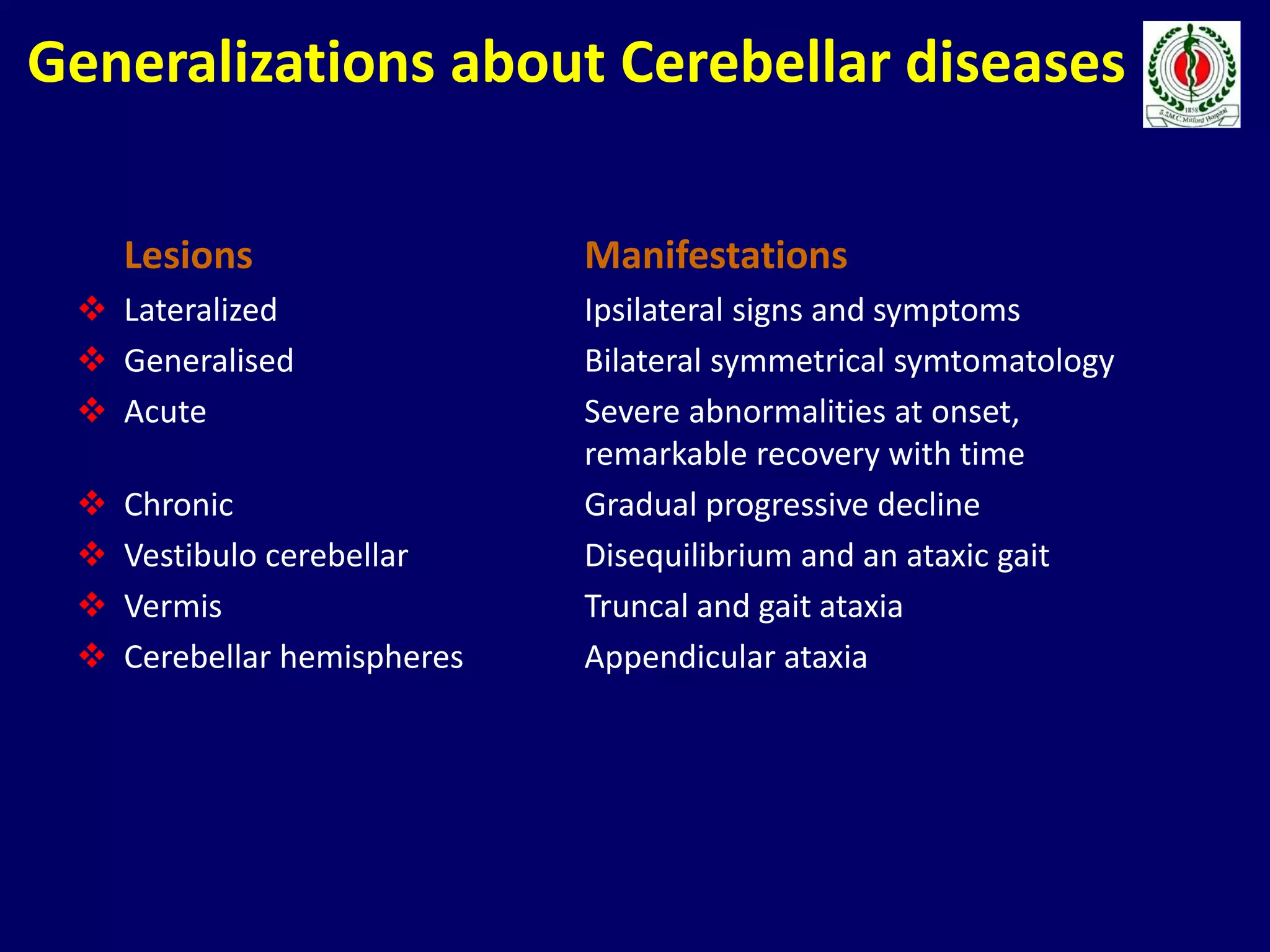
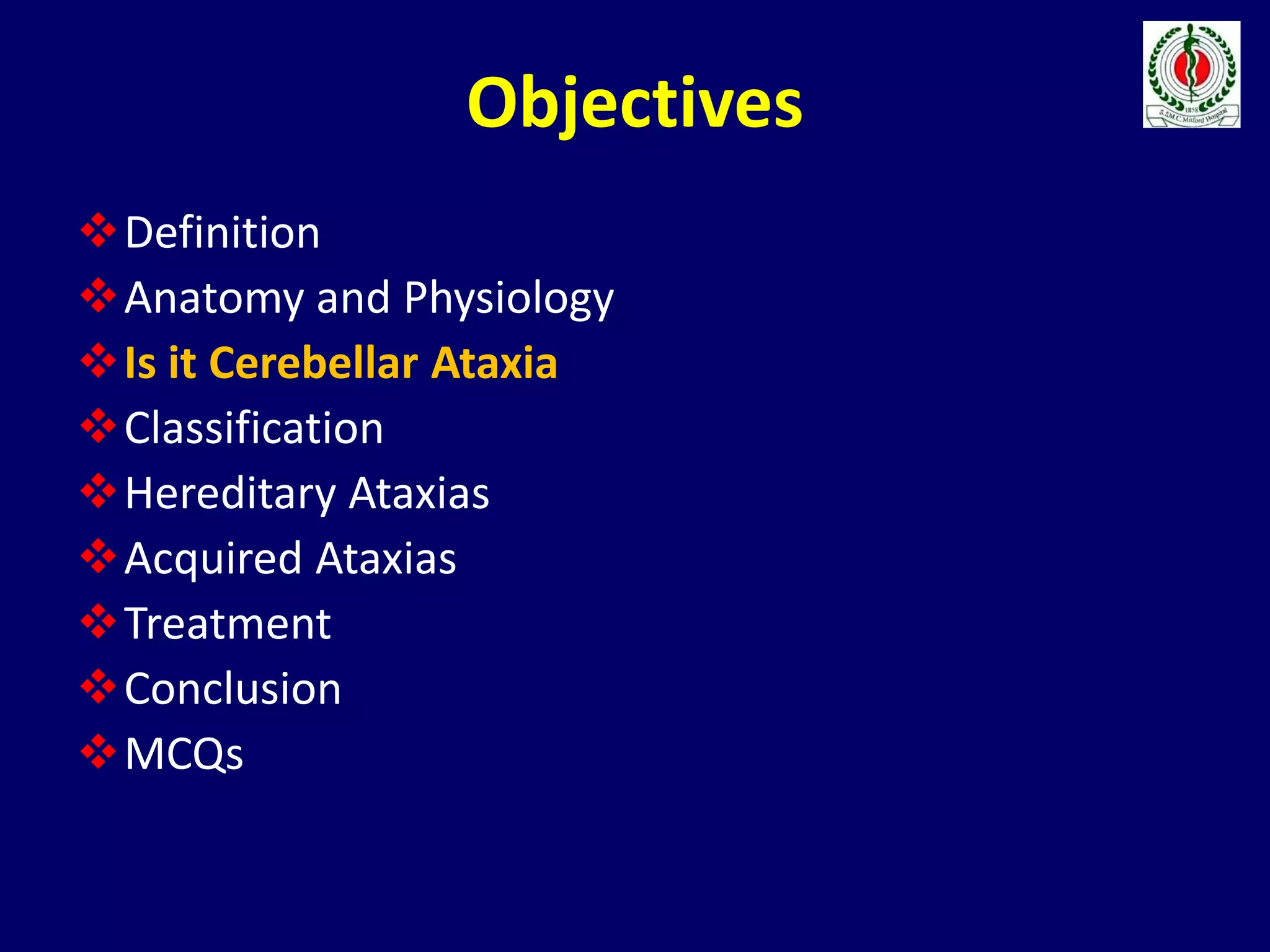
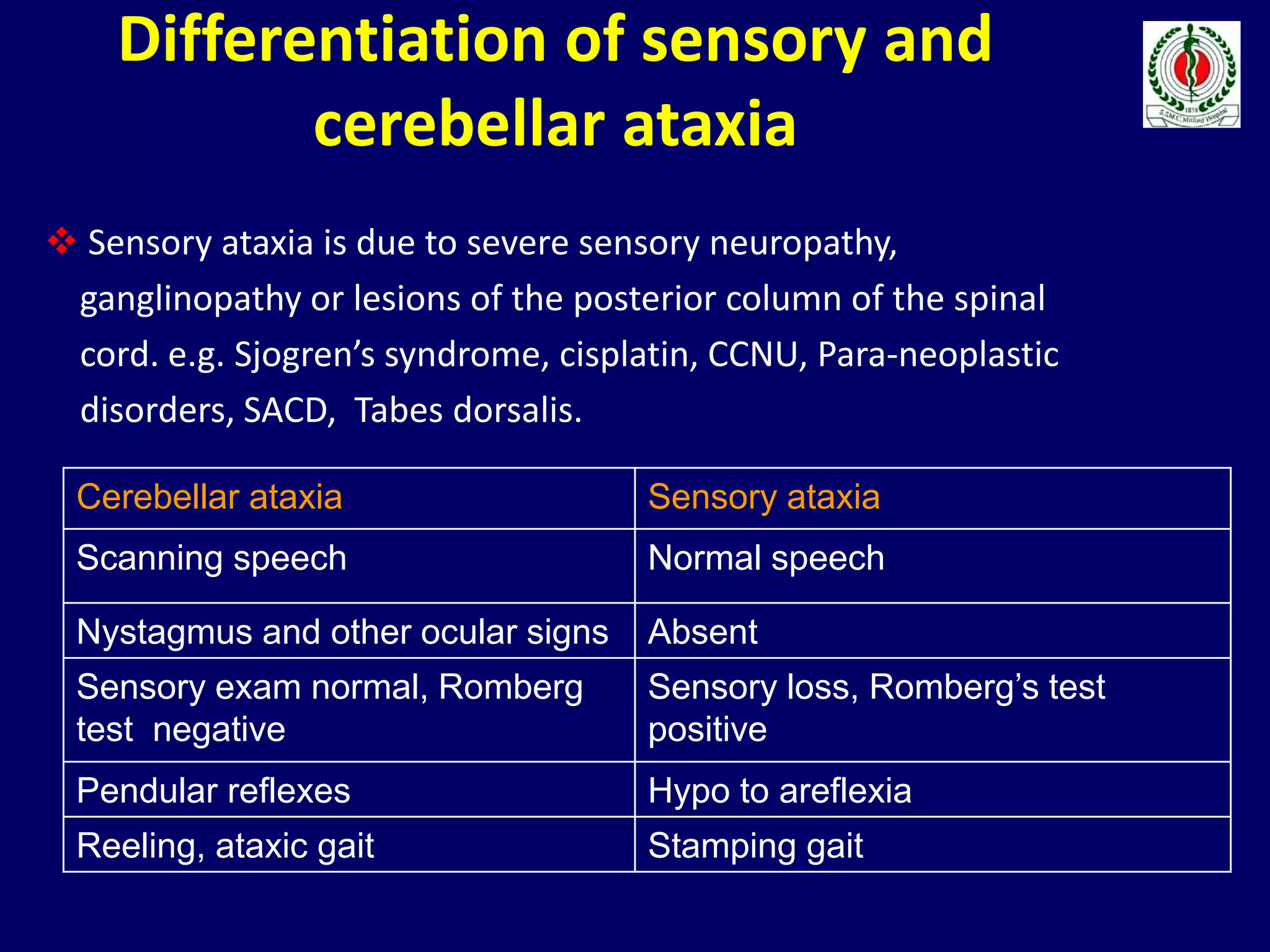
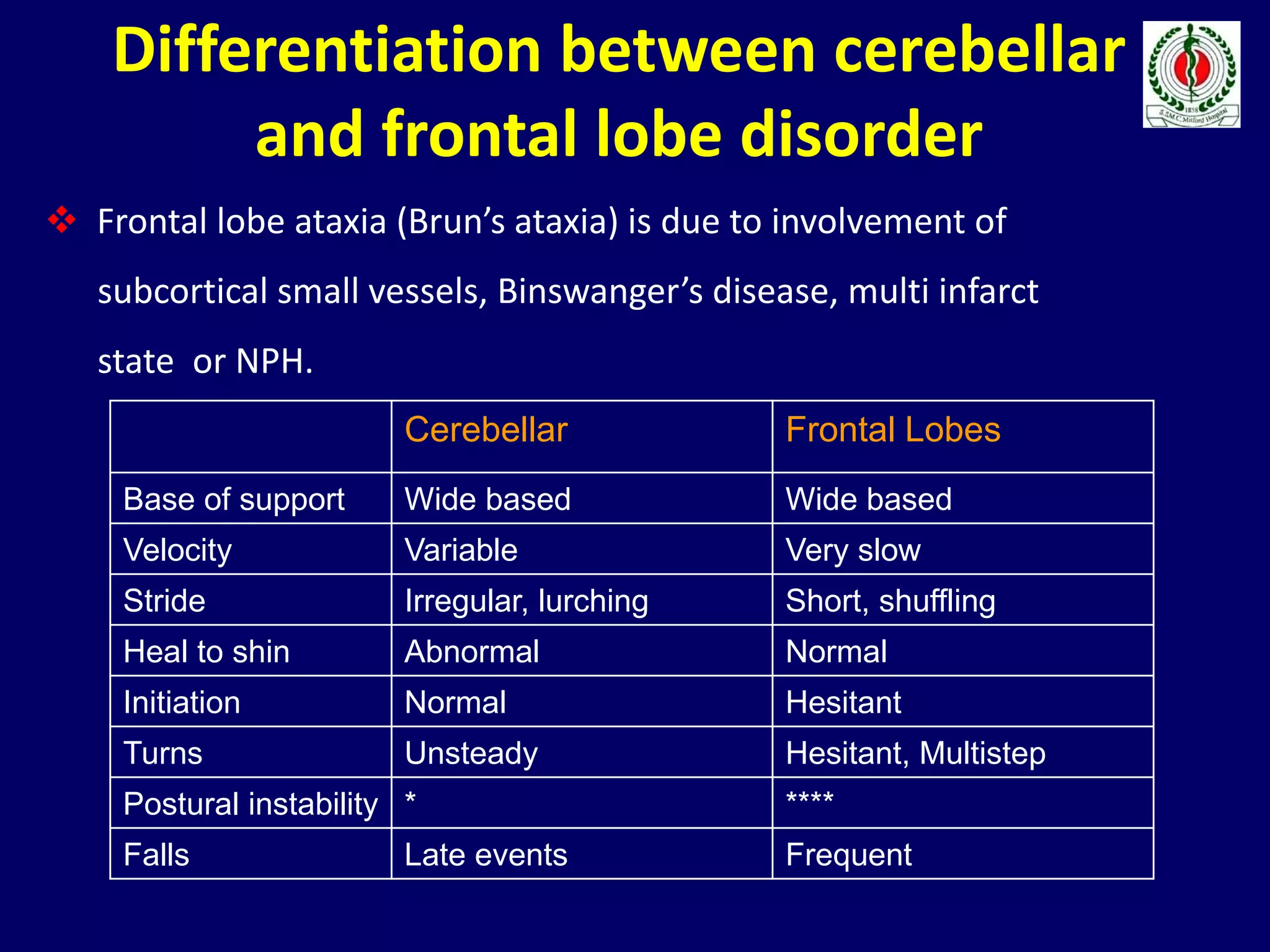
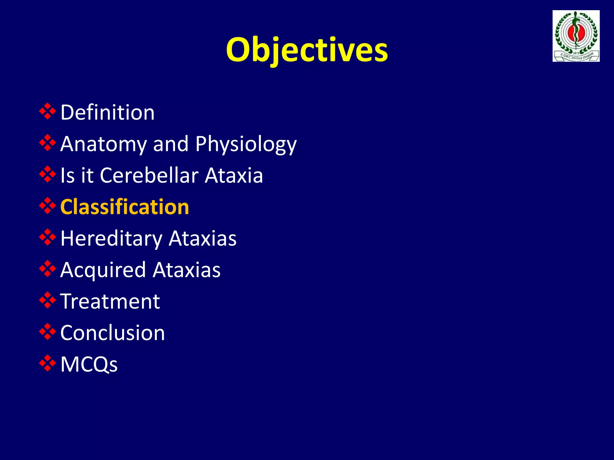
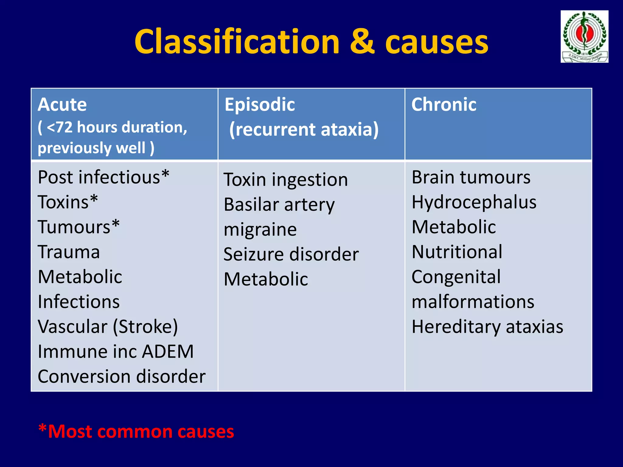
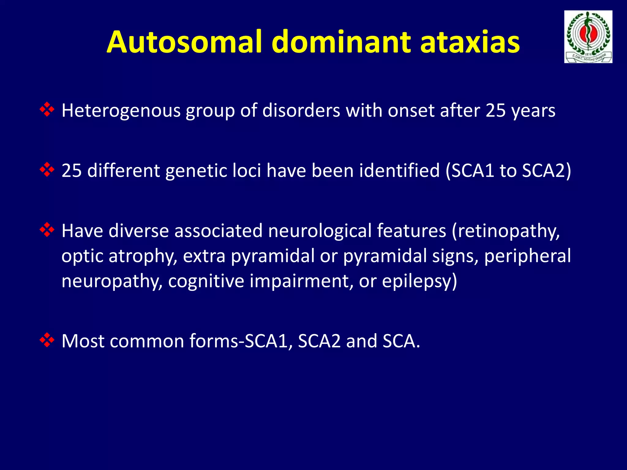
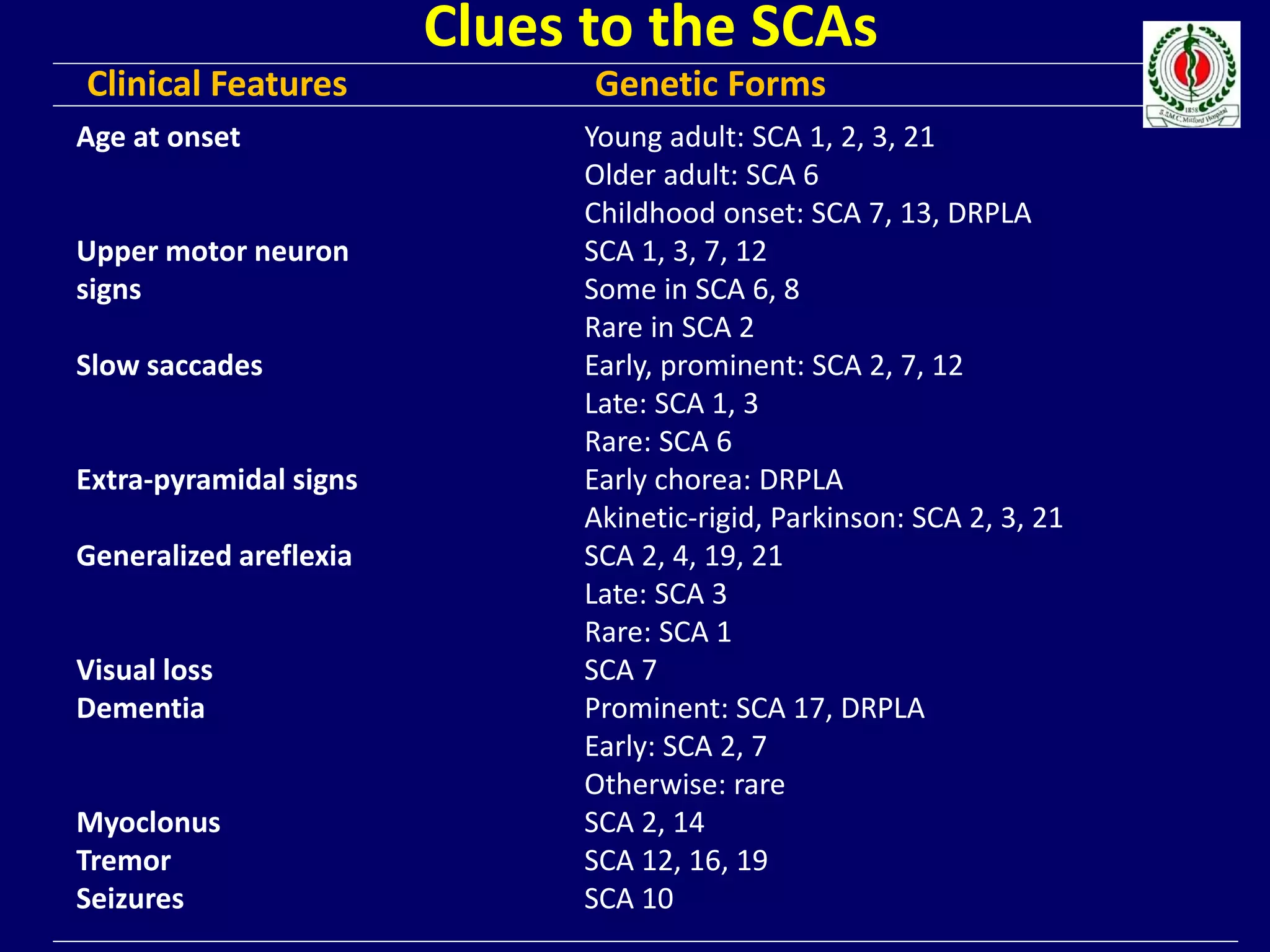
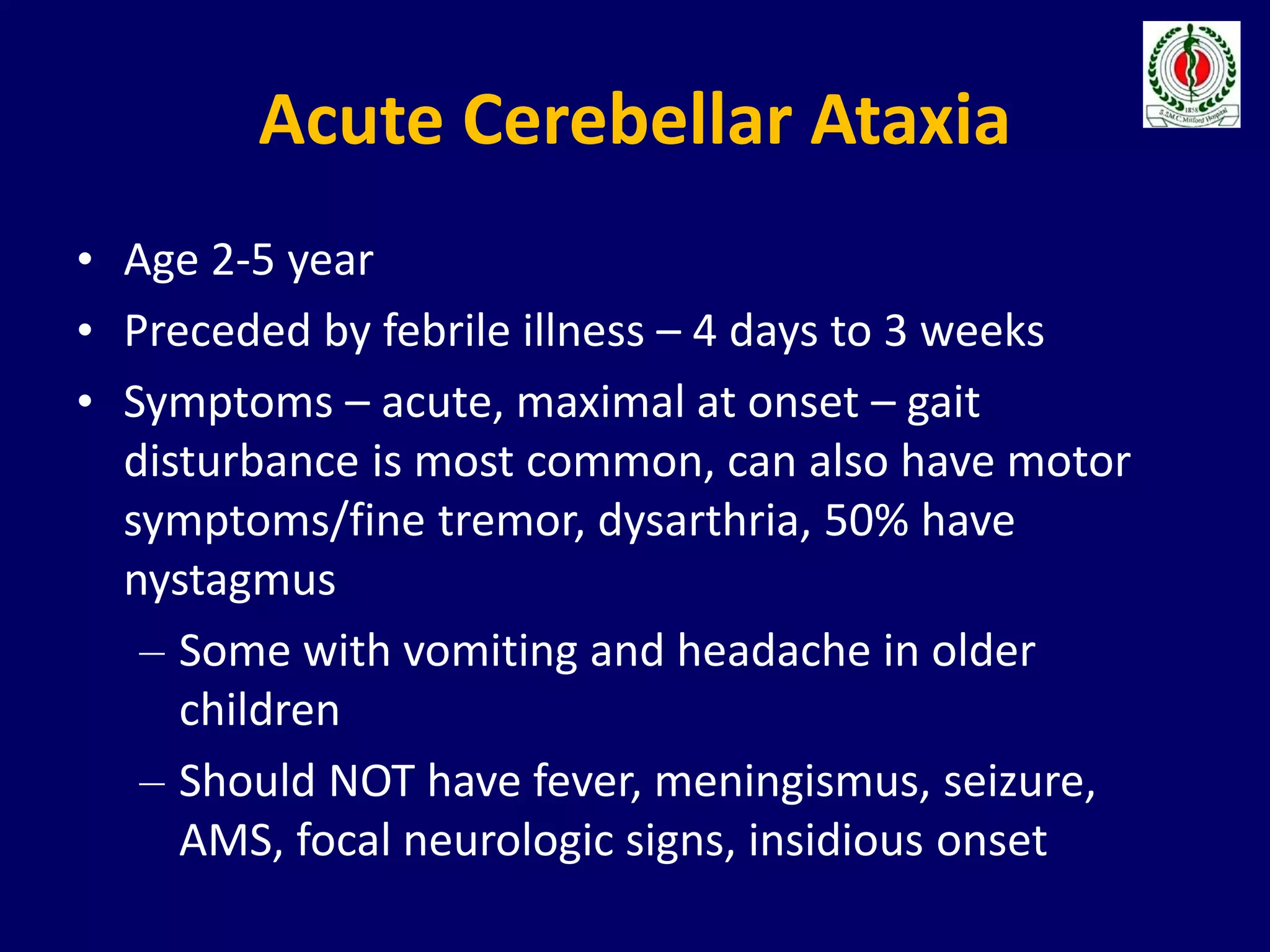
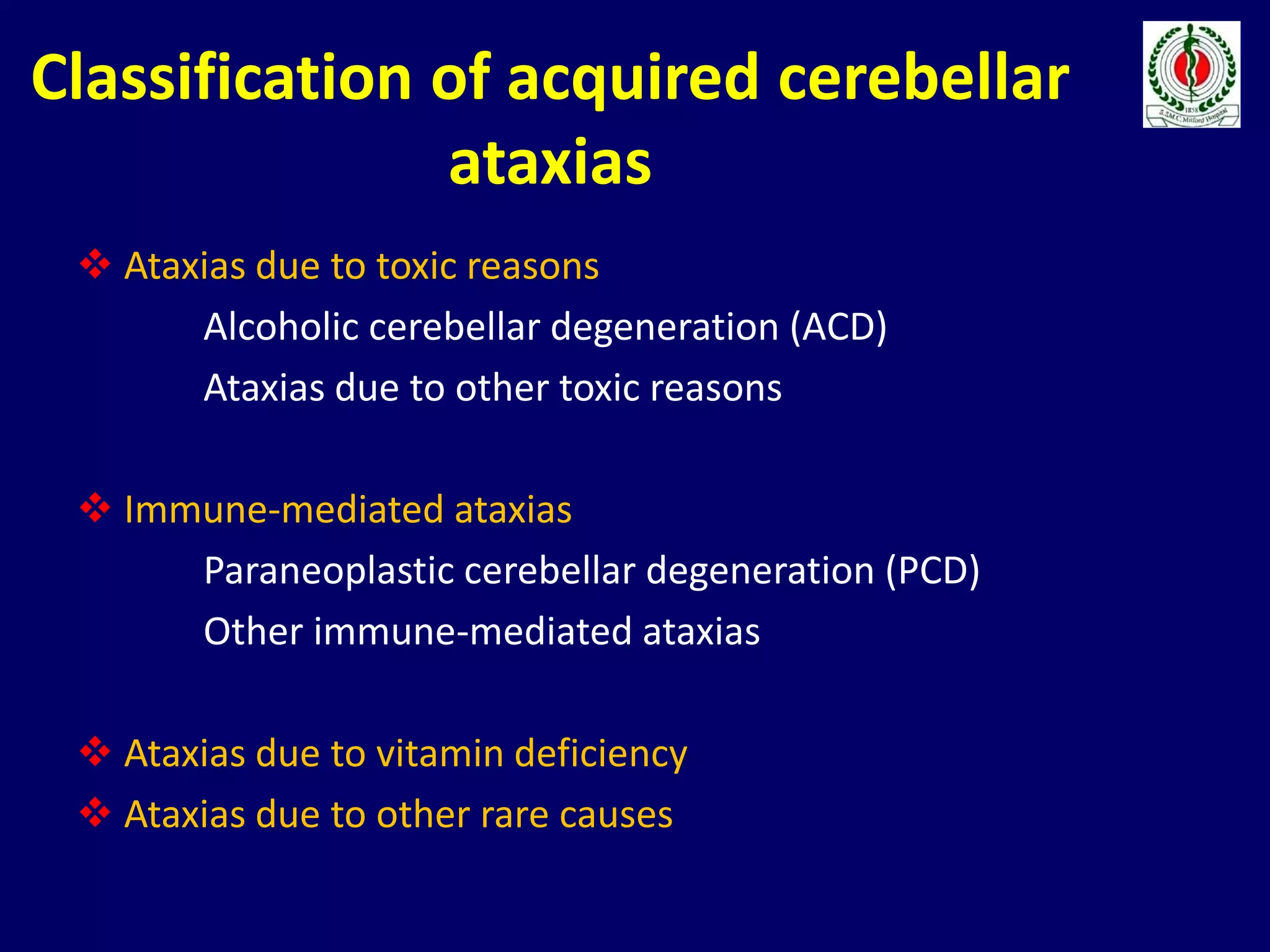
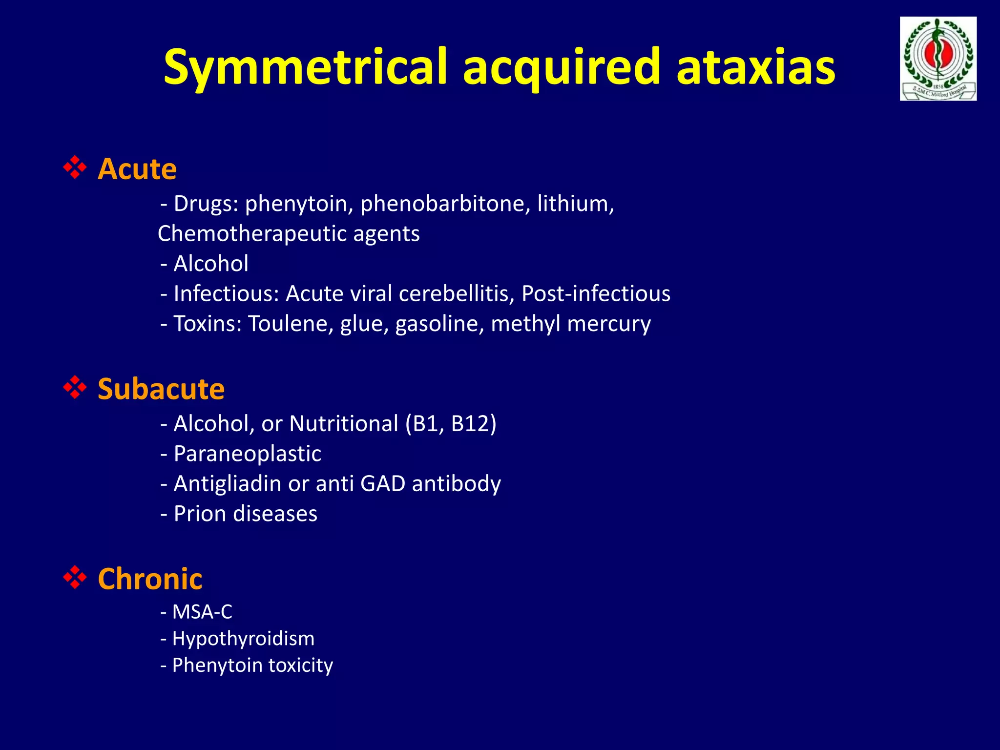
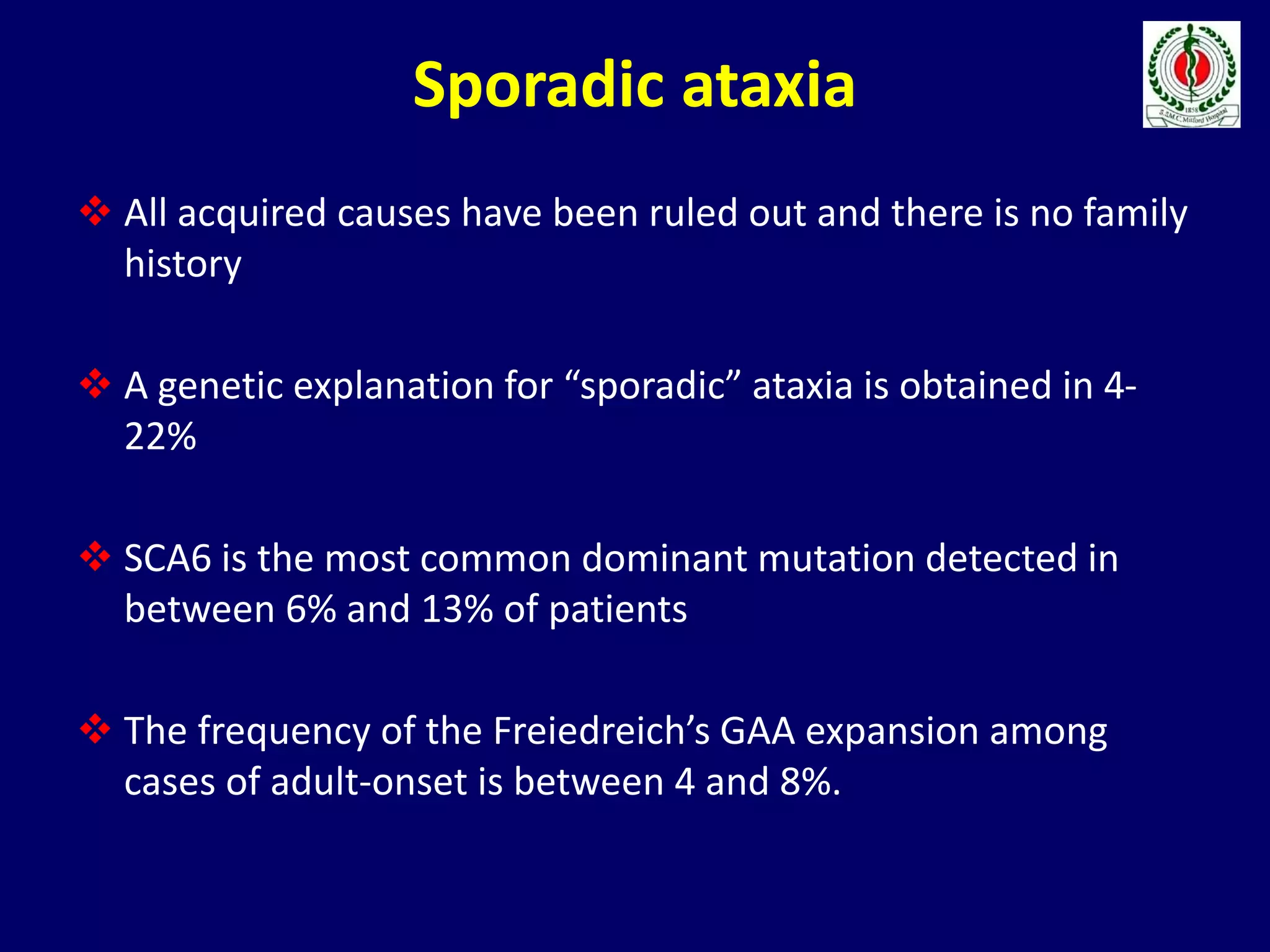
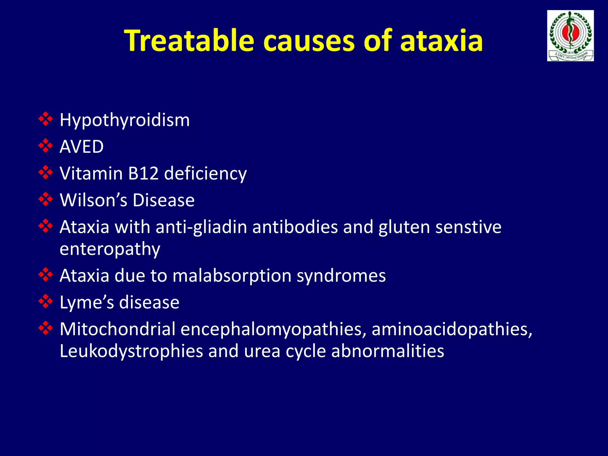
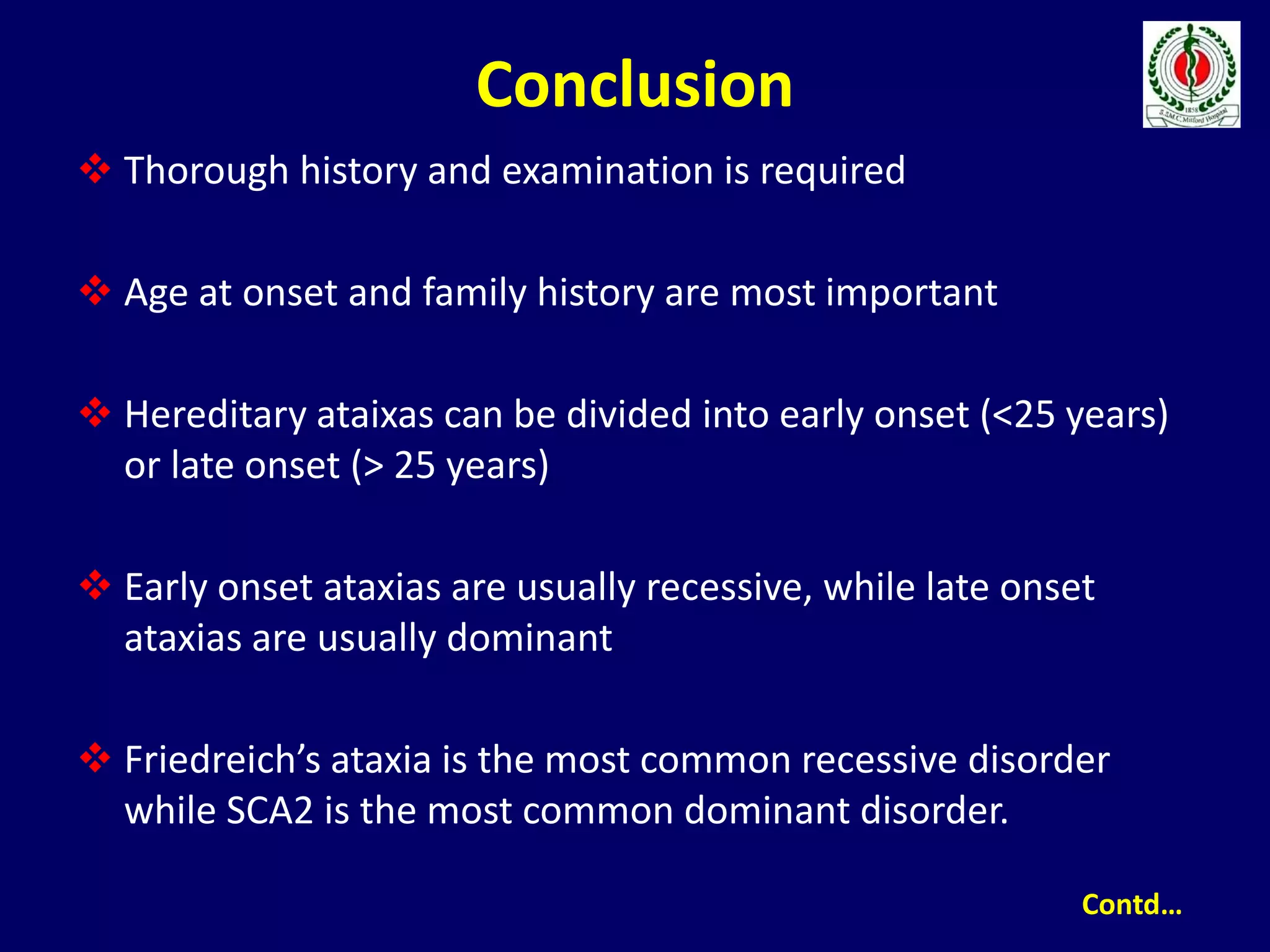
Approach to a Patient
with Ataxia
Dr. Aminur Rahman
FCPS (Med), MD(Neuro) ,FINR (Switzerland),
Member ACP (USA) Member AAN (USA)
Fellow Interventional Neuroradiology (Thailand)
Assistant Professor
Department of Neurology
Sir Salimullah Medical College
Objectives
Definition
Anatomy and Physiology
Is it Cerebellar Ataxia
Classification
Hereditary Ataxias
Acquired Ataxias
Treatment
Conclusion
Definition
 Ataxia (Gk. A Taxis = Order; means lack of order)
 Ataxia denotes a syndrome of imbalance and
incoordination involving gait, limbs, and speech and
usually results from the disorder of the cerebellum or its
connections
 It is characterized by dyssynergia, dysmetria,
dysdiadochokinesia (Joseph Babinski).
 It is a disorder of rate, range, direction and force of
movements (Gordon Holmes).
Objectives
Definition
Anatomy and Physiology
Is it Cerebellar Ataxia
Classification
Hereditary Ataxias
Acquired Ataxias
Treatment
Conclusion
MCQs
 Cerebellum has been recognised as a distinct division of the brain since
Herophilus (335-280 BC) and Galen (131-200 AD)
 Angevine et al., (1961) listed 24 different nomenclatures, terminology used
by Larsell (1972) is used commonly at present.
 It has a volume of about 144 cm3 and weighs 150g (~10% of brain weight),
but its surface area is about 40% that of the cerebellar cortex.
Dorsal view of the cerebellum
Cerebellar function/Ataxia
• Vestibulocerebellum (flocculonodular lobe)- Balance,
reflexive head/eye movements
• Spinocerebellum (vermis, paravermis)- Posture and
limb movements
• Cerebrocerebellum - Movement planning and motor
learning
Cerebellar Anatomy (Function)
Vestibulocerellum – (Archicerebellum)
• Abnormal gate
Abasia - wide based, lurching, staggering
Alcohol impairs cerebellum
• Titubations –
Trunk/head tremor - Vermis lesions
• Tandem gait
Fall or deviate toward lesion - Hemisphere lesions
Vestibulocerellum – (Archicerebellum)
• Ocular dysmetria
– Saccades over/undershoot target
– Jerky saccadic movements during smooth pursuit
• Nystagmus with peripheral gaze
– Slow toward primary, fast toward target
– Horizontal or vertical
– May change direction
– Does not extinguish with fixation
• Impaired suppression of VOR
Spinocerebellum - Paleocerebellum
• Dysmetria
– Over/undershoot, past-pointing
• Dysrhythmia/Asynergia
– Decomposition of complex movements
• Dysdiadochokinesis
– Impaired rate/regularity of alternating movements
• Tremor
– Intention/action- during movement toward target
• Impaired check/excessive rebound
Cerebrocerebellum - Neocerebellum
• Speech -Articulation and prosody
– Scanning or explosive
Irregular fluctuations rate and volume
- Dysarthria
Slurred Speech – extreme = mutism
• Limb ataxia
• Hypotonia
• Intention tremor
• Cognitive affective syndrome
Development of cerebellum
 Vestibular proprioceptors provide information about the
movement of head and its position. Having no limbs, primitive
animals have only the flocculonodular lobe which coordinates
the axial muscles that position the eyes, head and trunk
 All higher animals having limbs have the anterior lobe to
coordinate proprioceptive input from limbs and trunk.
Emergence of vertical bipedal from the quadripedal posture
places particular demands on gait coordination
 The third and newest cerebral lobe (posterior lobe) expands in
equal measure with the cerebrum, motor cortex, pyramidal
tract, basis pontis and inferior olivary nuclei
Cerebellar Anatomy (Lobes)
Homunculus
Cerebellar Peduncles
Cerebro-cerebello-cerebral circuit
Generalizations about Cerebellar diseases
Lesions Manifestations
 Lateralized Ipsilateral signs and symptoms
 Generalised Bilateral symmetrical symtomatology
 Acute Severe abnormalities at onset,
remarkable recovery with time
 Chronic Gradual progressive decline
 Vestibulo cerebellar Disequilibrium and an ataxic gait
 Vermis Truncal and gait ataxia
 Cerebellar hemispheres Appendicular ataxia
Objectives
Definition
Anatomy and Physiology
Is it Cerebellar Ataxia
Classification
Hereditary Ataxias
Acquired Ataxias
Treatment
Conclusion
MCQs
Differentiation of sensory and
cerebellar ataxia
 Sensory ataxia is due to severe sensory neuropathy,
ganglinopathy or lesions of the posterior column of the spinal
cord. e.g. Sjogren’s syndrome, cisplatin, CCNU, Para-neoplastic
disorders, SACD, Tabes dorsalis.
Cerebellar ataxia Sensory ataxia
Scanning speech Normal speech
Nystagmus and other ocular signs Absent
Sensory exam normal, Romberg
test negative
Sensory loss, Romberg’s test
positive
Pendular reflexes Hypo to areflexia
Reeling, ataxic gait Stamping gait
Differentiation of vestibular and
cerebellar ataxia
 Vestibular ataxia is due to lesion of vestibular pathways resulting
in impairment & imbalance of vestibular inputs. e.g. vestibular,
neuronitis, streptomycin toxicity.
Cerebellar ataxia Vestibular disorders
Sense of imbalance Vertigo and associated tinnitus and
hearing loss
Past pointing is in the ipsilateral limb
and in the direction of the lesion
Present in both the limbs and in the
direction of the lesion
Gaze evoked nystagmus Direction of the nystagmus in away from
the lesion
Scanning speech, intention tremors,
dysdiadochokinesia, rebound
phenomena, hypotonia and pendular
reflexes
Absent
Differentiation between cerebellar
and frontal lobe disorder
 Frontal lobe ataxia (Brun’s ataxia) is due to involvement of
subcortical small vessels, Binswanger’s disease, multi infarct
state or NPH.
Cerebellar Frontal Lobes
Base of support Wide based Wide based
Velocity Variable Very slow
Stride Irregular, lurching Short, shuffling
Heal to shin Abnormal Normal
Initiation Normal Hesitant
Turns Unsteady Hesitant, Multistep
Postural instability * ****
Falls Late events Frequent
Objectives
Definition
Anatomy and Physiology
Is it Cerebellar Ataxia
Classification
Hereditary Ataxias
Acquired Ataxias
Treatment
Conclusion
MCQs
Classification & causes
Acute
( <72 hours duration,
previously well )
Episodic
(recurrent ataxia)
Chronic
Post infectious*
Toxins*
Tumours*
Trauma
Metabolic
Infections
Vascular (Stroke)
Immune inc ADEM
Conversion disorder
Toxin ingestion
Basilar artery
migraine
Seizure disorder
Metabolic
Brain tumours
Hydrocephalus
Metabolic
Nutritional
Congenital
malformations
Hereditary ataxias
*Most common causes
Individual with
progressive ataxia
Negative FHAutosomal recessive or
uncertain inheritance
Autosomal dominant
inheritance
Exclude secondary
causes
>25 years
old
<25 years
old
ARCAs, X-linked and
mitochondrial inherited
Consider
ADCAs
Test for:
FRDA (GAA); AT (α-fetoprotein);
AVED (vit. E level); Refsum’s
(phytanic acid); Wilson’s
Harding’s classification
Negative ADCA I
(ataxia + CNS signs)
SCA 1, 2, 3, 4, 8, 12,
17, and FGF 14
ADCA II
(cerebellar syndrome +
pigmentary maculopathy)
SCA 7
ADCA III
(“pure” cerebellar
syndrome)
SCA 5, 6, 10, 11,
14, 15, and 22
Test for other
recessive
ataxias
Objectives
Anatomy and Physiology
Is it Cerebellar Ataxia
Classification
Hereditary Ataxias
Acquired Ataxias
Treatment
Conclusion
MCQs
Hereditary Ataxias
Diagnosis of hereditary ataxia
 Insidious onset, symmetrical, and progressive
 Age at onset
 Early onset ataxia (age at onset below 25 years) is more likely to be
consistent with autosomal recessive inheritance
Exceptions Friedreich’s ataxia, Tay Sachs disease
 Late onset ataxia (age at onset over 25 years) is usual for those
ataxias with dominant inheritance.
Exceptions, SCA7, DRPLA, EA-1, EA-2
 Family history:
- Direct questioning of patient and relatives.
- H/o consanguinity
- Pedigree charting
- Negative family history does not exclude the diagnosis
Causes Of negative F/H
 May be seen in
- Adoption
- Genetic non paternity
- Anticipation
- De novo mutation
- Small family size
- In X-linked inheritance only males are affected.
- In mitochondrial disorders matrilineal mode of
inheritance may be apparent although penetrance is
variable
Freidrich Ataxia
• AR
• Most common hereditary ataxia – 1:50,000
Caucasian
• Frataxin gene – 9q13 – trinculeotide repeat
• Affects brain, heart, pancreas. Slow onset and
progressive.
• Onset before age 25 (often by age 5), ataxia of all 4
limbs, cerebellar dysarthria, absence of LE reflexes,
pyramidal signs (weakness, extensor planters), optic
atrophy, brainstem
Clinical features of Friedreich’s Ataxia
Autosomal recessive inheritance
Onset before 25 years
Progressive limb and gait ataxia
Absent DTR in legs
Electrophysiological evidence of axonal sensory
neuropathy
Dysarthria*
Areflexia in all four limbs*
 Early loss of position/vibration without pain/temperature
loss*
Extensor plantar responses*
Pyramidal weakness of the legs*
*Develop within 5 years of onset of disease
Freidrich Ataxia
• Involvement (swallowing dysfunction),
• Sensineuronal hearing loss
• Cardiomyopathy – 66% of patients – hypertrophic –
main cause of death is arrhythmia and heart failure
• Diabetes mellitus – 33% of patients
Autosomal dominant ataxias
 Heterogenous group of disorders with onset after 25 years
 25 different genetic loci have been identified (SCA1 to SCA2)
 Have diverse associated neurological features (retinopathy,
optic atrophy, extra pyramidal or pyramidal signs, peripheral
neuropathy, cognitive impairment, or epilepsy)
 Most common forms-SCA1, SCA2 and SCA.
Bedside differentiation of SCAs
Large study n=526 patients (17 centres) with SCA 1,2, 3 or 6:
- Pyramidal signs (67%) and brainstem oculomotor
sign (74%) were most frequent in SCA 1
- PN involvement was most frequent in SCA 2 (68%)
- 24% of patients with SCA3 had dystonia
A decrease in visual (83%) and auditory (24%) acuity was
the predominant sign in SCA7
No clinician can accurately distinguish between different
polyglutamine expansion SCAs; but this form of SCA can be
distinguished from other SCA types
David G, et al., Human Med Genet 1998; 7: 165-70
Maschke M, et al., Mov Disord 2005; 20: 1405-12
Schmitz-Hubsch T, et al., Neurology 2008; 71: 982-9
Clues to the SCAs
Clinical Features Genetic Forms
Age at onset Young adult: SCA 1, 2, 3, 21
Older adult: SCA 6
Childhood onset: SCA 7, 13, DRPLA
Upper motor neuron SCA 1, 3, 7, 12
signs Some in SCA 6, 8
Rare in SCA 2
Slow saccades Early, prominent: SCA 2, 7, 12
Late: SCA 1, 3
Rare: SCA 6
Extra-pyramidal signs Early chorea: DRPLA
Akinetic-rigid, Parkinson: SCA 2, 3, 21
Generalized areflexia SCA 2, 4, 19, 21
Late: SCA 3
Rare: SCA 1
Visual loss SCA 7
Dementia Prominent: SCA 17, DRPLA
Early: SCA 2, 7
Otherwise: rare
Myoclonus SCA 2, 14
Tremor SCA 12, 16, 19
Seizures SCA 10
Objectives
Definition
Anatomy and Physiology
Is it Cerebellar Ataxia
Classification
Hereditary Ataxias
Acquired Ataxias
Treatment
Conclusion
MCQs
Acquired Ataxias
Acute Cerebellar Ataxia
• Age 2-5 year
• Preceded by febrile illness – 4 days to 3 weeks
• Symptoms – acute, maximal at onset – gait
disturbance is most common, can also have motor
symptoms/fine tremor, dysarthria, 50% have
nystagmus
– Some with vomiting and headache in older
children
– Should NOT have fever, meningismus, seizure,
AMS, focal neurologic signs, insidious onset
Acute Cerebellar Ataxia
• Differential Diagnoses –
• Toxic/metabolic,
• Infectious—meningitis, encephalitis, ADEM,
labrynthitis,
• Structural,
• Metabolic,
• Neurodegenerative,
• Episodic ataxic syndrome.
Acute Cerebellar Ataxia
• Work-up: CSF if fever, meningitic signs, AMS; imaging if
trauma, ICP signs, posterior fossa signs, asymmetric focal
findings, ADEM
• Usually resolves within 2-3 weeks; if worsening or relapse
than reconsider diagnosis
• Treatment – sometimes steroids, IVIG in severe/refractory
cases
• 10% will have some long-term effects, but most recover
completely
– older age and EBV associated with worse prognosis.
Ataxia-telangectasia
• AR; ATM gene; 11q22.3
• 1:20-100,000
• ATM gene involved in detecting DNA damage, plays role in cell
cycle progression
• Neurologic effects
– Progressive cerebellar ataxia (earliest feature; children
usual begin walking, then have problems with fluid
walking); articulation problems
– abnormal eye movements – oculmotor apraxia is earliest
– Can also have dystonia, chorea, peripheral axonal
neuropathy
Ataxia-telangectasia
• Oculocutaneous telangectasias – oculobulbar, nose,
face, ear, neck. Appear age 3-5 yo. Also can have
cafe-au-lait macules.
• Immune deficiency – recurrent sinopulmonary
infections
• Pulmonary disease – major cause of death –
recurrent infections, interstitial lung disease,
neuromuscular abnormalities (dysphagia, aspiration,
respiratory muscle weakness)
• Malignancy – 10-20% will develop cancer.
Opsoclonus-Myoclonus-Ataxia
Syndrome
• Dancing eyes – dancing feet
• Mean age – 2 yr.
• Opsoclonus – uncontrolled, frequent,
conjugate, saccidic movement of eyes in all
directions.
• irritability, sleep problems
• Auto-immune: Paraneoplastic –
neuroblastoma (unknown Ab); parainfectious
Opsoclonus-Myoclonus-Ataxia Syndrome
• Differential: other causes of myoclonus, ataxia;
toxic/metabolic dz, structural
• Work-up – Look for neuroblastoma – CAP MRI,
VMA/HVA, MIBG scan. If no neuroblastoma, then
MRI brain to look for structural, toxic/metabolic, med
review, infections – viral (hep C, Lyme, EBV, HIV,
coxsackie, rota, mycoplasma, GAS).
• Tx - Treat movements with immunosuppression-
steroids, IVIG, rituximab.
Classification of acquired cerebellar
ataxias
 Ataxias due to toxic reasons
Alcoholic cerebellar degeneration (ACD)
Ataxias due to other toxic reasons
 Immune-mediated ataxias
Paraneoplastic cerebellar degeneration (PCD)
Other immune-mediated ataxias
 Ataxias due to vitamin deficiency
 Ataxias due to other rare causes
Symmetrical acquired ataxias
 Acute
- Drugs: phenytoin, phenobarbitone, lithium,
Chemotherapeutic agents
- Alcohol
- Infectious: Acute viral cerebellitis, Post-infectious
- Toxins: Toulene, glue, gasoline, methyl mercury
 Subacute
- Alcohol, or Nutritional (B1, B12)
- Paraneoplastic
- Antigliadin or anti GAD antibody
- Prion diseases
 Chronic
- MSA-C
- Hypothyroidism
- Phenytoin toxicity
Asymmetrical acquired ataxias
 Acute
- Vascular: Cerebellar infarction or hemorrhage, Subdural
Haemotoma
- Infectious: Abscess
 Subacute
- Neoplastic : Glioma, metastates, lymphoma
- Demyelination : MS
- HIV related : Progressive multi-focal leuco-encephalopathy
 Chronic
- Congenital lesions: Arnold Chiari malformation, Dandy Walker
syndrome
Tumors that produce ataxia
Medulloblastoma
Astrocytoma
Ependymoma
Hemangioblastoma
Metastatic tumor
Meningioma
Cerebellopontine angle tumour: schwannoma
Sporadic ataxia
 All acquired causes have been ruled out and there is no family
history
 A genetic explanation for “sporadic” ataxia is obtained in 4-
22%
 SCA6 is the most common dominant mutation detected in
between 6% and 13% of patients
 The frequency of the Freiedreich’s GAA expansion among
cases of adult-onset is between 4 and 8%.
Objectives
Definition
Anatomy and Physiology
Is it Cerebellar Ataxia
Classification
Hereditary Ataxias
Acquired Ataxias
Treatment
Conclusion
MCQs
Treatment
 Identify treatable causes of ataxia
 No proven therapy for SCAs
 Some patients with parameoplastic cerebellar syndrome
improve following removal of tumour and immunotherapy
 Preliminary evidence suggests that idebenone, a free radical
scavenger improves myocardial hypertrophy
 Genetic counselling can reduce risk in future generations
Treatable causes of ataxia
 Hypothyroidism
 AVED
 Vitamin B12 deficiency
 Wilson’s Disease
 Ataxia with anti-gliadin antibodies and gluten senstive
enteropathy
 Ataxia due to malabsorption syndromes
 Lyme’s disease
 Mitochondrial encephalomyopathies, aminoacidopathies,
Leukodystrophies and urea cycle abnormalities
Objectives
Definition
Anatomy and Physiology
Is it Cerebellar Ataxia
Classification
Hereditary Ataxias
Acquired Ataxias
Treatment
Conclusion
MCQs
Conclusion
 Thorough history and examination is required
 Age at onset and family history are most important
 Hereditary ataixas can be divided into early onset (<25 years)
or late onset (> 25 years)
 Early onset ataxias are usually recessive, while late onset
ataxias are usually dominant
 Friedreich’s ataxia is the most common recessive disorder
while SCA2 is the most common dominant disorder.
Contd…
No single physical sign is specific for a single disorder
Investigations should be performed in a logical order
Treatable causes should be excluded
Conclusion contd…
Objectives
Definition
Anatomy and Physiology
Is it Cerebellar Ataxia
Classification
Hereditary Ataxias
Acquired Ataxias
Treatment
Conclusion
MCQs
MCQs
1. A horizontal gaze evoked nystagmus in which the direction of the
fast phase reverses with sustained lateral gaze or beats transiently in
the opposite direction when the eyes return to primary position is
called
A. Periodic alternating nystagmus
B. Seesaw nystagmus
C. Rebound nystagmus
D. Dysconjugate nystagmus
2. The typical signs of cerebellar herniation include the following
EXCEPT
A. Stiff neck
B. Alteration of consciousness
C. Ptosis and pupillary abnormality
D. Cardiac and respiratory abnormalities
2. The typical signs of cerebellar herniation include the following
EXCEPT
A. Stiff neck
B. Alteration of consciousness
C. Ptosis and pupillary abnormality
D. Cardiac and respiratory abnormalities
3. Romberg’s sign is positive in which type of lesion :
A. Cerebellar
B. Posterior column
C. Hysterical
D. Vestibular
3. Romberg’s sign is positive in which type of lesion :
A. Cerebellar
B. Posterior column
C. Hysterical
D. Vestibular
4. Harding’s classification of cerebellar ataxia is based upon :
A. Mode of inheritance
B. Site of involvement
C. Chromosomal abnormality
D. Metabolic abnormality
4. Harding’s classification of cerebellar ataxia is based upon :
A. Mode of inheritance
B. Site of involvement
C. Chromosomal abnormality
D. Metabolic abnormality
5. Which statement is not true of Friedreich’s ataxia?
A. Recessive inheritance
B.Dysarthria
C. Flexor plantar response
D. Absent ankle jerk
5. Which statement is not true of Friedreich’s ataxia?
A. Recessive inheritance
B.Dysarthria
C. Flexor plantar response
D. Absent ankle jerk
6. Alcoholic cerebellar degeneration is characterized by :
A. Gaze evoked nystagmus
B. Limb ataxia
C. Gait ataxia
D. Action myoclonus
6. Alcoholic cerebellar degeneration is characterized by :
A. Gaze evoked nystagmus
B. Limb ataxia
C. Gait ataxia
D. Action myoclonus
7. Cerebellar ataxia can result from intoxication with :
A. Lead
B. Mercury
C. Manganese
D. Gold
7. Cerebellar ataxia can result from intoxication with :
A. Lead
B. Mercury
C. Manganese
D. Gold
8. Wadia’s type of spinocerebellar degeneration is characterized by
A. Limitation of downgaze
B. Limitation of upgaze
C. Slowing of saccadic movements
D. Broken smooth pursuit
8. Wadia’s type of spinocerebellar degeneration is characterized by
A. Limitation of downgaze
B. Limitation of upgaze
C. Slowing of saccadic movements
D. Broken smooth pursuit
9. Which of the following spinocerebellar ataxias (SCAs) has a similar
ion channel, genetic and chromosomal abnormality as familial
hemiplegic migraine
A. SCA type 2
B. SCA type 4
C. SCA type 6
D. SCA type 8
9. Which of the following spinocerebellar ataxias (SCAs) has a similar
ion channel, genetic and chromosomal abnormality as familial
hemiplegic migraine
A. SCA type 2
B. SCA type 4
C. SCA type 6
D. SCA type 8
10. A combination of ipsilateral oculomotor palsy and ipsilateral
cerebellar ataxia is seen in which of the brainstem syndromes
A. Claude
B. Nothnagel
C. Weber
D. Benedikt
10. A combination of ipsilateral oculomotor palsy and ipsilateral
cerebellar ataxia is seen in which of the brainstem syndromes
A. Claude
B. Nothnagel
C. Weber
D. Benedikt
THANK YOU

Approach to a Patient with Ataxia

  • 1.
    Approach to aPatient with Ataxia Dr. Aminur Rahman FCPS (Med), MD(Neuro) ,FINR (Switzerland), Member ACP (USA) Member AAN (USA) Fellow Interventional Neuroradiology (Thailand) Assistant Professor Department of Neurology Sir Salimullah Medical College
  • 2.
    Objectives Definition Anatomy and Physiology Isit Cerebellar Ataxia Classification Hereditary Ataxias Acquired Ataxias Treatment Conclusion
  • 3.
    Definition  Ataxia (Gk.A Taxis = Order; means lack of order)  Ataxia denotes a syndrome of imbalance and incoordination involving gait, limbs, and speech and usually results from the disorder of the cerebellum or its connections  It is characterized by dyssynergia, dysmetria, dysdiadochokinesia (Joseph Babinski).  It is a disorder of rate, range, direction and force of movements (Gordon Holmes).
  • 5.
    Objectives Definition Anatomy and Physiology Isit Cerebellar Ataxia Classification Hereditary Ataxias Acquired Ataxias Treatment Conclusion MCQs
  • 6.
     Cerebellum hasbeen recognised as a distinct division of the brain since Herophilus (335-280 BC) and Galen (131-200 AD)  Angevine et al., (1961) listed 24 different nomenclatures, terminology used by Larsell (1972) is used commonly at present.  It has a volume of about 144 cm3 and weighs 150g (~10% of brain weight), but its surface area is about 40% that of the cerebellar cortex.
  • 7.
    Dorsal view ofthe cerebellum
  • 8.
    Cerebellar function/Ataxia • Vestibulocerebellum(flocculonodular lobe)- Balance, reflexive head/eye movements • Spinocerebellum (vermis, paravermis)- Posture and limb movements • Cerebrocerebellum - Movement planning and motor learning
  • 9.
  • 10.
    Vestibulocerellum – (Archicerebellum) •Abnormal gate Abasia - wide based, lurching, staggering Alcohol impairs cerebellum • Titubations – Trunk/head tremor - Vermis lesions • Tandem gait Fall or deviate toward lesion - Hemisphere lesions
  • 11.
    Vestibulocerellum – (Archicerebellum) •Ocular dysmetria – Saccades over/undershoot target – Jerky saccadic movements during smooth pursuit • Nystagmus with peripheral gaze – Slow toward primary, fast toward target – Horizontal or vertical – May change direction – Does not extinguish with fixation • Impaired suppression of VOR
  • 12.
    Spinocerebellum - Paleocerebellum •Dysmetria – Over/undershoot, past-pointing • Dysrhythmia/Asynergia – Decomposition of complex movements • Dysdiadochokinesis – Impaired rate/regularity of alternating movements • Tremor – Intention/action- during movement toward target • Impaired check/excessive rebound
  • 13.
    Cerebrocerebellum - Neocerebellum •Speech -Articulation and prosody – Scanning or explosive Irregular fluctuations rate and volume - Dysarthria Slurred Speech – extreme = mutism • Limb ataxia • Hypotonia • Intention tremor • Cognitive affective syndrome
  • 14.
    Development of cerebellum Vestibular proprioceptors provide information about the movement of head and its position. Having no limbs, primitive animals have only the flocculonodular lobe which coordinates the axial muscles that position the eyes, head and trunk  All higher animals having limbs have the anterior lobe to coordinate proprioceptive input from limbs and trunk. Emergence of vertical bipedal from the quadripedal posture places particular demands on gait coordination  The third and newest cerebral lobe (posterior lobe) expands in equal measure with the cerebrum, motor cortex, pyramidal tract, basis pontis and inferior olivary nuclei
  • 15.
  • 16.
  • 17.
  • 19.
  • 20.
    Generalizations about Cerebellardiseases Lesions Manifestations  Lateralized Ipsilateral signs and symptoms  Generalised Bilateral symmetrical symtomatology  Acute Severe abnormalities at onset, remarkable recovery with time  Chronic Gradual progressive decline  Vestibulo cerebellar Disequilibrium and an ataxic gait  Vermis Truncal and gait ataxia  Cerebellar hemispheres Appendicular ataxia
  • 21.
    Objectives Definition Anatomy and Physiology Isit Cerebellar Ataxia Classification Hereditary Ataxias Acquired Ataxias Treatment Conclusion MCQs
  • 22.
    Differentiation of sensoryand cerebellar ataxia  Sensory ataxia is due to severe sensory neuropathy, ganglinopathy or lesions of the posterior column of the spinal cord. e.g. Sjogren’s syndrome, cisplatin, CCNU, Para-neoplastic disorders, SACD, Tabes dorsalis. Cerebellar ataxia Sensory ataxia Scanning speech Normal speech Nystagmus and other ocular signs Absent Sensory exam normal, Romberg test negative Sensory loss, Romberg’s test positive Pendular reflexes Hypo to areflexia Reeling, ataxic gait Stamping gait
  • 23.
    Differentiation of vestibularand cerebellar ataxia  Vestibular ataxia is due to lesion of vestibular pathways resulting in impairment & imbalance of vestibular inputs. e.g. vestibular, neuronitis, streptomycin toxicity. Cerebellar ataxia Vestibular disorders Sense of imbalance Vertigo and associated tinnitus and hearing loss Past pointing is in the ipsilateral limb and in the direction of the lesion Present in both the limbs and in the direction of the lesion Gaze evoked nystagmus Direction of the nystagmus in away from the lesion Scanning speech, intention tremors, dysdiadochokinesia, rebound phenomena, hypotonia and pendular reflexes Absent
  • 24.
    Differentiation between cerebellar andfrontal lobe disorder  Frontal lobe ataxia (Brun’s ataxia) is due to involvement of subcortical small vessels, Binswanger’s disease, multi infarct state or NPH. Cerebellar Frontal Lobes Base of support Wide based Wide based Velocity Variable Very slow Stride Irregular, lurching Short, shuffling Heal to shin Abnormal Normal Initiation Normal Hesitant Turns Unsteady Hesitant, Multistep Postural instability * **** Falls Late events Frequent
  • 25.
    Objectives Definition Anatomy and Physiology Isit Cerebellar Ataxia Classification Hereditary Ataxias Acquired Ataxias Treatment Conclusion MCQs
  • 27.
    Classification & causes Acute (<72 hours duration, previously well ) Episodic (recurrent ataxia) Chronic Post infectious* Toxins* Tumours* Trauma Metabolic Infections Vascular (Stroke) Immune inc ADEM Conversion disorder Toxin ingestion Basilar artery migraine Seizure disorder Metabolic Brain tumours Hydrocephalus Metabolic Nutritional Congenital malformations Hereditary ataxias *Most common causes
  • 28.
    Individual with progressive ataxia NegativeFHAutosomal recessive or uncertain inheritance Autosomal dominant inheritance Exclude secondary causes >25 years old <25 years old ARCAs, X-linked and mitochondrial inherited Consider ADCAs Test for: FRDA (GAA); AT (α-fetoprotein); AVED (vit. E level); Refsum’s (phytanic acid); Wilson’s Harding’s classification Negative ADCA I (ataxia + CNS signs) SCA 1, 2, 3, 4, 8, 12, 17, and FGF 14 ADCA II (cerebellar syndrome + pigmentary maculopathy) SCA 7 ADCA III (“pure” cerebellar syndrome) SCA 5, 6, 10, 11, 14, 15, and 22 Test for other recessive ataxias
  • 29.
    Objectives Anatomy and Physiology Isit Cerebellar Ataxia Classification Hereditary Ataxias Acquired Ataxias Treatment Conclusion MCQs
  • 30.
  • 31.
    Diagnosis of hereditaryataxia  Insidious onset, symmetrical, and progressive  Age at onset  Early onset ataxia (age at onset below 25 years) is more likely to be consistent with autosomal recessive inheritance Exceptions Friedreich’s ataxia, Tay Sachs disease  Late onset ataxia (age at onset over 25 years) is usual for those ataxias with dominant inheritance. Exceptions, SCA7, DRPLA, EA-1, EA-2  Family history: - Direct questioning of patient and relatives. - H/o consanguinity - Pedigree charting - Negative family history does not exclude the diagnosis
  • 32.
    Causes Of negativeF/H  May be seen in - Adoption - Genetic non paternity - Anticipation - De novo mutation - Small family size - In X-linked inheritance only males are affected. - In mitochondrial disorders matrilineal mode of inheritance may be apparent although penetrance is variable
  • 33.
    Freidrich Ataxia • AR •Most common hereditary ataxia – 1:50,000 Caucasian • Frataxin gene – 9q13 – trinculeotide repeat • Affects brain, heart, pancreas. Slow onset and progressive. • Onset before age 25 (often by age 5), ataxia of all 4 limbs, cerebellar dysarthria, absence of LE reflexes, pyramidal signs (weakness, extensor planters), optic atrophy, brainstem
  • 34.
    Clinical features ofFriedreich’s Ataxia Autosomal recessive inheritance Onset before 25 years Progressive limb and gait ataxia Absent DTR in legs Electrophysiological evidence of axonal sensory neuropathy Dysarthria* Areflexia in all four limbs*  Early loss of position/vibration without pain/temperature loss* Extensor plantar responses* Pyramidal weakness of the legs* *Develop within 5 years of onset of disease
  • 35.
    Freidrich Ataxia • Involvement(swallowing dysfunction), • Sensineuronal hearing loss • Cardiomyopathy – 66% of patients – hypertrophic – main cause of death is arrhythmia and heart failure • Diabetes mellitus – 33% of patients
  • 36.
    Autosomal dominant ataxias Heterogenous group of disorders with onset after 25 years  25 different genetic loci have been identified (SCA1 to SCA2)  Have diverse associated neurological features (retinopathy, optic atrophy, extra pyramidal or pyramidal signs, peripheral neuropathy, cognitive impairment, or epilepsy)  Most common forms-SCA1, SCA2 and SCA.
  • 37.
    Bedside differentiation ofSCAs Large study n=526 patients (17 centres) with SCA 1,2, 3 or 6: - Pyramidal signs (67%) and brainstem oculomotor sign (74%) were most frequent in SCA 1 - PN involvement was most frequent in SCA 2 (68%) - 24% of patients with SCA3 had dystonia A decrease in visual (83%) and auditory (24%) acuity was the predominant sign in SCA7 No clinician can accurately distinguish between different polyglutamine expansion SCAs; but this form of SCA can be distinguished from other SCA types David G, et al., Human Med Genet 1998; 7: 165-70 Maschke M, et al., Mov Disord 2005; 20: 1405-12 Schmitz-Hubsch T, et al., Neurology 2008; 71: 982-9
  • 38.
    Clues to theSCAs Clinical Features Genetic Forms Age at onset Young adult: SCA 1, 2, 3, 21 Older adult: SCA 6 Childhood onset: SCA 7, 13, DRPLA Upper motor neuron SCA 1, 3, 7, 12 signs Some in SCA 6, 8 Rare in SCA 2 Slow saccades Early, prominent: SCA 2, 7, 12 Late: SCA 1, 3 Rare: SCA 6 Extra-pyramidal signs Early chorea: DRPLA Akinetic-rigid, Parkinson: SCA 2, 3, 21 Generalized areflexia SCA 2, 4, 19, 21 Late: SCA 3 Rare: SCA 1 Visual loss SCA 7 Dementia Prominent: SCA 17, DRPLA Early: SCA 2, 7 Otherwise: rare Myoclonus SCA 2, 14 Tremor SCA 12, 16, 19 Seizures SCA 10
  • 39.
    Objectives Definition Anatomy and Physiology Isit Cerebellar Ataxia Classification Hereditary Ataxias Acquired Ataxias Treatment Conclusion MCQs
  • 40.
  • 41.
    Acute Cerebellar Ataxia •Age 2-5 year • Preceded by febrile illness – 4 days to 3 weeks • Symptoms – acute, maximal at onset – gait disturbance is most common, can also have motor symptoms/fine tremor, dysarthria, 50% have nystagmus – Some with vomiting and headache in older children – Should NOT have fever, meningismus, seizure, AMS, focal neurologic signs, insidious onset
  • 42.
    Acute Cerebellar Ataxia •Differential Diagnoses – • Toxic/metabolic, • Infectious—meningitis, encephalitis, ADEM, labrynthitis, • Structural, • Metabolic, • Neurodegenerative, • Episodic ataxic syndrome.
  • 43.
    Acute Cerebellar Ataxia •Work-up: CSF if fever, meningitic signs, AMS; imaging if trauma, ICP signs, posterior fossa signs, asymmetric focal findings, ADEM • Usually resolves within 2-3 weeks; if worsening or relapse than reconsider diagnosis • Treatment – sometimes steroids, IVIG in severe/refractory cases • 10% will have some long-term effects, but most recover completely – older age and EBV associated with worse prognosis.
  • 44.
    Ataxia-telangectasia • AR; ATMgene; 11q22.3 • 1:20-100,000 • ATM gene involved in detecting DNA damage, plays role in cell cycle progression • Neurologic effects – Progressive cerebellar ataxia (earliest feature; children usual begin walking, then have problems with fluid walking); articulation problems – abnormal eye movements – oculmotor apraxia is earliest – Can also have dystonia, chorea, peripheral axonal neuropathy
  • 45.
    Ataxia-telangectasia • Oculocutaneous telangectasias– oculobulbar, nose, face, ear, neck. Appear age 3-5 yo. Also can have cafe-au-lait macules. • Immune deficiency – recurrent sinopulmonary infections • Pulmonary disease – major cause of death – recurrent infections, interstitial lung disease, neuromuscular abnormalities (dysphagia, aspiration, respiratory muscle weakness) • Malignancy – 10-20% will develop cancer.
  • 46.
    Opsoclonus-Myoclonus-Ataxia Syndrome • Dancing eyes– dancing feet • Mean age – 2 yr. • Opsoclonus – uncontrolled, frequent, conjugate, saccidic movement of eyes in all directions. • irritability, sleep problems • Auto-immune: Paraneoplastic – neuroblastoma (unknown Ab); parainfectious
  • 47.
    Opsoclonus-Myoclonus-Ataxia Syndrome • Differential:other causes of myoclonus, ataxia; toxic/metabolic dz, structural • Work-up – Look for neuroblastoma – CAP MRI, VMA/HVA, MIBG scan. If no neuroblastoma, then MRI brain to look for structural, toxic/metabolic, med review, infections – viral (hep C, Lyme, EBV, HIV, coxsackie, rota, mycoplasma, GAS). • Tx - Treat movements with immunosuppression- steroids, IVIG, rituximab.
  • 48.
    Classification of acquiredcerebellar ataxias  Ataxias due to toxic reasons Alcoholic cerebellar degeneration (ACD) Ataxias due to other toxic reasons  Immune-mediated ataxias Paraneoplastic cerebellar degeneration (PCD) Other immune-mediated ataxias  Ataxias due to vitamin deficiency  Ataxias due to other rare causes
  • 49.
    Symmetrical acquired ataxias Acute - Drugs: phenytoin, phenobarbitone, lithium, Chemotherapeutic agents - Alcohol - Infectious: Acute viral cerebellitis, Post-infectious - Toxins: Toulene, glue, gasoline, methyl mercury  Subacute - Alcohol, or Nutritional (B1, B12) - Paraneoplastic - Antigliadin or anti GAD antibody - Prion diseases  Chronic - MSA-C - Hypothyroidism - Phenytoin toxicity
  • 50.
    Asymmetrical acquired ataxias Acute - Vascular: Cerebellar infarction or hemorrhage, Subdural Haemotoma - Infectious: Abscess  Subacute - Neoplastic : Glioma, metastates, lymphoma - Demyelination : MS - HIV related : Progressive multi-focal leuco-encephalopathy  Chronic - Congenital lesions: Arnold Chiari malformation, Dandy Walker syndrome
  • 51.
    Tumors that produceataxia Medulloblastoma Astrocytoma Ependymoma Hemangioblastoma Metastatic tumor Meningioma Cerebellopontine angle tumour: schwannoma
  • 52.
    Sporadic ataxia  Allacquired causes have been ruled out and there is no family history  A genetic explanation for “sporadic” ataxia is obtained in 4- 22%  SCA6 is the most common dominant mutation detected in between 6% and 13% of patients  The frequency of the Freiedreich’s GAA expansion among cases of adult-onset is between 4 and 8%.
  • 53.
    Objectives Definition Anatomy and Physiology Isit Cerebellar Ataxia Classification Hereditary Ataxias Acquired Ataxias Treatment Conclusion MCQs
  • 54.
    Treatment  Identify treatablecauses of ataxia  No proven therapy for SCAs  Some patients with parameoplastic cerebellar syndrome improve following removal of tumour and immunotherapy  Preliminary evidence suggests that idebenone, a free radical scavenger improves myocardial hypertrophy  Genetic counselling can reduce risk in future generations
  • 55.
    Treatable causes ofataxia  Hypothyroidism  AVED  Vitamin B12 deficiency  Wilson’s Disease  Ataxia with anti-gliadin antibodies and gluten senstive enteropathy  Ataxia due to malabsorption syndromes  Lyme’s disease  Mitochondrial encephalomyopathies, aminoacidopathies, Leukodystrophies and urea cycle abnormalities
  • 56.
    Objectives Definition Anatomy and Physiology Isit Cerebellar Ataxia Classification Hereditary Ataxias Acquired Ataxias Treatment Conclusion MCQs
  • 57.
    Conclusion  Thorough historyand examination is required  Age at onset and family history are most important  Hereditary ataixas can be divided into early onset (<25 years) or late onset (> 25 years)  Early onset ataxias are usually recessive, while late onset ataxias are usually dominant  Friedreich’s ataxia is the most common recessive disorder while SCA2 is the most common dominant disorder. Contd…
  • 58.
    No single physicalsign is specific for a single disorder Investigations should be performed in a logical order Treatable causes should be excluded Conclusion contd…
  • 59.
    Objectives Definition Anatomy and Physiology Isit Cerebellar Ataxia Classification Hereditary Ataxias Acquired Ataxias Treatment Conclusion MCQs
  • 60.
  • 61.
    1. A horizontalgaze evoked nystagmus in which the direction of the fast phase reverses with sustained lateral gaze or beats transiently in the opposite direction when the eyes return to primary position is called A. Periodic alternating nystagmus B. Seesaw nystagmus C. Rebound nystagmus D. Dysconjugate nystagmus
  • 62.
    1. A horizontalgaze evoked nystagmus in which the direction of the fast phase reverses with sustained lateral gaze or beats transiently in the opposite direction when the eyes return to primary position is called A. Periodic alternating nystagmus B. Seesaw nystagmus C. Rebound nystagmus D. Dysconjugate nystagmus
  • 63.
    2. The typicalsigns of cerebellar herniation include the following EXCEPT A. Stiff neck B. Alteration of consciousness C. Ptosis and pupillary abnormality D. Cardiac and respiratory abnormalities
  • 64.
    2. The typicalsigns of cerebellar herniation include the following EXCEPT A. Stiff neck B. Alteration of consciousness C. Ptosis and pupillary abnormality D. Cardiac and respiratory abnormalities
  • 65.
    3. Romberg’s signis positive in which type of lesion : A. Cerebellar B. Posterior column C. Hysterical D. Vestibular
  • 66.
    3. Romberg’s signis positive in which type of lesion : A. Cerebellar B. Posterior column C. Hysterical D. Vestibular
  • 67.
    4. Harding’s classificationof cerebellar ataxia is based upon : A. Mode of inheritance B. Site of involvement C. Chromosomal abnormality D. Metabolic abnormality
  • 68.
    4. Harding’s classificationof cerebellar ataxia is based upon : A. Mode of inheritance B. Site of involvement C. Chromosomal abnormality D. Metabolic abnormality
  • 69.
    5. Which statementis not true of Friedreich’s ataxia? A. Recessive inheritance B.Dysarthria C. Flexor plantar response D. Absent ankle jerk
  • 70.
    5. Which statementis not true of Friedreich’s ataxia? A. Recessive inheritance B.Dysarthria C. Flexor plantar response D. Absent ankle jerk
  • 71.
    6. Alcoholic cerebellardegeneration is characterized by : A. Gaze evoked nystagmus B. Limb ataxia C. Gait ataxia D. Action myoclonus
  • 72.
    6. Alcoholic cerebellardegeneration is characterized by : A. Gaze evoked nystagmus B. Limb ataxia C. Gait ataxia D. Action myoclonus
  • 73.
    7. Cerebellar ataxiacan result from intoxication with : A. Lead B. Mercury C. Manganese D. Gold
  • 74.
    7. Cerebellar ataxiacan result from intoxication with : A. Lead B. Mercury C. Manganese D. Gold
  • 75.
    8. Wadia’s typeof spinocerebellar degeneration is characterized by A. Limitation of downgaze B. Limitation of upgaze C. Slowing of saccadic movements D. Broken smooth pursuit
  • 76.
    8. Wadia’s typeof spinocerebellar degeneration is characterized by A. Limitation of downgaze B. Limitation of upgaze C. Slowing of saccadic movements D. Broken smooth pursuit
  • 77.
    9. Which ofthe following spinocerebellar ataxias (SCAs) has a similar ion channel, genetic and chromosomal abnormality as familial hemiplegic migraine A. SCA type 2 B. SCA type 4 C. SCA type 6 D. SCA type 8
  • 78.
    9. Which ofthe following spinocerebellar ataxias (SCAs) has a similar ion channel, genetic and chromosomal abnormality as familial hemiplegic migraine A. SCA type 2 B. SCA type 4 C. SCA type 6 D. SCA type 8
  • 79.
    10. A combinationof ipsilateral oculomotor palsy and ipsilateral cerebellar ataxia is seen in which of the brainstem syndromes A. Claude B. Nothnagel C. Weber D. Benedikt
  • 80.
    10. A combinationof ipsilateral oculomotor palsy and ipsilateral cerebellar ataxia is seen in which of the brainstem syndromes A. Claude B. Nothnagel C. Weber D. Benedikt
  • 81.