The document discusses appendicitis, including:
- The appendix's anatomy and functions. Appendicitis occurs when the appendix becomes blocked and inflamed.
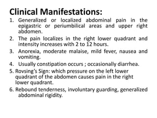
- Appendicitis symptoms include abdominal pain that starts in the center and moves to the lower right side. Other symptoms are nausea, vomiting, and fever.
- Risk factors include infections, foreign objects blocking the appendix, and inflammatory bowel disease.
- Diagnosis involves physical exam, blood tests, and imaging scans like ultrasound or CT.
- Treatment is an appendectomy to remove the appendix. Antibiotics are also given to prevent infection.
- Complications can include infection, abscess, or rupture of the appendix if not treated. Nursing


![APPENDICITIS Nursing managment[Autosaved].pptx](https://cdn.slidesharecdn.com/ss_thumbnails/appendicitisautosaved-250207063037-951fd6a3-thumbnail.jpg?width=640&height=640&fit=bounds)


