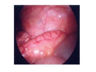
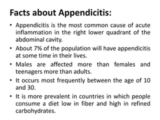
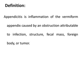
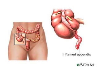
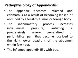
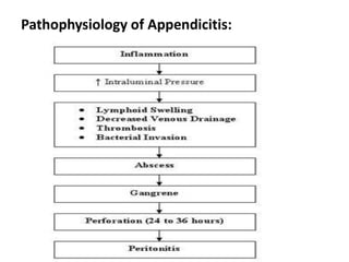
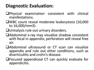
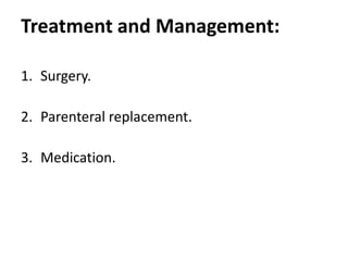
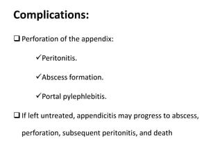
Appendicitis
Fadi Jehad Zaben RN MSN
Indroduction:
• The appendix is a small fingerlike appendage
about 10 cm (4 in) long, attached to the
cecum just below the ileocecal valve.
• No definite functions can be assigned to it in
humans.
• The appendix fills with food and empties as
regularly as does the cecum.
• It is prone to become obstructed and is
particularly vulnerable to infection
(appendicitis) because it is small.
Facts about Appendicitis:
• Appendicitis is the most common cause of acute
inflammation in the right lower quadrant of the
abdominal cavity.
• About 7% of the population will have appendicitis
at some time in their lives.
• Males are affected more than females and
teenagers more than adults.
• It occurs most frequently between the age of 10
and 30.
• It is more prevalent in countries in which people
consume a diet low in fiber and high in refined
carbohydrates.
Definition:
Appendicitis is inflammation of the vermiform
appendix caused by an obstruction attributable
to infection, structure, fecal mass, foreign
body, or tumor.
Pathophysiology of Appendicitis:
• The appendix becomes inflamed and
edematous as a result of becoming kinked or
occluded by a fecalith, tumor, or foreign body.
• The inflammatory process increases
intraluminal pressure, initiating a
progressively severe, generalized or
periumbilical pain that become localized to
the right lower quadrant of the abdomen
within few hour.
• The inflamed appendix fills with pus.
Pathophysiology of Appendicitis:
Risk Factors:
• Age.
• Gender.
Clinical Manifestations:
1. Generalized or localized abdominal pain in the
epigastric or periumbilical areas and upper right
abdomen.
2. The pain localizes in the right lower quadrant and
intensity increases with 2 to 12 hours.
3. Anorexia, moderate malaise, mild fever, nausea and
vomiting.
4. Usually constipation occurs ; occasionally diarrhea.
5. Rovsing’s Sign: which pressure on the left lower
quadrant of the abdomen causes pain in the right
lower quadrant.
6. Rebound tenderness, involuntary guarding, generalized
abdominal rigidity.
Diagnostic Evaluation:
Physical examination consistent with clinical
manifestations.
WBC count reveal moderate leukocytosis (10,000
to 16,000/mm3).
Urinalysis rule out urinary disorders.
Abdominal x-ray may visualize shadow consistent
with fecal in appendix; perforation will reveal free
air.
Abdominal ultrasound or CT scan can visualize
appendix and rule out other conditions, such as
diverticulitis and crohn’s disease.
Focused appendiceal CT can quickly evaluate for
appendicitis.
Treatment and Management:
1. Surgery.
2. Parenteral replacement.
3. Medication.
Continue……… Surgery:
Appendectomy:
It is a surgery to remove of the appendix.
It is the effective treatment.
It is performed if appendicitis is diagnosed as
soon as possible to decrease the risk of
perforation.
Appendectomy may be performed under a
general or spinal anesthetics with a low
abdominal incisions or by laparoscopy which
is recently highly effective method.
Continue…….. Treatment
Administration of IV fluids and antibiotic to
correct or prevent fluid and electrolyte
imbalance, dehydration and sepsis until
surgery is performed.
Administration of Antibiotics.
Analgesics can be administered after the
diagnosed is made.
Complications:
Perforation of the appendix:
Peritonitis.
Abscess formation.
Portal pylephlebitis.
If left untreated, appendicitis may progress to abscess,
perforation, subsequent peritonitis, and death
Nursing Interventions:
Monitor frequently for signs and symptoms of
worsening condition, indicating perforation, abscess,
or peritonitis (increasing severity of pain, tenderness,
rigidity, distention, absent bowel sounds, fever,
malaise, and tachycardia).
Notify health care provider immediately if pain
suddenly ceases, this indicates perforation, which is a
medical emergency.
Assist patient to position of comfort such as semi-
fowlers with knees are flexed.
Continue………..Nursing Interventions
Apply ice bag to abdomen for comfort.
Avoid indiscriminate palpation of the abdomen to avoid
increasing the patients discomfort.
Promptly prepare patient for surgery once diagnosis is
established.
Explain signs and symptoms of postoperative
complications to report-elevated temperature, nausea
and vomiting, or abdominal distention; these may
indicate infection.
Do not give analgesics/antipyretics to mask fever, and
do not administer cathartics because they may cause
rupture.
Restrict activity that may aggravate pain, such as
coughing and ambulation.
Instruct patient on turning, coughing, or deep
breathing, use of incentive spirometer, and
ambulation. Discuss purpose and continued
importance of these maneuvers during recovery
period.
Teach incisional care and avoidance of heavy
lifting or driving until advised by the surgeon.
Advise avoidance of enemas or harsh laxatives;
increased fluids and stool softeners may be used
for postoperative constipation.
Continue………..Nursing Interventions
Discharge Planning:
Antibiotics for infection and analgesic agent can
be given for pain after the surgery.
Within 12 hrs of surgery you may get up and
move around.
Within 2-3 week usually can return to normal
activities s after laparoscopic surgery.
To care wound perform dressing changes and
irrigations as prescribe avoid taking laxative or
applying heat to abdomen when abdominal pain
of unknown cause is experienced.
Reinforce need for follow-up appointment with
the surgeon and to call the physician if the pain
increase at the incision site .
Continue……………Discharge Planning
Document bowel sounds and the passing of
flatus or bowel movements (these are signs of
the return of peristalsis.
Watch for surgical complications such as
continuing pain or fever, which indicate an
abscess .
Stitches removed between fifth and seventh
day.
Liquid or soft diet until the infection subsides
Soft diet is low in fiber and easily breaks down
in the gastrointestinal tract.
Nursing Responsibilities:
Relieving Pain.
Preventing Fluid Volume Deficit.
Reducing Anxiety.
Eliminating Infection.
Maintaining Skin Integrity.
Attaining Optimal Nutrition
Thanks

appendicitis in human body an overview ..

  • 1.
  • 3.
    Indroduction: • The appendixis a small fingerlike appendage about 10 cm (4 in) long, attached to the cecum just below the ileocecal valve. • No definite functions can be assigned to it in humans. • The appendix fills with food and empties as regularly as does the cecum. • It is prone to become obstructed and is particularly vulnerable to infection (appendicitis) because it is small.
  • 4.
    Facts about Appendicitis: •Appendicitis is the most common cause of acute inflammation in the right lower quadrant of the abdominal cavity. • About 7% of the population will have appendicitis at some time in their lives. • Males are affected more than females and teenagers more than adults. • It occurs most frequently between the age of 10 and 30. • It is more prevalent in countries in which people consume a diet low in fiber and high in refined carbohydrates.
  • 5.
    Definition: Appendicitis is inflammationof the vermiform appendix caused by an obstruction attributable to infection, structure, fecal mass, foreign body, or tumor.
  • 7.
    Pathophysiology of Appendicitis: •The appendix becomes inflamed and edematous as a result of becoming kinked or occluded by a fecalith, tumor, or foreign body. • The inflammatory process increases intraluminal pressure, initiating a progressively severe, generalized or periumbilical pain that become localized to the right lower quadrant of the abdomen within few hour. • The inflamed appendix fills with pus.
  • 8.
  • 9.
  • 10.
    Clinical Manifestations: 1. Generalizedor localized abdominal pain in the epigastric or periumbilical areas and upper right abdomen. 2. The pain localizes in the right lower quadrant and intensity increases with 2 to 12 hours. 3. Anorexia, moderate malaise, mild fever, nausea and vomiting. 4. Usually constipation occurs ; occasionally diarrhea. 5. Rovsing’s Sign: which pressure on the left lower quadrant of the abdomen causes pain in the right lower quadrant. 6. Rebound tenderness, involuntary guarding, generalized abdominal rigidity.
  • 11.
    Diagnostic Evaluation: Physical examinationconsistent with clinical manifestations. WBC count reveal moderate leukocytosis (10,000 to 16,000/mm3). Urinalysis rule out urinary disorders. Abdominal x-ray may visualize shadow consistent with fecal in appendix; perforation will reveal free air. Abdominal ultrasound or CT scan can visualize appendix and rule out other conditions, such as diverticulitis and crohn’s disease. Focused appendiceal CT can quickly evaluate for appendicitis.
  • 12.
    Treatment and Management: 1.Surgery. 2. Parenteral replacement. 3. Medication.
  • 13.
    Continue……… Surgery: Appendectomy: It isa surgery to remove of the appendix. It is the effective treatment. It is performed if appendicitis is diagnosed as soon as possible to decrease the risk of perforation. Appendectomy may be performed under a general or spinal anesthetics with a low abdominal incisions or by laparoscopy which is recently highly effective method.
  • 14.
    Continue…….. Treatment Administration ofIV fluids and antibiotic to correct or prevent fluid and electrolyte imbalance, dehydration and sepsis until surgery is performed. Administration of Antibiotics. Analgesics can be administered after the diagnosed is made.
  • 15.
    Complications: Perforation of theappendix: Peritonitis. Abscess formation. Portal pylephlebitis. If left untreated, appendicitis may progress to abscess, perforation, subsequent peritonitis, and death
  • 17.
    Nursing Interventions: Monitor frequentlyfor signs and symptoms of worsening condition, indicating perforation, abscess, or peritonitis (increasing severity of pain, tenderness, rigidity, distention, absent bowel sounds, fever, malaise, and tachycardia). Notify health care provider immediately if pain suddenly ceases, this indicates perforation, which is a medical emergency. Assist patient to position of comfort such as semi- fowlers with knees are flexed.
  • 18.
    Continue………..Nursing Interventions Apply icebag to abdomen for comfort. Avoid indiscriminate palpation of the abdomen to avoid increasing the patients discomfort. Promptly prepare patient for surgery once diagnosis is established. Explain signs and symptoms of postoperative complications to report-elevated temperature, nausea and vomiting, or abdominal distention; these may indicate infection. Do not give analgesics/antipyretics to mask fever, and do not administer cathartics because they may cause rupture.
  • 19.
    Restrict activity thatmay aggravate pain, such as coughing and ambulation. Instruct patient on turning, coughing, or deep breathing, use of incentive spirometer, and ambulation. Discuss purpose and continued importance of these maneuvers during recovery period. Teach incisional care and avoidance of heavy lifting or driving until advised by the surgeon. Advise avoidance of enemas or harsh laxatives; increased fluids and stool softeners may be used for postoperative constipation. Continue………..Nursing Interventions
  • 20.
    Discharge Planning: Antibiotics forinfection and analgesic agent can be given for pain after the surgery. Within 12 hrs of surgery you may get up and move around. Within 2-3 week usually can return to normal activities s after laparoscopic surgery. To care wound perform dressing changes and irrigations as prescribe avoid taking laxative or applying heat to abdomen when abdominal pain of unknown cause is experienced. Reinforce need for follow-up appointment with the surgeon and to call the physician if the pain increase at the incision site .
  • 21.
    Continue……………Discharge Planning Document bowelsounds and the passing of flatus or bowel movements (these are signs of the return of peristalsis. Watch for surgical complications such as continuing pain or fever, which indicate an abscess . Stitches removed between fifth and seventh day. Liquid or soft diet until the infection subsides Soft diet is low in fiber and easily breaks down in the gastrointestinal tract.
  • 22.
    Nursing Responsibilities: Relieving Pain. PreventingFluid Volume Deficit. Reducing Anxiety. Eliminating Infection. Maintaining Skin Integrity. Attaining Optimal Nutrition
  • 23.