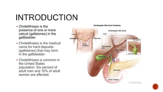
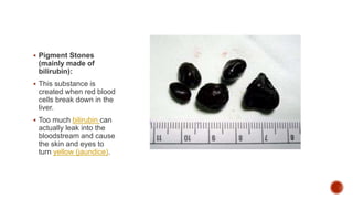
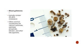
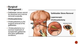
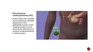
CHOLITHEASIS
BY – SHEHNAZ KHAN
BSC NURSING 2ND YEAR III
SEMESTER
 Cholelithiasis is the
presence of one or more
calculi (gallstones) in the
gallbladder.
 Cholelithiasis is the medical
name for hard deposits
(gallstones) that may form
in the gallbladder.
 Cholelithiasis is common in
the United States
population. Six percent of
adult men and 10% of adult
women are affected.
 In cholelithiasis calculi
(gallstone) usually form in
gall bladder from solid
constituents of bile and
vary gently in size and
shape and composition.
 Gall stone is result of bile
supersaturation with
cholesterol due to
increases synthesis of
cholesterol and decrease
synthesis of bile acids that
dissolve cholesterol.
 Typically, there are three
types:
 Cholesterol Stones :
Made up of fatty
substances in the
blood, cholesterol is found
throughout the body. These
are the most common type
of gallstones.
 Pigment Stones
(mainly made of
bilirubin):
 This substance is
created when red blood
cells break down in the
liver.
 Too much bilirubin can
actually leak into the
bloodstream and cause
the skin and eyes to
turn yellow (jaundice).
 Mixed gallstones
:
 typically contain
20–80%
cholesterol.
 Other common
constituents are
calcium carbonate,
palmitate
phosphate,
bilirubin, and other
bile pigments.
 Your bile contains too much cholesterol. Normally, your bile contains enough
chemicals to dissolve the cholesterol excreted by your liver. But if your liver excretes
more cholesterol than your bile can dissolve, the excess cholesterol may form into
crystals and eventually into stones.
 Your bile contains too much bilirubin. Bilirubin is a chemical that’s produced when your
body breaks down red blood cells. The excess bilirubin contributes to gallstone
formation.
 Your gallbladder doesn’t empty correctly. If your gallbladder doesn’t empty completely
or often enough, bile may become very concentrated, contributing to the formation of
gallstones.
 Women
 Birth control pills
 Pregnancy
 A family history
 Obesity
 Diabetes
 Sedentary life style
 Liver disease
 Rapid weight loss.
 Symptoms may include:
 Pain in your upper belly, often on the right, just under your ribs
 Pain in your right shoulder or back
 An upset stomach
 Vomiting
 Other digestive problems, including indigestion, heartburn, and gas
 See your doctor or go to the hospital if you have signs of a serious infection
or inflammation:
 Belly pain that lasts several hours
 Fever and chills
 Yellow skin or eyes
 Dark urine and light-colored Stool.
 Ultrasound Most sensitive and specific test for gallstones
 Computerized tomography (CT) scan May show gallstones or complications, such
as infection and rupture of GB or bile ducts
 Cholescintigraphy (HIDA scan) Used to diagnose abnormal contraction of
gallbladder or obstruction of bile ducts
 Endoscopic retrograde cholangiopancreatography (ERCP) Used to locate and
remove stones in bile ducts
 Blood tests Performed to look for signs of infection, obstruction, pancreatitis, or
jaundice
 Inflammation of the gallbladder (acute cholecystitis): If a bile duct becomes
permanently blocked, it can lead to a build-up of bile inside the gallbladder. This can
cause the gallbladder to become infected and inflamed.
 Jaundice : You can get jaundice if a gallstone passes out of the gallbladder into the
bile duct and blocks the flow of bile.
 Infection of the bile ducts (acute cholangitis): If the bile ducts become blocked,
they're vulnerable to infection by bacteria. The medical term for a bile duct infection is
acute cholangitis.
 Acute pancreatitis may develop when a gallstone moves out of the gallbladder and
blocks the opening (duct) of the pancreas, causing it to become inflamed.
 Cancer of the gallbladder
 Medical Manegment :
 IV fluids
 NPO with NG tube later progressing to low fat diets
 Antiemetics to prevent nausea and vomiting
 Analgesics to relieve pain
 Anticholinergic to decrease secretions which prevents biliary contraction
 Antibiotics
Surgical
Manegment :
 Gallbladder stones cannot
be dissolved and must be
removed surgically.
 Cholecystectomy :
 Cholecystectomy is the
surgical removal of the
gallbladder.
 Cholecystectomy is a
common treatment of
symptomatic gallstones
and other gallbladder
conditions.
 Percutaneous
cholecystostomy (PC):
 A technique that consists
of percutaneous catheter
placement in the
gallbladder lumen under
imaging guidance, has
become an alternative to
surgical cholecystostomy
in recent years.
Assisment :
 Assess health history : Note history of smoking or prior respiratory problems
 Assess respiratory status: Note shallow respirations, persistent cough, or ineffective or
adventitious breath sounds
 Evaluate nutritional status ( dietary history, general examination and laboratory study
results).
 Acute pain & discomfort related to surgical incision.
 Impaired gas exchange related to the high abdominal surgical incision
 Impaired skin integrity related to altered biliary drainage after surgical intervention
 Imbalanced nutrition, less than body requirements, related to inadequate bile
secretion
 Deficient knowledge about self-care activities related to incision care, dietary
modifications (if needed), medications, reportable signs or symptoms (eg, fever,
bleeding, vomiting)
 Postoperative:
 Place the patient in the low Fowler’s position.
 Provide Intravenous fluids.
 Provide water and other fluids and soft diet is started when bowel sounds return.
 Relieving pain:
 Administer analgesic agents as prescribed to relieve the pain.
 Help the patient to turn, cough, breathe deeply, and ambulate as indicated.
 Use of a pillow or binder over the incision during these maneuvers.
 Improving respiratory status
 Remind patients to take deep breaths and cough every hour to expand the lungs fully
and prevent atelectasis.
 Promote early ambulation. Early ambulation prevents pulmonary complications as well
 Monitor elderly and obese patients must closely for respiratory problem.
 Promoting skin care and biliary drainage:
 observed for indications of infection, leakage of bile into the peritoneal cavity, and
obstruction of bile drainage, clay colored stool and vital sign.
 note and report right upper quadrant abdominal, pain, nausea and vomiting,
 Observe for jaundice.
 changes frequently the outer dressings and protection of the skin from irritation.
 Maintain a careful record of fluid intake and output
Cholelithiasis ( gallstones)

Cholelithiasis ( gallstones)

  • 1.
    CHOLITHEASIS BY – SHEHNAZKHAN BSC NURSING 2ND YEAR III SEMESTER
  • 2.
     Cholelithiasis isthe presence of one or more calculi (gallstones) in the gallbladder.  Cholelithiasis is the medical name for hard deposits (gallstones) that may form in the gallbladder.  Cholelithiasis is common in the United States population. Six percent of adult men and 10% of adult women are affected.
  • 3.
     In cholelithiasiscalculi (gallstone) usually form in gall bladder from solid constituents of bile and vary gently in size and shape and composition.  Gall stone is result of bile supersaturation with cholesterol due to increases synthesis of cholesterol and decrease synthesis of bile acids that dissolve cholesterol.
  • 4.
     Typically, thereare three types:  Cholesterol Stones : Made up of fatty substances in the blood, cholesterol is found throughout the body. These are the most common type of gallstones.
  • 5.
     Pigment Stones (mainlymade of bilirubin):  This substance is created when red blood cells break down in the liver.  Too much bilirubin can actually leak into the bloodstream and cause the skin and eyes to turn yellow (jaundice).
  • 6.
     Mixed gallstones : typically contain 20–80% cholesterol.  Other common constituents are calcium carbonate, palmitate phosphate, bilirubin, and other bile pigments.
  • 7.
     Your bilecontains too much cholesterol. Normally, your bile contains enough chemicals to dissolve the cholesterol excreted by your liver. But if your liver excretes more cholesterol than your bile can dissolve, the excess cholesterol may form into crystals and eventually into stones.  Your bile contains too much bilirubin. Bilirubin is a chemical that’s produced when your body breaks down red blood cells. The excess bilirubin contributes to gallstone formation.  Your gallbladder doesn’t empty correctly. If your gallbladder doesn’t empty completely or often enough, bile may become very concentrated, contributing to the formation of gallstones.
  • 8.
     Women  Birthcontrol pills  Pregnancy  A family history  Obesity  Diabetes  Sedentary life style  Liver disease  Rapid weight loss.
  • 10.
     Symptoms mayinclude:  Pain in your upper belly, often on the right, just under your ribs  Pain in your right shoulder or back  An upset stomach  Vomiting  Other digestive problems, including indigestion, heartburn, and gas  See your doctor or go to the hospital if you have signs of a serious infection or inflammation:  Belly pain that lasts several hours  Fever and chills  Yellow skin or eyes  Dark urine and light-colored Stool.
  • 11.
     Ultrasound Mostsensitive and specific test for gallstones  Computerized tomography (CT) scan May show gallstones or complications, such as infection and rupture of GB or bile ducts  Cholescintigraphy (HIDA scan) Used to diagnose abnormal contraction of gallbladder or obstruction of bile ducts  Endoscopic retrograde cholangiopancreatography (ERCP) Used to locate and remove stones in bile ducts  Blood tests Performed to look for signs of infection, obstruction, pancreatitis, or jaundice
  • 12.
     Inflammation ofthe gallbladder (acute cholecystitis): If a bile duct becomes permanently blocked, it can lead to a build-up of bile inside the gallbladder. This can cause the gallbladder to become infected and inflamed.  Jaundice : You can get jaundice if a gallstone passes out of the gallbladder into the bile duct and blocks the flow of bile.  Infection of the bile ducts (acute cholangitis): If the bile ducts become blocked, they're vulnerable to infection by bacteria. The medical term for a bile duct infection is acute cholangitis.  Acute pancreatitis may develop when a gallstone moves out of the gallbladder and blocks the opening (duct) of the pancreas, causing it to become inflamed.  Cancer of the gallbladder
  • 13.
     Medical Manegment:  IV fluids  NPO with NG tube later progressing to low fat diets  Antiemetics to prevent nausea and vomiting  Analgesics to relieve pain  Anticholinergic to decrease secretions which prevents biliary contraction  Antibiotics
  • 14.
    Surgical Manegment :  Gallbladderstones cannot be dissolved and must be removed surgically.  Cholecystectomy :  Cholecystectomy is the surgical removal of the gallbladder.  Cholecystectomy is a common treatment of symptomatic gallstones and other gallbladder conditions.
  • 15.
     Percutaneous cholecystostomy (PC): A technique that consists of percutaneous catheter placement in the gallbladder lumen under imaging guidance, has become an alternative to surgical cholecystostomy in recent years.
  • 16.
    Assisment :  Assesshealth history : Note history of smoking or prior respiratory problems  Assess respiratory status: Note shallow respirations, persistent cough, or ineffective or adventitious breath sounds  Evaluate nutritional status ( dietary history, general examination and laboratory study results).
  • 17.
     Acute pain& discomfort related to surgical incision.  Impaired gas exchange related to the high abdominal surgical incision  Impaired skin integrity related to altered biliary drainage after surgical intervention  Imbalanced nutrition, less than body requirements, related to inadequate bile secretion  Deficient knowledge about self-care activities related to incision care, dietary modifications (if needed), medications, reportable signs or symptoms (eg, fever, bleeding, vomiting)
  • 18.
     Postoperative:  Placethe patient in the low Fowler’s position.  Provide Intravenous fluids.  Provide water and other fluids and soft diet is started when bowel sounds return.  Relieving pain:  Administer analgesic agents as prescribed to relieve the pain.  Help the patient to turn, cough, breathe deeply, and ambulate as indicated.  Use of a pillow or binder over the incision during these maneuvers.
  • 19.
     Improving respiratorystatus  Remind patients to take deep breaths and cough every hour to expand the lungs fully and prevent atelectasis.  Promote early ambulation. Early ambulation prevents pulmonary complications as well  Monitor elderly and obese patients must closely for respiratory problem.
  • 20.
     Promoting skincare and biliary drainage:  observed for indications of infection, leakage of bile into the peritoneal cavity, and obstruction of bile drainage, clay colored stool and vital sign.  note and report right upper quadrant abdominal, pain, nausea and vomiting,  Observe for jaundice.  changes frequently the outer dressings and protection of the skin from irritation.  Maintain a careful record of fluid intake and output