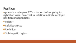
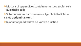
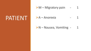
Appendix
• Appendix
• Caecum
• mesoappendix
CT
Remember
•Length of appendices –
•2 – 20 cm
•average – 9 cm
•Development –
•8th week of gestation
•as outpouching of cecal wall
Position
•appendix undergoes 270◦ rotation before going to
right iliac fossa. So arrest in rotation indicates ectopic
position of appendices
•Region –
Left ileac fossa
Umbilicus
Sub-hepatic region
Position
Retro coecal – 70%
Pelvic – 30%
Retro peritoneal – 7%
Mucosa of appendices contain numerous goblet cells
– kulchitsky cells
Sub-mucosa contain numerous lymphoid follicles –
called abdominal tonsil
In adult appendix have no known function
Blood supply
•appendicular artery- branch of Inferior division of Ilea-
colic artery
•pass behind the terminal part of ileum, enter into the
meso-appendix upto tip
•It is end-artery
•Accessory appendicular artery branch of Posterior cecal
artery (called Artery of seshachalam)
Appendicular carcinoid
It is a neuro-endocrinal tumour
 It secretes S100
 <1 cm – Appendectomy suffice
aggressive behavior / >1cm / at the base – right
hemicolectomy
Pathogenesis
Clinical feature
 Few patients present with anorexia,diarrhea,frequency of micturition
On examination
i. Patient looks ill,lying still in bed
ii. Tenderness-max at MacBurney’s point
iii. Rebound Tenderness-always pathognomic
iv. Voluntary muscle guarding
In pelvic type of appendicitis,due to irritation of
bladder,patient may produce with diarrhea and
micturition.
Rofsin’s sign-palpation of left iliac fossa produce pain in rt
iliac fossa
Psoas test-Extension of hip produce pain at rt iliac
fossa(more in retrocaecal position)
Obturator’s sign-internal rotation of hip produce pain.
seen in pelvic type of appendicitis
Alvardo scoring system
• MNEMONIC---MANTRELS
• TOTAL 10 POINT
• IF > 7 POINT – ACUTE APPENDICITIS
M – Migratory pain - 1
A – Anorexia - 1
N – Nausea, Vomiting - 1
PATIENT
DOCTOR
T – Tenderness at Right iliac
fossa - 2
R – Rebound tenderness - 1
E – Elevated temperature - 1
LAB
L – Leukocytosis - 2
S – Left shift [progenitor WBC
cell in periphery] - 1
Diagnosis
Peripheral Blood Count
URINE
1. PUS CELL
2. MICROSCOPIC HEMATURIA
X-ray
Plain X-ray abdomen
shows :---
fecolith / appendicolith
Ureteric stone / Intestine obstruction, perforation
USG
1) Specificity > 90%
2) AP diameter > 7 mm
3) Non-compressibility
4) Target lesion
5) Appendicolith
6) Periappendiceal fluid collection
CT
Specifity- 85-90%
Above +
1. + Appendicolith
2. + Abscess
3. + Any mass
Indication of proplylactic appendicectomy
When doubtful, diagnostic laproscopic
Barium meal X-ray – replaced by CT
MANAGEMENT
Complications / fate
1. Resolution
2. Appendicular lump
3. Appendicular abscess
4. Perforation
5. Gangrenous
6. Peritonitis
7. Portal pyemia
8. Sepsis
9. Mucocoele of appendicitis

Appendicitis

  • 1.
  • 3.
  • 4.
  • 5.
    Remember •Length of appendices– •2 – 20 cm •average – 9 cm •Development – •8th week of gestation •as outpouching of cecal wall
  • 6.
    Position •appendix undergoes 270◦rotation before going to right iliac fossa. So arrest in rotation indicates ectopic position of appendices •Region – Left ileac fossa Umbilicus Sub-hepatic region
  • 8.
    Position Retro coecal –70% Pelvic – 30% Retro peritoneal – 7%
  • 9.
    Mucosa of appendicescontain numerous goblet cells – kulchitsky cells Sub-mucosa contain numerous lymphoid follicles – called abdominal tonsil In adult appendix have no known function
  • 10.
    Blood supply •appendicular artery-branch of Inferior division of Ilea- colic artery •pass behind the terminal part of ileum, enter into the meso-appendix upto tip •It is end-artery •Accessory appendicular artery branch of Posterior cecal artery (called Artery of seshachalam)
  • 11.
    Appendicular carcinoid It isa neuro-endocrinal tumour  It secretes S100  <1 cm – Appendectomy suffice aggressive behavior / >1cm / at the base – right hemicolectomy
  • 13.
  • 14.
    Clinical feature  Fewpatients present with anorexia,diarrhea,frequency of micturition
  • 15.
    On examination i. Patientlooks ill,lying still in bed ii. Tenderness-max at MacBurney’s point iii. Rebound Tenderness-always pathognomic iv. Voluntary muscle guarding In pelvic type of appendicitis,due to irritation of bladder,patient may produce with diarrhea and micturition.
  • 16.
    Rofsin’s sign-palpation ofleft iliac fossa produce pain in rt iliac fossa Psoas test-Extension of hip produce pain at rt iliac fossa(more in retrocaecal position) Obturator’s sign-internal rotation of hip produce pain. seen in pelvic type of appendicitis
  • 17.
    Alvardo scoring system •MNEMONIC---MANTRELS • TOTAL 10 POINT • IF > 7 POINT – ACUTE APPENDICITIS
  • 18.
    M – Migratorypain - 1 A – Anorexia - 1 N – Nausea, Vomiting - 1 PATIENT
  • 19.
    DOCTOR T – Tendernessat Right iliac fossa - 2 R – Rebound tenderness - 1 E – Elevated temperature - 1
  • 20.
    LAB L – Leukocytosis- 2 S – Left shift [progenitor WBC cell in periphery] - 1
  • 21.
  • 22.
    Peripheral Blood Count URINE 1.PUS CELL 2. MICROSCOPIC HEMATURIA
  • 23.
    X-ray Plain X-ray abdomen shows:--- fecolith / appendicolith Ureteric stone / Intestine obstruction, perforation
  • 24.
    USG 1) Specificity >90% 2) AP diameter > 7 mm 3) Non-compressibility 4) Target lesion 5) Appendicolith 6) Periappendiceal fluid collection
  • 25.
    CT Specifity- 85-90% Above + 1.+ Appendicolith 2. + Abscess 3. + Any mass Indication of proplylactic appendicectomy When doubtful, diagnostic laproscopic Barium meal X-ray – replaced by CT
  • 26.
  • 33.
    Complications / fate 1.Resolution 2. Appendicular lump 3. Appendicular abscess 4. Perforation 5. Gangrenous 6. Peritonitis 7. Portal pyemia 8. Sepsis 9. Mucocoele of appendicitis