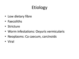
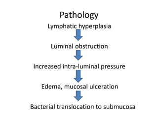
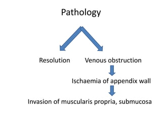
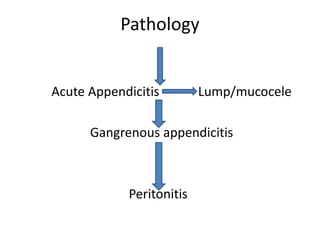
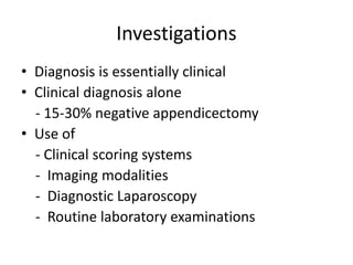
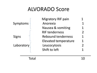
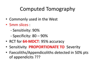
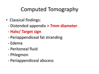
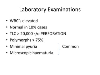
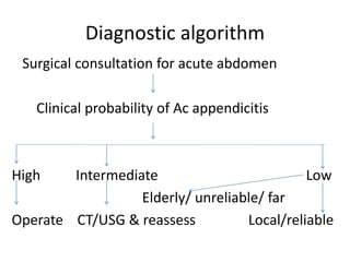
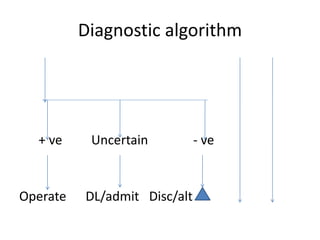
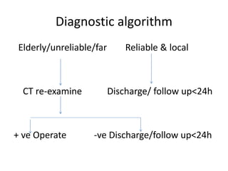
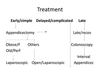
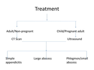
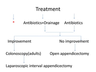
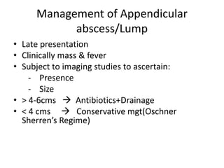
ACUTE APPENDICITIS
Resident: Dr SD Sanyal
Moderator: Brig SR Ghosh
Consultant Surgery and GI Surgeon
Introduction
• Vestigeal organ
• Surgical importance: Propensity for
inflammation
• Most important cause of “Acute Abdomen” in
young adults
Anatomy
• Present only in humans
• At birth: Short & broad at its jn with the
Caecum
• Typical tubular structure produced by 02 years
of age
• Results from differential growth of caecum
Anatomy
• Position: constant  at the confluence of
the 03 taenia coli of caecum
• Mesoappendix : Arises from the lower surface
of the mesentery of terminal ileum
• Appendicular Artery: Branch of Ileo-colic
artery – End Artery
• 04-06 Lymphatic channels traverse
Mesoappendix ----------> Ileo-caecal LNs
Microscopic anatomy
• Layers:
- Mucosa
- Submucosa
- Muscularis
- Serosa
Microscopic anatomy
• Lumen has longitudinal folds of mucous
membrane
• Lining: Columnar cells of colonic type
• Crypts: Argentaffin ( Kulchitsky cells )
• Submucosa: Lymphatic aggregations/Follicles
Anatomical Positions
RETROCAECAL 74%
PELVIC 21%
PARACAECAL 2%
SUBCAECAL 1.5%
PREILEAL 1%
POSTILEAL 0.5%
Anatomical positions
Etiology
• Low dietary fibre
• Faecoliths
• Stricture
• Worm infestations: Oxyuris vermicularis
• Neoplasms: Ca caecum, carcinoids
• Viral
Pathology
Lymphatic hyperplasia
Luminal obstruction
Increased intra-luminal pressure
Edema, mucosal ulceration
Bacterial translocation to submucosa
Pathology
Resolution Venous obstruction
Ischaemia of appendix wall
Invasion of muscularis propria, submucosa
Pathology
Acute Appendicitis Lump/mucocele
Gangrenous appendicitis
Peritonitis
Bacteriology of perforated appendicitis
TYPE OF BACTERIA PATIENTS (%)
ANAEROBIC
B. fragilis 80
B. thetiaotaomicron 61
Bilophila wadsworthia 55
Peptostrptococcus spp 46
AEROBIC
E.coli 77
S.viridans 43
Group D streptococcus 27
P.aeruginosa 18
Clinical features
• Symptoms:
1. Periumbilical pain 50% cases
2. Pain shifts to RIF
3. Anorexia
4. Nausea/vomiting
Clinical features
• Signs:
1. Pyrexia
2. Localized tenderness in RIF
3. Muscle guarding
4. Rebound tenderness
5. Rovsing’s sign
6. Pointing sign
7. Psoas sign
8. Obturator sign
Clinical features
• Risk factors for perforation:
1. Extremes of age
2. Immunosuppression
3. Diabetes mellitus
4. Pelvic appendix
5. Previous abdominal surgery
Special clinical scenarios
• According to position:
1. Retro-caecal
- Silent appendix
- Quadratus lumborum rigidity
- Psoas sign
- Loin tenderness
Special clinical scenarios
2. Pelvic
- Early diarhoea
- Increased urinary frequency
- Deep tenderness over symphysis pubis
- DRE: Rectovesical pouch/POD tenderness
- Obturator/Psoas sign +ve
Special clinical scenarios
3. Post-ileal
- Diarrhoea
- Marked retching
- Ill defined tenderness to rt of umbilicus
Special clinical scenarios
• As per age:
1. Infants
- Uncommon <36 mths
- Difficult to diagnose
- Diffuse peritonitis common
- High incidence of perforation
Special clinical scenarios
2. Children
- Vomiting
- Marked anorexia
3. Elderly
- High incidence of gangrene & perforation
- Features of SAIO
4. Obese
- Diminished signs/ delayed dignosis
- Midline/ Laparoscopic approach
Special clinical scenarios
5. Pregnancy
- Most common extra-uterine cause of acute
abdomen
- Delayed presentation
- Fetal loss
• 3-5%
• Upto 20% : Perforation
Differential Diagnosis
1. Children
- Gastroenteritis
- Mesenteric adenitis
- Meckel’s diverticulitis
- Intussusception
- HS purpura
- Lobar pneumonia
Differential Diagnosis
2. Adults
- Regional enteritis
- Ureteric colic
- Perforated peptic ulcer
- Torsion of testis
- Pancreatitis
- Rectus sheath haematoma
Differential Diagnosis
3. Adult female
- Mittelschmerz
- PID
- Pyelonephritis
- Ectopic pregnancy
- Torsion/ rupture of ovarian cyst
- Endometriosis
Differential Diagnosis
4. Elderly
- Diverticulitis
- Intestinal obstruction
- Ca colon
- Mesenteric infarction
- Torsion of appendix epiploicae
- Leaking aortic aneurysm
Differential Diagnosis
5. Rare
- Tabetic crisis
- Spinal conditions
- Porphyria
- Diabetes
- Typhilitis
Investigations
• Diagnosis is essentially clinical
• Clinical diagnosis alone
- 15-30% negative appendicectomy
• Use of
- Clinical scoring systems
- Imaging modalities
- Diagnostic Laparoscopy
- Routine laboratory examinations
ALVORADO Score
Migratory RIF pain 1
Symptoms Anorexia 1
Nausea & vomiting 1
RIF tenderness 2
Signs Rebound tenderness 1
Elevated temperature 1
Laboratory Leucocytosis 2
Shift to left 1
_______________________________________
Total 10
ALVORADO Score
• < 4: Excludes diagnosis
• 5-6: Equivocal
• >7 : Strongly s/o appendicitis
• Modified Alvorado Score:
- 9 points
- Differential count not done
• PAS:
- Total : 10
- Rebound tenderness excluded
- Cough/percussion/hopping tendeness = 2
- Leucocytosis > 10,000  1
Tzanakis Score
1. Rt lower abdominal tenderness = 4
2. Rebound tenderness = 3
3. WBC’s> 12,000 in the blood = 2
4. Positive USS findings of appendicitis = 6
• Total score = 15
• > 8 = 96% chances
Computed Tomography
• Commonly used in the West
• 5mm slices :
- Sensitivity: 90%
- Specificity: 80 – 90%
• RCT for 64-MDCT: 95% accuracy
• Sensitivity PROPORTIONATE TO Severity
• Faecoliths/Appendicoliths detected in 50% pts
of appendicits ???
Computed Tomography
• Classical findings:
- Distended appendix > 7mm diameter
- Halo/ Target sign
- Periappendiceal fat stranding
- Edema
- Peritoneal fluid
- Phlegmon
- Periappendiceal abscess
Computed Tomography
• Rational use:
- Elderly
- Atypical presentations
- Neoplasms
- Acute diverticulits
- Intestinal obstruction
• MRI: ??
US Scans
• Sensitivity = 85% Operator based
• Specificity > 90%
• AP dia appendix > 7mm
• CROSS SECTIONAL VIEW:
- Thick walled
- Non compressible luminal structure : Target Lesion
• Periappendiceal fluid/ Mass
Plain Abdominal X-Rays
• Low sensitivity
• Appendicoliths picked up in only 10-15% cases
• Can be combined with Barium enema
• Failure of appendix to “Fill up”
• Low specificity  20% of normal Appendices
do not fill up
Diagnostic Laparoscopy
• Small fraction of pts
• Women of child bearing age
• Prompt intervention ------- Implications on
future fertility
Laboratory Examinations
• WBC’s elevated
• Normal in 10% cases
• TLC > 20,000 s/o PERFORATION
• Polymorphs > 75%
• Minimal pyuria Common
• Microscopic haematuria
Diagnostic algorithm
Surgical consultation for acute abdomen
Clinical probability of Ac appendicitis
High Intermediate Low
Elderly/ unreliable/ far
Operate CT/USG & reassess Local/reliable
Diagnostic algorithm
+ ve Uncertain - ve
Operate DL/admit Disc/alt
Diagnostic algorithm
Elderly/unreliable/far Reliable & local
CT re-examine Discharge/ follow up<24h
+ ve Operate -ve Discharge/follow up<24h
Treatment
Early/simple Delayed/complicated Late
Appendicectomy * Late/recov
Obese/F Others Colonoscopy
Old/Perf
Interval
Laparoscopic Open/Laparoscopic Appendicec
Treatment
Adult/Non-pregnant Child/Pregnant adult
CT Scan Ultrasound
Simple Large abscess Phlegmon/small
appendicitis abscess
Treatment
* Antibiotics+Drainage Antibiotics
Improvement No improvement
Colonoscopy(adults) Open appendicectomy
Laparoscopic interval appendicectomy
Management of Appendicular
abscess/Lump
• Late presentation
• Clinically mass & fever
• Subject to imaging studies to ascertain:
- Presence
- Size
• > 4-6cms  Antibiotics+Drainage
• < 4 cms  Conservative mgt(Oschner
Sherren’s Regime)
Management of Appendicular
abscess/Lump
• Criteriae for stopping conservative mgt:
- Rising pulse rate
- Increase in the size of the mass
- Increasing/ spreading abdominal pain
Problems Intra-operatively
1. Normal appendix – Exclude:
- Meckel’s
- Terminal ilietis
- Tubo-ovarian causes
- Perform appendicectomy in Open approach
- Appendix may be left behind during
laparoscopy
Problems Intra-operatively
2. Appendix not found:
- Mobilise caecum
- Taenia coli to be traced to their confluence
3. Appendicular tumour:
- <2cms: Appendicectomy
- >2cms: Rt hemicolectomy
Complications
1. Wound infection
2. Intra-abdominal abscess
3. Ileus
4. Portal pyaemia( pyelephlebitis)
5. Faecal fistula
6. Adhesive intestinal obstruction
Discussion
• TAC test in a peripheral set-up???

acuteappendicitis-151202182202-lva1-app6891.pdf