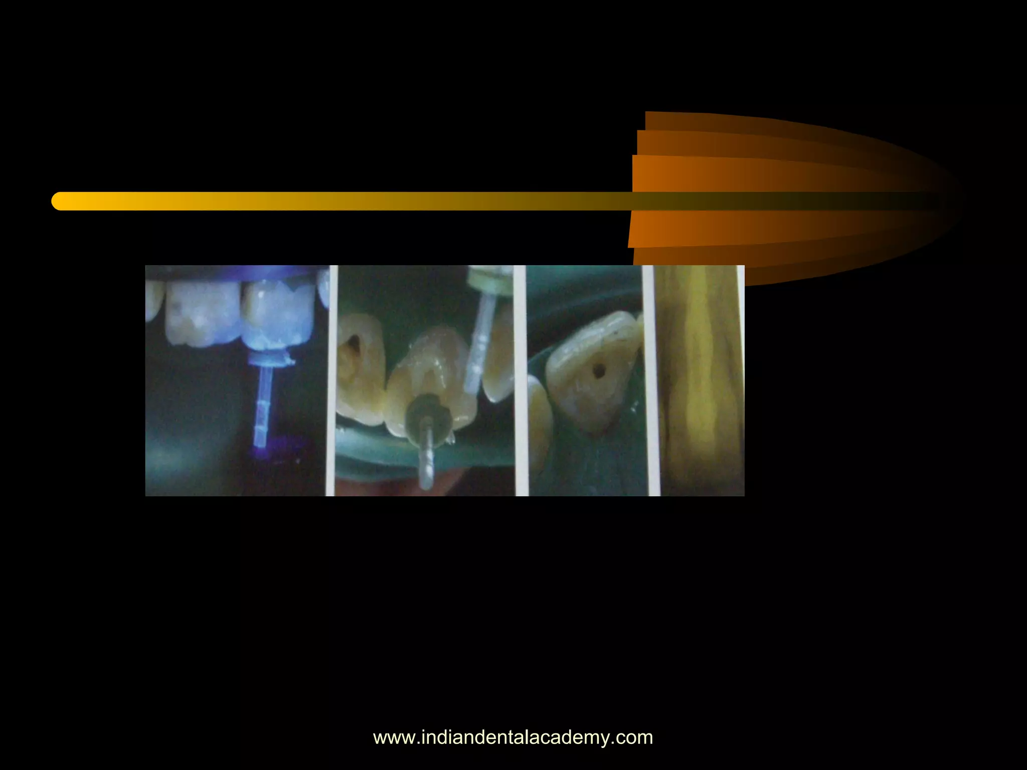
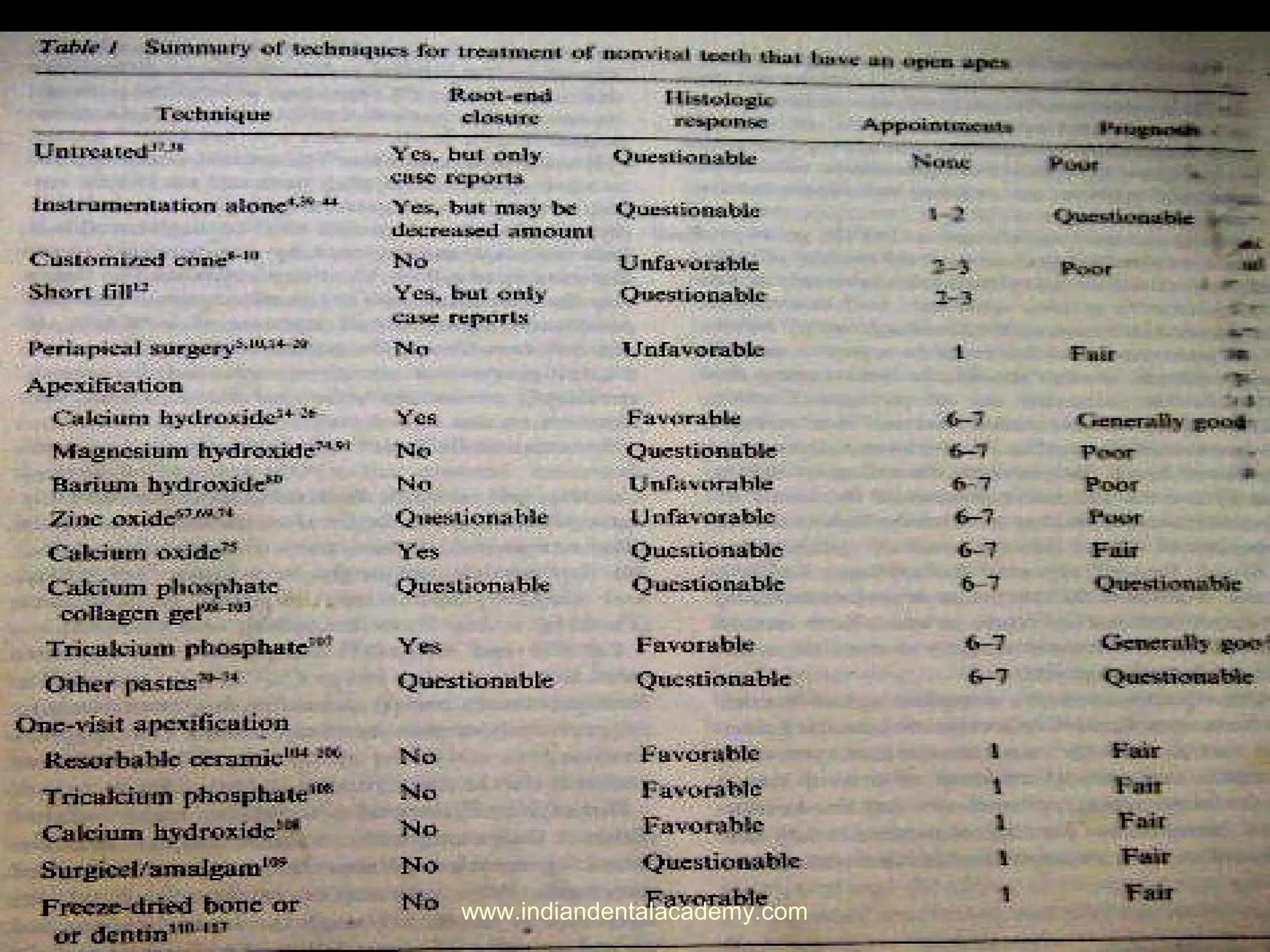
APEXIFICATION : A REVIEW
INDIAN DENTAL ACADEMY
Leader in continuing Dental Education
www.indiandentalacademy.com
APEXIFICATION : A REVIEW
• INDIAN DENTAL ACADEMY
• Leader in continuing Dental Education
www.indiandentalacademy.com
• CONTENTS
• NEED FOR APEXIFICATION
• DIAGNOSIS AND CASE ASSESSMENT
• TREATMENT MODALITIES FOR TEETH WITH OPEN
APEX
• APEXIFICATION
• ONE VISIT APEXIFICATION
• TOOTH RESTORATION FOLLOWING
APEXIFICATION
• CONCLUSIONS
• REFERENCES
www.indiandentalacademy.com
Indian Dental academy
• www.indiandentalacademy.com
• Leader continuing dental education
• Offer both online and offline dental courses
Root development
• Inner & outer enamel epithelia fuse at cervix of the crown to form
Hertwig’s epithelial root sheath
-template for outlining the shape and size of the root
-Differentiation of new odontoblasts from cells of dental papilla
or newly developed pulp
-Epithelial cells of the root sheath loose their continuity,
permitting the ingrowth of CT cells from the surrounding dental
follicle
-Cells of the follicle nearest to the dentin – cementoblasts
-Cells of the follicle farthest to the dentin-osteoblasts
-Intermediate cells forms the Collagen
www.indiandentalacademy.com
Pulpal injury in teeth with developing roots
• Root formation can continue even in the presence of
pulpal inflamation and necrosis
• Complete destruction of HERS results in cessation of
normal root development
• Hard tissue forms by cementoblasts & fibroblasts
www.indiandentalacademy.com
DIAGNOSIS AND CASE ASSESSMENT
• History of subjective symptoms
• Careful clinical and radiographic examination
• Diagnostic tests
www.indiandentalacademy.com
• APEXOGENESIS
‘A vital pulp therapy procedure performed to
encourage continued physiological development and
formation of the root end’
• Goals (webber) :
-sustaining a viable HERS
-Maintaining the pulpal vitality
-Promoting root end closure
-Generating a dentinal bridge at the site of the pulpotomy
www.indiandentalacademy.com
• APEXIFICATION
‘A method to induce a calcified barrier in a root
with an open apex or the continued apical development of
an incomplete root in teeth with necrotic pulp’
Reason:
-To eliminate the periapical infection
-To induce the apical closure, if possible continued root
growth
www.indiandentalacademy.com
• INDICATIONS:
-Immature nonvital permanent tooth with open apex
-Open apex in matured teeth due to periapical osteoclastic
action
www.indiandentalacademy.com
DIFFERENT METHODS
1.Large(blunt) end of gutta-percha cone or customized gutta-
percha cones with sealer
2.Filling the root canal well short of the apex with gutta-
percha and sealer
3.Periapical surgery with or without reverse seal
4.Inducing apical closure by formation of apical stop
5.Placing a biologically acceptable substance in the apical
portion (One visit apexification)
www.indiandentalacademy.com
• Blunt end or rolled cone :
-Apical foramen is wider
than orifice
www.indiandentalacademy.com
• Short fill (Moodnick)
-Filling the root canal short of the apex
- No Healing due to incomplete obturation
• Periapical surgery:
-Reduction in crown : root
-Physical and psychological trauma
-Less possibility of further root development
-Apical walls are thin and shatter when touched by rotating bur,
Retrograde filling difficult
-Periapical tissues may not adapt to the wide and irregular surface of
amalgam
www.indiandentalacademy.com
• Nygard-ostby:Laceration of the periapical tissues inducing
apical closure
• Moller et al: Removal of the infected pulp tissue
• Mc Cormick: Debridement of the root canal, removal of
the necrotic pulp tissue and microorganisms
• Cooke and Robotham:Remnants of HERS
• Antibiotic pastes, antiseptic pastes
www.indiandentalacademy.com
APICAL CLOSURE INDUCTION BY Ca(OH)2:
APEXIFICATTION TECHNIQUE:
Access:
-Straight line access
Instrumentation:
-Working length is determined
-Carefull debridement is a primary factor to induce
apical closure
-Canals enlarged upto a size 120-140
www.indiandentalacademy.com
• Filing motion is needed
Avoid over instrumentation
www.indiandentalacademy.com
• Drying of the canals:
-An inverted coarse paper point is desirable
-A dry pre fitted paper points
• Introduction of paste:
- A Carrier with a teflon or plastic sleeve is recommend
• Condensation:
-A plugger that occludes the canal at a distance of 2-
3mm short of the radiographic apex is selected
www.indiandentalacademy.com
APICAL BARRIERS
www.indiandentalacademy.com
APEXIFICATTION TECHNIQUE:
www.indiandentalacademy.com
APICAL CLOSURE INDUCTION BY Ca(OH)2:
-Kaiser (1964), Ca(OH)2 + CMCP
-Frank (1966)
-Apex closure with definite , although minimal canal
recession
-Apex closure without root space change
-Radiographically apparent bridge formation
immediately coronal to apex
-No radiographic evidence of apical closure,
however, upon instrumentation, definite apical stop
www.indiandentalacademy.com
APICAL CLOSURE INDUCTION BY Ca(OH)2:
• Klein and Levy
-Ca(OH)2 + Cresatin (minimal inflammatory potential)
-less toxic than CMCP
• Ca (OH)2 + Saline
Distilled water
L A Solution with out vasoconstrictor
• Heithersay & others
-Ca (OH)2 + Methyl cellulose
- solubility & a firm physical consistency
www.indiandentalacademy.com
APICAL CLOSURE INDUCTION BY Ca(OH)2:
• Mitchel & Shankwalker
-Osteogenic potential by forming heterotopic bone
• Holland etal
-Multilayered necrosis with subjacent mineralization
• Schroder and Granath
-Layer of firm necrosis generates a low-grade irritation
www.indiandentalacademy.com
• Javelet etal
-Compared the ability of Ca(OH)2 (PH 11.8)
• Antibacterial efficacy :
-Hydroxyl ions are highly oxidant , show extreme
reactivity
-Damage to the bacterial cytoplasmic membrane,
protein denaturation, damage to bacterial DNA
www.indiandentalacademy.com
• Heithersay
-Ca(OH)2 increases the Ca concentration at the pre
capillary sphincter, reducing the plasma flow
-Stimulates the enzyme pyrophosphatase (collagen
synthesis)
• Hard tissue Barriers:
Ghose et al:
-Cap, bridge or ingrown wedge and may be composed
of cementum , dentin , bone or ‘osteodentine’
www.indiandentalacademy.com
• Hard tissue barriers:
Torneck et al
- Deposition of bone like material
Steiner and van Hassel
-Calcific bridge (cementum)
• Cementum formation proceeds from the periphery of the
original apex towards the center in decreasing concentric
circles
• Histologically barrier is porous
www.indiandentalacademy.com
• SEM and Histologic study:
- Outer surface of the bridge extended in a ‘cap’ like
fashion over the root apex , displaying irregular
topography with indentations and convexities
- The outer layer appear to be composed of a dense
acellular cementum like tissue , surrounding a central mix
of irregular dense fibrocollagenous CT containing foreign
material with irregular fragments of highly mineralized
calcIfications
www.indiandentalacademy.com
• How often the dressing should be changed?
Chawla : Place the paste only once and wait for
radiographic evidence of barrier formation
Chosak et al:Ca (OH)2 only required to initiate the healing
reactions
Others:
-Should be replaced only when symptoms develop or the
material appears to have washed out of the canal
Abbots :Regular replacement is advantageous
www.indiandentalacademy.com
• Time required for apical barrier formation:
Sheey and Roberts : 5- 20 mnths
Finucane and Kinirons: 34.2 weeks ( range :13-67 weeks)
Cvek : presence of infection delays the closure
Kleier and barr : 5 – 15.9 mnths delay in the prescence of
infection
www.indiandentalacademy.com
• Disadvantages of apexification:
- Long time span with multiple appointments
-Patient compliance may be poor
-Reinfection and prolongation or failure of treatment
www.indiandentalacademy.com
One visit Apexification :
• One visit Apexification :
-Non surgical condensation of a biocompatible material into the
apical end of the root canal( Morse et al)
Materials:
-Tricalcium phosphate
-Ca (OH)2
-Freeze dried bone and Freeze dried Dentin
-A Resorbable ceramic (Ca Po4 ) Koings etal
-Matrix for invasion of blastic cells
-Allows for cellular defferentiation and proliferation
- Permits deposition of hard tissues
www.indiandentalacademy.com
One visit Apexification :
Surgicel ,Ethicon ltd ( Dimashkieh):
-Oxidised regeneration cellulose , non irritating and
resorbable
Dentinal shavings or chips:
Gollmer (1937), Tronstad : Osteo cementum
Collagen- calcium phosphate gel: Nevins et al
www.indiandentalacademy.com
One visit Apexification
• Mineral trioxide aggregate: 1993, 1998 (FDA Approval)
- Fine hydrophilic particles of Tricalcium silicate, Tri
calcium aluminate,Tricalcium oxide and silicate oxide
-PH of 12.5, Antibacterial properties
-low solubility, radiopacity greater than dentin
-good sealability, biocompatibility
-Stimulate the cytokine release and the production of
interleukins
www.indiandentalacademy.com
Dovgan carrier
Buchanan pluggers
MTA KIT
www.indiandentalacademy.com
• Clinical procedure:
-Access cavity preparation and irrigation with Na O Cl
EDTA(17%), Citric acid(7%)
-Working length is determined, dry the canals by using
paper points
-Canal is dressed with non setting Ca (0H)2 for one to
two weeks.
Re-access, Ca (OH)2 is removed
-MTA is mixed with sterile water to a thick slurry,
placed and condensed into the canal
www.indiandentalacademy.com
- Radiographically position of MTA is
checked
-A moist cotton pellet is placed in
canal,Temporary dressing is given(settin
time-3-4 hr)
www.indiandentalacademy.com
• Advantages :
- Time saving
-Better apical seal, nonresorbable
-Better strengthening of the roots
www.indiandentalacademy.com
Tooth restoration following apexification:
• Strengthening the immature root by using newer dentin
bonding techniques
• Goldbreg et al : Reinforcing effect of RMGIC
• Katebzadeth et al : access is restored with composite
restoration
www.indiandentalacademy.com
Tooth restoration following apexification
• Clinical procedure for internal rehabilitation of root canal:
-Select the proper light transmitting post
-Etch , Apply DBA into root canal
- Composite resin is packed into the canal
-Post is coated with vaseline, inserted centrally
into the canal and cured
www.indiandentalacademy.com
References
• DCNA 1973,17(1):125-134
• DCNA 1974,18(2):297-308
• DCNA 1982,26(3):481-504
• DCNA 1984,28(4):669-697
• IJPD 2004,14:376-379
• QUINT INT 1990;21:589-598
• INT ENDO J 1981;14:173-178
• BDJ 1997;183:241-246
• DENT TRAUMATOL 2002;18:217-221
• PED DENT 2000;23:326-330
• JADA 1981;103:417-420
• BDJ 2005;198:609-617
www.indiandentalacademy.com

Apexification /prosthodontic courses

  • 1.
    APEXIFICATION : AREVIEW INDIAN DENTAL ACADEMY Leader in continuing Dental Education www.indiandentalacademy.com
  • 2.
    APEXIFICATION : AREVIEW • INDIAN DENTAL ACADEMY • Leader in continuing Dental Education www.indiandentalacademy.com
  • 3.
    • CONTENTS • NEEDFOR APEXIFICATION • DIAGNOSIS AND CASE ASSESSMENT • TREATMENT MODALITIES FOR TEETH WITH OPEN APEX • APEXIFICATION • ONE VISIT APEXIFICATION • TOOTH RESTORATION FOLLOWING APEXIFICATION • CONCLUSIONS • REFERENCES www.indiandentalacademy.com
  • 4.
    www.indiandentalacademy.com Indian Dental academy •www.indiandentalacademy.com • Leader continuing dental education • Offer both online and offline dental courses
  • 5.
    Root development • Inner& outer enamel epithelia fuse at cervix of the crown to form Hertwig’s epithelial root sheath -template for outlining the shape and size of the root -Differentiation of new odontoblasts from cells of dental papilla or newly developed pulp -Epithelial cells of the root sheath loose their continuity, permitting the ingrowth of CT cells from the surrounding dental follicle -Cells of the follicle nearest to the dentin – cementoblasts -Cells of the follicle farthest to the dentin-osteoblasts -Intermediate cells forms the Collagen www.indiandentalacademy.com
  • 6.
    Pulpal injury inteeth with developing roots • Root formation can continue even in the presence of pulpal inflamation and necrosis • Complete destruction of HERS results in cessation of normal root development • Hard tissue forms by cementoblasts & fibroblasts www.indiandentalacademy.com
  • 7.
    DIAGNOSIS AND CASEASSESSMENT • History of subjective symptoms • Careful clinical and radiographic examination • Diagnostic tests www.indiandentalacademy.com
  • 8.
    • APEXOGENESIS ‘A vitalpulp therapy procedure performed to encourage continued physiological development and formation of the root end’ • Goals (webber) : -sustaining a viable HERS -Maintaining the pulpal vitality -Promoting root end closure -Generating a dentinal bridge at the site of the pulpotomy www.indiandentalacademy.com
  • 9.
    • APEXIFICATION ‘A methodto induce a calcified barrier in a root with an open apex or the continued apical development of an incomplete root in teeth with necrotic pulp’ Reason: -To eliminate the periapical infection -To induce the apical closure, if possible continued root growth www.indiandentalacademy.com
  • 10.
    • INDICATIONS: -Immature nonvitalpermanent tooth with open apex -Open apex in matured teeth due to periapical osteoclastic action www.indiandentalacademy.com
  • 11.
    DIFFERENT METHODS 1.Large(blunt) endof gutta-percha cone or customized gutta- percha cones with sealer 2.Filling the root canal well short of the apex with gutta- percha and sealer 3.Periapical surgery with or without reverse seal 4.Inducing apical closure by formation of apical stop 5.Placing a biologically acceptable substance in the apical portion (One visit apexification) www.indiandentalacademy.com
  • 12.
    • Blunt endor rolled cone : -Apical foramen is wider than orifice www.indiandentalacademy.com
  • 13.
    • Short fill(Moodnick) -Filling the root canal short of the apex - No Healing due to incomplete obturation • Periapical surgery: -Reduction in crown : root -Physical and psychological trauma -Less possibility of further root development -Apical walls are thin and shatter when touched by rotating bur, Retrograde filling difficult -Periapical tissues may not adapt to the wide and irregular surface of amalgam www.indiandentalacademy.com
  • 14.
    • Nygard-ostby:Laceration ofthe periapical tissues inducing apical closure • Moller et al: Removal of the infected pulp tissue • Mc Cormick: Debridement of the root canal, removal of the necrotic pulp tissue and microorganisms • Cooke and Robotham:Remnants of HERS • Antibiotic pastes, antiseptic pastes www.indiandentalacademy.com
  • 15.
    APICAL CLOSURE INDUCTIONBY Ca(OH)2: APEXIFICATTION TECHNIQUE: Access: -Straight line access Instrumentation: -Working length is determined -Carefull debridement is a primary factor to induce apical closure -Canals enlarged upto a size 120-140 www.indiandentalacademy.com
  • 16.
    • Filing motionis needed Avoid over instrumentation www.indiandentalacademy.com
  • 17.
    • Drying ofthe canals: -An inverted coarse paper point is desirable -A dry pre fitted paper points • Introduction of paste: - A Carrier with a teflon or plastic sleeve is recommend • Condensation: -A plugger that occludes the canal at a distance of 2- 3mm short of the radiographic apex is selected www.indiandentalacademy.com
  • 18.
  • 19.
  • 20.
  • 21.
  • 22.
  • 23.
    APICAL CLOSURE INDUCTIONBY Ca(OH)2: -Kaiser (1964), Ca(OH)2 + CMCP -Frank (1966) -Apex closure with definite , although minimal canal recession -Apex closure without root space change -Radiographically apparent bridge formation immediately coronal to apex -No radiographic evidence of apical closure, however, upon instrumentation, definite apical stop www.indiandentalacademy.com
  • 24.
    APICAL CLOSURE INDUCTIONBY Ca(OH)2: • Klein and Levy -Ca(OH)2 + Cresatin (minimal inflammatory potential) -less toxic than CMCP • Ca (OH)2 + Saline Distilled water L A Solution with out vasoconstrictor • Heithersay & others -Ca (OH)2 + Methyl cellulose - solubility & a firm physical consistency www.indiandentalacademy.com
  • 25.
    APICAL CLOSURE INDUCTIONBY Ca(OH)2: • Mitchel & Shankwalker -Osteogenic potential by forming heterotopic bone • Holland etal -Multilayered necrosis with subjacent mineralization • Schroder and Granath -Layer of firm necrosis generates a low-grade irritation www.indiandentalacademy.com
  • 26.
    • Javelet etal -Comparedthe ability of Ca(OH)2 (PH 11.8) • Antibacterial efficacy : -Hydroxyl ions are highly oxidant , show extreme reactivity -Damage to the bacterial cytoplasmic membrane, protein denaturation, damage to bacterial DNA www.indiandentalacademy.com
  • 27.
    • Heithersay -Ca(OH)2 increasesthe Ca concentration at the pre capillary sphincter, reducing the plasma flow -Stimulates the enzyme pyrophosphatase (collagen synthesis) • Hard tissue Barriers: Ghose et al: -Cap, bridge or ingrown wedge and may be composed of cementum , dentin , bone or ‘osteodentine’ www.indiandentalacademy.com
  • 28.
    • Hard tissuebarriers: Torneck et al - Deposition of bone like material Steiner and van Hassel -Calcific bridge (cementum) • Cementum formation proceeds from the periphery of the original apex towards the center in decreasing concentric circles • Histologically barrier is porous www.indiandentalacademy.com
  • 29.
    • SEM andHistologic study: - Outer surface of the bridge extended in a ‘cap’ like fashion over the root apex , displaying irregular topography with indentations and convexities - The outer layer appear to be composed of a dense acellular cementum like tissue , surrounding a central mix of irregular dense fibrocollagenous CT containing foreign material with irregular fragments of highly mineralized calcIfications www.indiandentalacademy.com
  • 30.
    • How oftenthe dressing should be changed? Chawla : Place the paste only once and wait for radiographic evidence of barrier formation Chosak et al:Ca (OH)2 only required to initiate the healing reactions Others: -Should be replaced only when symptoms develop or the material appears to have washed out of the canal Abbots :Regular replacement is advantageous www.indiandentalacademy.com
  • 31.
    • Time requiredfor apical barrier formation: Sheey and Roberts : 5- 20 mnths Finucane and Kinirons: 34.2 weeks ( range :13-67 weeks) Cvek : presence of infection delays the closure Kleier and barr : 5 – 15.9 mnths delay in the prescence of infection www.indiandentalacademy.com
  • 32.
  • 33.
    • Disadvantages ofapexification: - Long time span with multiple appointments -Patient compliance may be poor -Reinfection and prolongation or failure of treatment www.indiandentalacademy.com
  • 34.
    One visit Apexification: • One visit Apexification : -Non surgical condensation of a biocompatible material into the apical end of the root canal( Morse et al) Materials: -Tricalcium phosphate -Ca (OH)2 -Freeze dried bone and Freeze dried Dentin -A Resorbable ceramic (Ca Po4 ) Koings etal -Matrix for invasion of blastic cells -Allows for cellular defferentiation and proliferation - Permits deposition of hard tissues www.indiandentalacademy.com
  • 35.
    One visit Apexification: Surgicel ,Ethicon ltd ( Dimashkieh): -Oxidised regeneration cellulose , non irritating and resorbable Dentinal shavings or chips: Gollmer (1937), Tronstad : Osteo cementum Collagen- calcium phosphate gel: Nevins et al www.indiandentalacademy.com
  • 36.
    One visit Apexification •Mineral trioxide aggregate: 1993, 1998 (FDA Approval) - Fine hydrophilic particles of Tricalcium silicate, Tri calcium aluminate,Tricalcium oxide and silicate oxide -PH of 12.5, Antibacterial properties -low solubility, radiopacity greater than dentin -good sealability, biocompatibility -Stimulate the cytokine release and the production of interleukins www.indiandentalacademy.com
  • 37.
    Dovgan carrier Buchanan pluggers MTAKIT www.indiandentalacademy.com
  • 38.
    • Clinical procedure: -Accesscavity preparation and irrigation with Na O Cl EDTA(17%), Citric acid(7%) -Working length is determined, dry the canals by using paper points -Canal is dressed with non setting Ca (0H)2 for one to two weeks. Re-access, Ca (OH)2 is removed -MTA is mixed with sterile water to a thick slurry, placed and condensed into the canal www.indiandentalacademy.com
  • 39.
    - Radiographically positionof MTA is checked -A moist cotton pellet is placed in canal,Temporary dressing is given(settin time-3-4 hr) www.indiandentalacademy.com
  • 40.
    • Advantages : -Time saving -Better apical seal, nonresorbable -Better strengthening of the roots www.indiandentalacademy.com
  • 41.
    Tooth restoration followingapexification: • Strengthening the immature root by using newer dentin bonding techniques • Goldbreg et al : Reinforcing effect of RMGIC • Katebzadeth et al : access is restored with composite restoration www.indiandentalacademy.com
  • 42.
    Tooth restoration followingapexification • Clinical procedure for internal rehabilitation of root canal: -Select the proper light transmitting post -Etch , Apply DBA into root canal - Composite resin is packed into the canal -Post is coated with vaseline, inserted centrally into the canal and cured www.indiandentalacademy.com
  • 43.
  • 44.
  • 45.
    References • DCNA 1973,17(1):125-134 •DCNA 1974,18(2):297-308 • DCNA 1982,26(3):481-504 • DCNA 1984,28(4):669-697 • IJPD 2004,14:376-379 • QUINT INT 1990;21:589-598 • INT ENDO J 1981;14:173-178 • BDJ 1997;183:241-246 • DENT TRAUMATOL 2002;18:217-221 • PED DENT 2000;23:326-330 • JADA 1981;103:417-420 • BDJ 2005;198:609-617 www.indiandentalacademy.com
  • 46.
  • 47.